Antigen-specific T cell–mediated gene therapy in collagen...
9
Introduction Rheumatoid arthritis (RA) is an autoimmune disease characterized by chronic inflammatory synovitis and sub- sequent progressive destruction of articular tissue. The etiologic cause of RA has not been clearly delineated, but cumulative evidence suggests that CD4 + T cell–mediated autoimmune responses play a critical role in the patho- genesis of RA (1). IFN-γ–producing Th1 cells appear to be pivotal in the development of autoimmune arthritis in both humans and animal models, whereas Th2 cells that secrete IL-4 or IL-10 are protective (2). Thus, recent ther- apeutic strategies have focused on modulating the response of CD4 + T cells. Depletion of CD4 + T cells was effective in treating a mouse model of RA, type II colla- gen–induced (CII-induced) arthritis (3). Unfortunately, the same treatment seemed less effective in RA (4). To increase the specificity of therapies for RA, empha- sis has shifted to targeting cytokines and their recep- tors. Neutralization of proinflammatory cytokines by mAb’s or soluble receptors can efficiently control RA, revealing the potential of modulating the cytokine bal- ance as a therapeutic strategy for controlling RA (5). However, systemic administration of anti-inflamma- tory cytokines or neutralizing mAb’s against proin- flammatory cytokines is antigen nonspecific and often results in systemic immune suppression. The local delivery of “regulatory proteins” that could modulate an autoimmune response would be a desirable new approach to the treatment of RA. Autoantigen-specific CD4 + T cells can transfer organ- specific autoimmune disease in mice, and CD4 + T cells can be found in target organs in both human and mouse models of autoimmunity; thus, autoantigen- specific CD4 + T cells have tissue-specific homing properties. These findings suggest that CII-reactive CD4 + T cells, retrovirally transduced to express regu- latory proteins, may be ideal candidates for the local delivery of gene therapy. We and others have demon- strated that expression of “immunoregulatory pro- teins” by autoantigen-specific CD4 + T cells could ameliorate the clinical signs of experimental autoim- mune encephalomyelitis (EAE) after adoptive transfer (6, 7). These previous reports also provided indirect evidence that homing to the site of inflammation was necessary for the therapeutic effect. Development of the Th1 subset during an immune response is influenced by the cytokines present during the initial phase of the immune response, where a bioac- tive cytokine, IL-12, plays a major role. IL-12 is a het- erodimeric protein composed of 35-kDa (p35) and 40- kDa (p40) subunits; the latter is responsible for receptor binding (8). It has been demonstrated that the expres- The Journal of Clinical Investigation | May 2001 | Volume 107 | Number 10 1293 Antigen-specific T cell–mediated gene therapy in collagen-induced arthritis Atsuo Nakajima, 1 Christine M. Seroogy, 1 Matthew R. Sandora, 1 Ingo H. Tarner, 1 Gina L. Costa, 1 Cariel Taylor-Edwards, 1 Michael H. Bachmann, 2 Christopher H. Contag, 2 and C. Garrison Fathman 1 1 Department of Medicine, Division of Immunology and Rheumatology, and 2 Department of Pediatrics, Stanford University, School of Medicine, Stanford, California, USA Address correspondence to: C. Garrison Fathman, Department of Medicine, Division of Immunology and Rheumatology, Stanford University, School of Medicine, CCSR Building, Room 2225, Stanford, California 94305-5111, USA. Phone: (650) 723-7887; Fax: (650) 725-1958; E-mail: [email protected]. Received for publication December 18, 2000, and accepted in revised form February 26, 2001. Autoantigen-specific T cells have tissue-specific homing properties, suggesting that these cells may be ideal vehicles for the local delivery of immunoregulatory molecules. We tested this hypothesis by using type II collagen–specific (CII-specific) CD4 + T hybridomas or primary CD4 + T cells after gene transfer, as vehicles to deliver an immunoregulatory protein for the treatment of collagen-induced arthritis (CIA), a mouse model of rheumatoid arthritis (RA). CII-specific T cells or hybridomas were transduced using retroviral vectors to constitutively express the IL-12 antagonist, IL-12 p40. Transfer of engineered CD4 + T cells after immunization significantly inhibited the development of CIA, while cells transduced with vector control had no effect. The beneficial effect on CIA of IL-12 p40-transduced T cells required TCR specificity against CII, since transfer of T cells specific for another antigen producing equivalent amounts of IL-12 p40 had no effect. In vivo cell detection using bioluminescent labels and RT-PCR showed that transferred CII-reactive T-cell hybridomas accumulated in inflamed joints in mice with CIA. These results indicate that the local delivery of IL-12 p40 by T cells inhibited CIA by suppressing autoimmune respons- es at the site of inflammation. Modifying antigen-specific T cells by retroviral transduction for local expression of immunoregulatory proteins thus offers a promising strategy for treating RA. J. Clin. Invest. 107:1293–1301 (2001).
Transcript of Antigen-specific T cell–mediated gene therapy in collagen...
IntroductionRheumatoid arthritis (RA) is an autoimmune diseasecharacterized by chronic inflammatory synovitis and sub-sequent progressive destruction of articular tissue. Theetiologic cause of RA has not been clearly delineated, butcumulative evidence suggests that CD4+ T cell–mediatedautoimmune responses play a critical role in the patho-genesis of RA (1). IFN-γ–producing Th1 cells appear to bepivotal in the development of autoimmune arthritis inboth humans and animal models, whereas Th2 cells thatsecrete IL-4 or IL-10 are protective (2). Thus, recent ther-apeutic strategies have focused on modulating theresponse of CD4+ T cells. Depletion of CD4+ T cells waseffective in treating a mouse model of RA, type II colla-gen–induced (CII-induced) arthritis (3). Unfortunately,the same treatment seemed less effective in RA (4).
To increase the specificity of therapies for RA, empha-sis has shifted to targeting cytokines and their recep-tors. Neutralization of proinflammatory cytokines bymAb’s or soluble receptors can efficiently control RA,revealing the potential of modulating the cytokine bal-ance as a therapeutic strategy for controlling RA (5).However, systemic administration of anti-inflamma-tory cytokines or neutralizing mAb’s against proin-flammatory cytokines is antigen nonspecific and oftenresults in systemic immune suppression.
The local delivery of “regulatory proteins” thatcould modulate an autoimmune response would be adesirable new approach to the treatment of RA.Autoantigen-specific CD4+ T cells can transfer organ-specific autoimmune disease in mice, and CD4+ Tcells can be found in target organs in both human andmouse models of autoimmunity; thus, autoantigen-specific CD4+ T cells have tissue-specific homingproperties. These findings suggest that CII-reactiveCD4+ T cells, retrovirally transduced to express regu-latory proteins, may be ideal candidates for the localdelivery of gene therapy. We and others have demon-strated that expression of “immunoregulatory pro-teins” by autoantigen-specific CD4+ T cells couldameliorate the clinical signs of experimental autoim-mune encephalomyelitis (EAE) after adoptive transfer(6, 7). These previous reports also provided indirectevidence that homing to the site of inflammation wasnecessary for the therapeutic effect.
Development of the Th1 subset during an immuneresponse is influenced by the cytokines present duringthe initial phase of the immune response, where a bioac-tive cytokine, IL-12, plays a major role. IL-12 is a het-erodimeric protein composed of 35-kDa (p35) and 40-kDa (p40) subunits; the latter is responsible for receptorbinding (8). It has been demonstrated that the expres-
The Journal of Clinical Investigation | May 2001 | Volume 107 | Number 10 1293
Antigen-specific T cell–mediated gene therapy in collagen-induced arthritis
Atsuo Nakajima,1 Christine M. Seroogy,1 Matthew R. Sandora,1 Ingo H. Tarner,1
Gina L. Costa,1 Cariel Taylor-Edwards,1 Michael H. Bachmann,2 Christopher H. Contag,2
and C. Garrison Fathman1
1Department of Medicine, Division of Immunology and Rheumatology, and2Department of Pediatrics, Stanford University, School of Medicine, Stanford, California, USA
Address correspondence to: C. Garrison Fathman, Department of Medicine, Division of Immunology and Rheumatology,Stanford University, School of Medicine, CCSR Building, Room 2225, Stanford, California 94305-5111, USA. Phone: (650) 723-7887; Fax: (650) 725-1958; E-mail: [email protected].
Received for publication December 18, 2000, and accepted in revised form February 26, 2001.
Autoantigen-specific T cells have tissue-specific homing properties, suggesting that these cells may beideal vehicles for the local delivery of immunoregulatory molecules. We tested this hypothesis by usingtype II collagen–specific (CII-specific) CD4+ T hybridomas or primary CD4+ T cells after gene transfer, asvehicles to deliver an immunoregulatory protein for the treatment of collagen-induced arthritis (CIA), amouse model of rheumatoid arthritis (RA). CII-specific T cells or hybridomas were transduced usingretroviral vectors to constitutively express the IL-12 antagonist, IL-12 p40. Transfer of engineered CD4+
T cells after immunization significantly inhibited the development of CIA, while cells transduced withvector control had no effect. The beneficial effect on CIA of IL-12 p40-transduced T cells required TCRspecificity against CII, since transfer of T cells specific for another antigen producing equivalent amountsof IL-12 p40 had no effect. In vivo cell detection using bioluminescent labels and RT-PCR showed thattransferred CII-reactive T-cell hybridomas accumulated in inflamed joints in mice with CIA. These resultsindicate that the local delivery of IL-12 p40 by T cells inhibited CIA by suppressing autoimmune respons-es at the site of inflammation. Modifying antigen-specific T cells by retroviral transduction for localexpression of immunoregulatory proteins thus offers a promising strategy for treating RA.
J. Clin. Invest. 107:1293–1301 (2001).
sion of p35 and p40 is differentially regulated and thatIL-12 p40 can be produced as a homodimer or amonomer in the absence of p35 and act as an IL-12antagonist in vitro and in vivo (9–11). It is thus possiblethat the development of Th1-mediated autoimmunearthritis might be inhibited by IL-12 p40 (12). In thestudy described below, we demonstrate that the consti-tutive delivery of IL-12 p40 by retroviral-transduced CII-specific T lymphocytes ameliorated collagen-inducedarthritis (CIA). By using bioluminescence real-timeimaging (13–16), we demonstrated that antigen-specif-ic therapeutic T cells migrated into and persisted in theinflamed joints. These data support our hypothesis thatthe therapeutic effect was the result of expression of theregulatory protein in the inflamed joints, not in region-al lymph nodes. These data further suggest that localdelivery of therapeutic proteins via antigen-specific Tcells will be a promising strategy for controlling RAlocally, at the site of inflammation.
MethodsMice. Male DBA/1 LacJ (H-2q) mice were purchasedfrom The Jackson Laboratory (Bar Harbor, Maine,USA) and were used at 7–10 weeks of age. CII-specificTCR transgenic (Tg) mice on the DBA/1 LacL back-ground, were kindly provided by Warren C. Ladiges(University of Washington, Seattle, Washington, USA;see ref. 17). Myelin basic protein–specific (MBP-specif-ic) TCR Tg mice have been described elsewhere (18).
Generation of T-cell hybridomas. Spleen cells from eitherCII-specific or MBP-specific TCR Tg mice were stimu-lated with specific peptide antigens (40 µg/ml for CIIand 30 µg/ml for MBP). Forty-eight hours after stim-ulation, CD4+ T cells were purified by magnetic acti-vated cell sorting using anti-CD4 MicroBeads (Mil-tenyi Biotec, Bergisch Gladbach, Germany). These cells(106) were then fused with the BW5147 TCR-αβ–neg-ative T-cell line (106) using 40% polyethylene glycol.Twenty-four hours after the fusion, the cells wereselected by sodium hypoxanthine, aminopterin, andthymidine–containing media. CII- or MBP-specificcells were further selected by staining with anti-TgTCR Vβ8.2 Ab by FACScan (Becton DickinsonImmunocytometry Systems, San Jose, California,USA), with purity above 98%.
Retrovirus construct, transfection, and infection. ThepGCIRES retroviral plasmid was constructed asdescribed previously (19). The gene for green fluores-cent protein (GFP; ref. 20) was PCR-amplified usingPfu-polymerase (Stratagene, La Jolla, California, USA)and primers MB.GFP5′: 5′ actagctgacgcggccgcccATGGT-GAGCAAGGGCGAGGAGCTGTTCA-3′, with uppercase let-ters corresponding to the GFP sequence; andMB.GFP3′: 5′-ttcagagatcatgagctcggatccacctccacctgatccaccgc-ctccggatccaccaccgccggccccCTTGTACAGCTCGTCCATGC-CGTGAGT-3′, the latter encoding for a poly-glycine-ser-ine, [G4S]3, linker (21). The PCR product was clonedinto the vector pGL3 upstream of the modified fireflyluciferase gene (Promega Corp., Madison, Wisconsin,
USA). The resulting GFP-Luc fusion gene was theninserted into the pGCIRES plasmid that was termedpGC-GFP-Luc. A pGCy retroviral plasmid (6,700 bp)was constructed as described (19), except a yellow flu-orescent protein (YFP; CLONTECH Laboratories, PaloAlto, California, USA) was used as the reporter protein(22). Murine IL-12 p40 cDNA was obtained from RikenGene Bank (Tsukuba, Japan) with the approval of H.Hamada (10). The IL-12 p40 fragment (1.0 kb) wasobtained and subcloned into pGCy (G.L. Costa et al.,manuscript submitted for publication) and termedpGCy-mIL-12 p40. Transfection of a producer line tomake viral supernatants and infection was performedas described previously (19). At 48 hours after infection,cells were analyzed for transduction and sorted by flowcytometric analysis of either the GFP or YFP protein.Primary CD4+ T cells were purified and transducedusing the same protocols as described (19).
Immunofluorescence and flow cytometry. The cells werefirst preincubated with anti-FcγR mAb (2.4G2;PharMingen, San Diego, California, USA) to blocknonspecific binding of mAb to FcγR and then incu-bated with phycoerythrin-conjugated and/or FITC-conjugated mAb for 30 minutes at 4°C. The mAb’sagainst CD4 (GK1.5) and TCR Vβ8.2 (MR5-2) wereobtained from PharMingen. The stained cells wereanalyzed on a FACScan (Becton Dickinson Immuno-cytometry Systems). The FACS data were analyzedusing FlowJo (Tree Star Software, San Carlos, Cali-fornia, USA).
Induction of CIA, treatment, and clinical assessment ofarthritis. CIA was induced as described previously (23).Briefly, DBA/1 LacJ mice were immunized intrader-mally at the tail base with 200 µg of bovine CII (Uni-versity of Utah, Salt Lake City, Utah, USA) in 0.05 Macetic acid, emulsified with an equal volume of CFAcontaining 100 µg of H37Ra Mycobacterium tuberculosis(Difco Laboratories, Detroit, Michigan, USA). On day21, the mice were boosted by intradermal injection with200 µg of bovine CII emulsified with incomplete Fre-und’s adjuvant (IFA) at the base of the tail.
Starting 1 day before the booster immunization (day20), groups of DBA/1 LacJ mice received intradermal orintravenous injection of 106 cells of pGCy-mIL-12p40–transduced CII-specific T-cell hybridomas or 5 ×10⁶ cells of CII-specific primary T cells. pGCy-trans-duced CII-specific T-cell hybridomas (CII-YFP) andpGCy-mIL-12 p40–transduced MBP-specific T-cellhybridomas were used as controls. For imaging experi-ments, prearthritic mice (day 21) or mice that hadsevere arthritis (day 35) were transferred with the samenumber of CII-specific T-cell hybridomas that weretransduced with the GFP-luciferase fusion gene with orwithout the mIL-12 p40 gene. The clinical develop-ment of CIA was scored by daily observation where theinflammation of all four paws was graded from 0 to 4as described (23). Each paw was graded, and the fourscores were totaled so that the maximal possible scoreper mouse was 16.
1294 The Journal of Clinical Investigation | May 2001 | Volume 107 | Number 10
T-cell stimulation assay. Draining lymph node (DLN)cells were isolated from three to four mice in each groupat either 3 or 7 days after the booster immunization.DLN cells (5 × 10⁵)were stimulated with bovine CII, andproliferative responses were determined as described(23). For assessment of cytokine production, culturesupernatants were collected after 48 hours and storedat –80°C until the ELISA assay was performed.
ELISA for serum anti-CII Ab’s and quantification ofcytokines. Sera from each group of mice was individual-ly collected at 7 days after booster immunization. CII-specific IgG1 and IgG2a were determined as describedpreviously (23). Cell-free culture supernatants wereassayed for IL-4, IL-10, IFN-γ, and IL-12 p40 concen-tration by using specific ELISAs according to the pro-tocol recommended by the manufacturer. All anticy-tokine mAb’s and cytokine standards were obtainedfrom PharMingen (10, 23).
Imaging of T-cell trafficking. Before imaging, mice wereanesthetized with Avertin (250 mg/kg; Sigma-Aldrich,Milwaukee, Wisconsin, USA). An aqueous solution ofthe substrate luciferin (126 mg/kg; Xenogen Corp.,Alameda, California, USA) was injected into the peri-toneal cavity 5 minutes before imaging. The animalswere then placed in the light-tight chamber of a low-light imaging system equipped with a cooled chargecoupled device (CCD) camera and a Navitar f 0.95 lens(IVIS; Xenogen Corp.). Then a gray-scale body-surfacereference image was collected under weak illumination.Photons emitted from luciferase within the animal andthen transmitted through the tissue were collectedusing the IVIS system with 5-minute integration times.A pseudocolor image representing light intensity (blueis least intense and red is most intense) was generatedusing LivingImage Software (Xenogen Corp.) as an over-lay on the IGOR image analysis package (WaveMetrics,Lake Oswego, Oregon, USA). Gray-scale referenceimages and pseudocolor images were superimposed bythe LivingImage software, and annotations were added.
RT-PCR. Total RNA was isolated from either DLNcells or cells from collagenase-digested ankle jointsusing RNeasy Mini Kit (QIAGEN Inc., Valencia, Cali-fornia, USA). First-strand cDNA was synthesized usingoligo-dT primers and SuperScript reverse transcriptase(Life Technologies Inc., Rockville, Maryland, USA)from 5 µg of each RNA sample. For PCR, cDNA prod-ucts were amplified in a reaction mixture containing 2µM each of 5′ and 3′ primers (sense 5′-TCGCCACCATG-GTGAGCAAGGGCG-3′ and antisense 5′-TCCTCCGGAT-CATTACTTGTACAGCTCGTCCAT-3′ for YFP, and sense5′-GTGGGCCGCTCTAGGCACCA-3′ and antisense 5′-CGGTTGGCCTTAGGGTTCAGGGGGG-3′ for β-actin;Operon Technologies Inc., Alameda, California, USA),225 µM each dNTP, 1 U of Taq DNA polymerase(Promega Corp.), and 1× PCR buffer. PCR was per-formed on a PTC-100 thermal controller (MJ ResearchInc., Waltham, Massachusetts, USA) for 40 cycles(95°C for 1 minute, 62°C for 90 seconds, and 72°C for1 minute) followed by a 5-minute extension at 72°C.
The PCR products were electrophoresed in 1.5%agarose gel and visualized by ultraviolet light.
Statistical analysis. Mann Whitney’s rank sum test wasperformed to determine the statistical significance. A Pvalue less than 0.05 was considered to be significant.
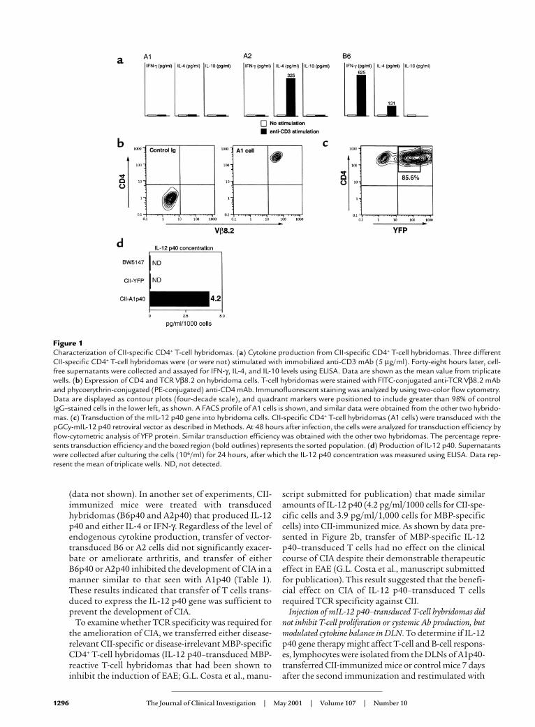
ResultsCharacterization of T-cell hybridomas. We generated severalCII-specific CD4+ T-cell hybridomas from CII-specificTCR Tg mice to use in attempts to deliver immunoreg-ulatory molecules locally. This Tg TCR recognized CII inan MHC-restricted, antigen-specific manner (ref. 17 andour unpublished data). We used three different lines inthis study (termed A1, A2, and B6) that displayed differ-ent profiles of endogenous cytokine expression (Figure1a). However, each of these T-cell lines expressed highlevels of CD4 and Tg TCR Vβ8.2 chain as determined byFACS analysis (Figure 1b). To generate regulatory pro-tein expressing CD4+ T cells, CII-specific CD4+ T-cellhybridomas from all three lines were transduced withthe pGCy-mIL-12 p40 retroviral vector. Greater than80% of the CII-specific CD4+ T-cell hybridomas weretransduced and expressed the mIL-12 p40 gene (Figure1c). We have demonstrated previously a direct correla-tion between expression of the fluorescent reporter geneand the gene upstream of the IRES sequence in these vec-tors (19), which allows control of the production of IL-12 p40 by the expression level of the reporter, YFP. YFP-positive cells were sorted and termed A1p40, A2p40, andB6p40, corresponding to the A1, A2, and B6 parentallines. Significant levels of IL-12 p40 were detected in theYFP-positive but not in the pGCy vector–transducedcells (CII-YFP) or the parental cells (Figure 1d). BioactiveIL-12 was not detected in any cells, and production of IL-12 p40 was confirmed using Western blot analysis (datanot shown). No significant differences in the growthrates in vitro nor alteration of cytokine profiles wasobserved following transduction (data not shown).
Transfer of mIL-12 p40–transduced T-cell hybridomas pre-vents the development of CIA. To examine if local deliveryof an immunoregulatory protein is sufficient to sup-press autoimmune arthritis, CII-specific T-cellhybridomas, either transduced with a retrovirus encod-ing the murine IL-12 p40 subunit and the marker pro-tein YFP, or only the marker protein, were transferredinto CII-immunized mice. In the first set of experi-ments, groups of mice received injections intraperi-toneally with cells from one of the CII-specific T-cellhybridomas (A1) that either expressed YFP or YFP andIL-12 p40. These cells were administered at day 20 afterprimary immunization, 1 day before the boosterimmunization. As shown by data presented in Figure2a, the mice given control vector-transduced cells (CII-YFP) developed severe arthritis, as did the untreatedmice. In contrast, the transfer of IL-12 p40–producingcells (A1p40) efficiently inhibited the development ofCIA (Figure 2a and Table 1). Injection either intraperi-toneally or intravenously with CII-specific IL-12p40–producing T cells inhibited CIA development
The Journal of Clinical Investigation | May 2001 | Volume 107 | Number 10 1295
(data not shown). In another set of experiments, CII-immunized mice were treated with transducedhybridomas (B6p40 and A2p40) that produced IL-12p40 and either IL-4 or IFN-γ. Regardless of the level ofendogenous cytokine production, transfer of vector-transduced B6 or A2 cells did not significantly exacer-bate or ameliorate arthritis, and transfer of eitherB6p40 or A2p40 inhibited the development of CIA in amanner similar to that seen with A1p40 (Table 1).These results indicated that transfer of T cells trans-duced to express the IL-12 p40 gene was sufficient toprevent the development of CIA.
To examine whether TCR specificity was required forthe amelioration of CIA, we transferred either disease-relevant CII-specific or disease-irrelevant MBP-specificCD4+ T-cell hybridomas (IL-12 p40–transduced MBP-reactive T-cell hybridomas that had been shown toinhibit the induction of EAE; G.L. Costa et al., manu-
script submitted for publication) that made similaramounts of IL-12 p40 (4.2 pg/ml/1000 cells for CII-spe-cific cells and 3.9 pg/ml/1,000 cells for MBP-specificcells) into CII-immunized mice. As shown by data pre-sented in Figure 2b, transfer of MBP-specific IL-12p40–transduced T cells had no effect on the clinicalcourse of CIA despite their demonstrable therapeuticeffect in EAE (G.L. Costa et al., manuscript submittedfor publication). This result suggested that the benefi-cial effect on CIA of IL-12 p40–transduced T cellsrequired TCR specificity against CII.
Injection of mIL-12 p40–transduced T-cell hybridomas didnot inhibit T-cell proliferation or systemic Ab production, butmodulated cytokine balance in DLN. To determine if IL-12p40 gene therapy might affect T-cell and B-cell respons-es, lymphocytes were isolated from the DLNs of A1p40-transferred CII-immunized mice or control mice 7 daysafter the second immunization and restimulated with
1296 The Journal of Clinical Investigation | May 2001 | Volume 107 | Number 10
Figure 1Characterization of CII-specific CD4+ T-cell hybridomas. (a) Cytokine production from CII-specific CD4+ T-cell hybridomas. Three differentCII-specific CD4+ T-cell hybridomas were (or were not) stimulated with immobilized anti-CD3 mAb (5 µg/ml). Forty-eight hours later, cell-free supernatants were collected and assayed for IFN-γ, IL-4, and IL-10 levels using ELISA. Data are shown as the mean value from triplicatewells. (b) Expression of CD4 and TCR Vβ8.2 on hybridoma cells. T-cell hybridomas were stained with FITC-conjugated anti-TCR Vβ8.2 mAband phycoerythrin-conjugated (PE-conjugated) anti-CD4 mAb. Immunofluorescent staining was analyzed by using two-color flow cytometry.Data are displayed as contour plots (four-decade scale), and quadrant markers were positioned to include greater than 98% of controlIgG–stained cells in the lower left, as shown. A FACS profile of A1 cells is shown, and similar data were obtained from the other two hybrido-mas. (c) Transduction of the mIL-12 p40 gene into hybridoma cells. CII-specific CD4+ T-cell hybridomas (A1 cells) were transduced with thepGCy-mIL-12 p40 retroviral vector as described in Methods. At 48 hours after infection, the cells were analyzed for transduction efficiency byflow-cytometric analysis of YFP protein. Similar transduction efficiency was obtained with the other two hybridomas. The percentage repre-sents transduction efficiency and the boxed region (bold outlines) represents the sorted population. (d) Production of IL-12 p40. Supernatantswere collected after culturing the cells (106/ml) for 24 hours, after which the IL-12 p40 concentration was measured using ELISA. Data rep-resent the mean of triplicate wells. ND, not detected.
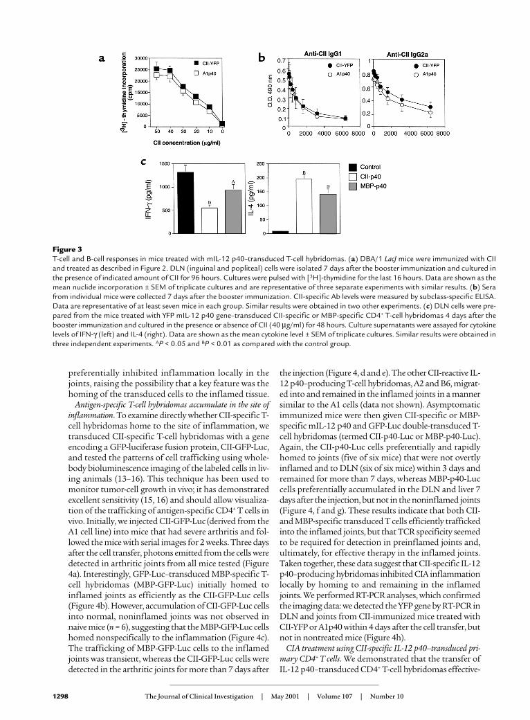
CII in vitro. As shown by data presented in Figure 3a,DLN cells from the mice treated with A1p40 prolifer-ated in response to antigen as well as those from thecontrol mice. Next, we measured CII-specific IgG1 andIgG2a Ab’s in the serum of treated mice 7 days after thesecond immunization. The treatment with IL-12p40–transduced T cells had no significant effect on theserum levels of anti-CII IgG1 or IgG2a Ab’s (Figure 3b).These results suggested that treatment with IL-12p40–transduced T cells did not affect the antigen-spe-cific T-cell activation or systemic Ab responses.
Since this treatment might affect the balance ofcytokine production, we examined cytokine produc-tion in the DLN lymphocytes of mice treated with var-ious transduced T cells. DLN cells from the CII-immu-nized mice treated with either A1p40 or MBP-p40 wererestimulated with CII in vitro, and IFN-γ and IL-4 lev-
els in the culture supernatants were measured usingELISA. Interestingly, transfer of MBP-p40 cells, whichhad no disease-ameliorating effects in CIA (Figure 2b),modulated cytokine production as efficiently as didA1p40: CII-specific IFN-γ production was suppressedin mice treated with A1p40 or MBP-p40, while IL-4production was augmented (Figure 3c). Therefore, IL-12 p40 produced by either antigen-specific or antigen-nonspecific T cells suppressed Th1-type immuneresponses and augmented Th2 responses in the DLN.Since the transfer of MBP-p40 cells did not affect theclinical signs of CIA (Figure 2b), these results indicat-ed that the modulation of cytokine production in thedraining lymph node did not play a major role in ame-lioration of CIA by the CII-specific IL-12 p40 gene ther-apy. These results suggested that the transfer of CII-specific IL-12 p40–producing T-cell hybridomas
The Journal of Clinical Investigation | May 2001 | Volume 107 | Number 10 1297
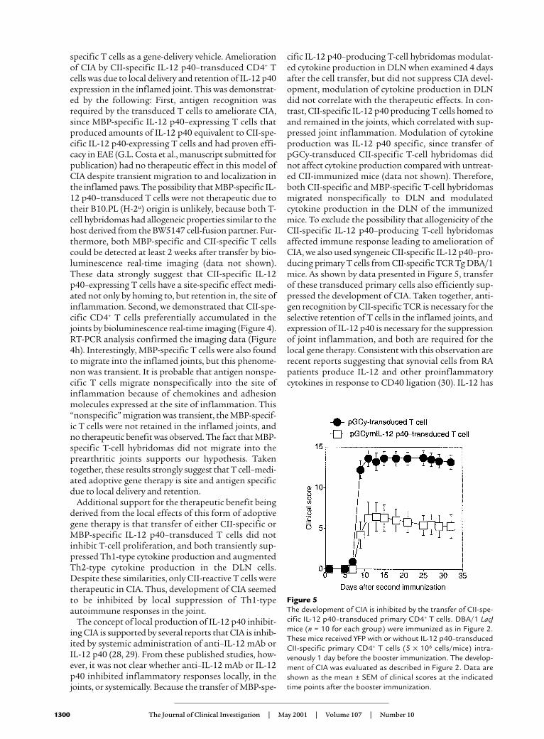
Figure 2Adoptive transfer of mIL-12 p40–transduced CII-specific CD4+ T-cell hybridomas prevents the development of CIA. DBA/1 LacJ mice wereimmunized with bovine CII (200 µg/mouse) in CFA. On day 21, the mice were boosted by intradermal injection with CII in IFA. Starting 1 daybefore the booster injection, the mice were given either pGCy-mIL-12 p40– or pGCy-transduced CII-specific CD4+ T-cell hybridomas (106/mice)intraperitoneally. Untreated CII-immunized DBA/1 LacJ mice were used as controls. The development of CIA was evaluated as described inMethods. Data are shown as the mean ± SEM of clinical scores at the indicated time points after the booster immunization. (b) Ameliorationof CIA by mIL-12 p40 gene therapy requires TCR specificity. DBA/1 LacJ mice (n = 7 for each group) were immunized, and the development ofCIA was evaluated as in a. These mice received either CII-specific or MBP-specific mIL-12 p40–transduced CD4+ T-cell hybridomas (106/mouse)intraperitoneally 1 day before the booster immunization. Similar results were obtained from two other experiments.
Table 1Incidence and severity of collagen-induced arthritis in mice treated with mIL-12 p40–transduced CII-reactive CD4+ T-cell hybridomas
Cell Transduced gene Production of IL-12 p40A Incidence Day of onsetB Maximal (sick mice)(pg/ml/1,000 cells) clinical scoreC
(–) (–) 0 5/5 6.0 ± 1.0 8.8 ± 1.6A1 pGCy 0 10/10 6.3 ± 1.1 9.1 ± 1.2
pGCymIL-12 p40 4.2 5/10 8.2 ± 1.9 2.8 ± 1.6D (6.2 ± 2.9)A2 pGCy 0 12/12 6.7 ± 1.6 8.2 ± 1.0
pGCymIL-12 p40 3.9 6/13 6.7 ± 1.2 3.7 ± 1.7E (7.3 ± 2.9)B6 pGCy 0 7/8 7.6 ± 1.4 7.3 ± 1.2 (8.3 ± 0.7)
pGCmIL-12 p40 4.5 2/8 8.0 ± 9.9 1.9 ± 1.9E (8.0 ± 7.0)
Data shown are the mean maximal clinical scores ± SEM for all mice in each group. Maximal clinical scores ± SEM for the mice with arthritis are indicated inparentheses. ASupernatants were collected after culturing the cells (106/ml) for 24 hours, and the amount of IL-12 p40 was measured using ELISA. Data rep-resent the mean values from triplicate wells. BMice were immunized with bovine CII in CFA. One day before the booster immunization, mice received mIL-12p40–transduced CD4+ T-cell hybridomas as described in Methods. The clinical signs of arthritis were evaluated daily from the day of booster immunization.Data shown are the mean day of onset ± SEM for arthritic mice only. CThe maximal clinical score of arthritis was assessed as described in Methods. DP < 0.01and EP < 0.05 as compared with the group of mice treated with pGCy-transduced control CD4+ T-cell hybridomas.
preferentially inhibited inflammation locally in thejoints, raising the possibility that a key feature was thehoming of the transduced cells to the inflamed tissue.
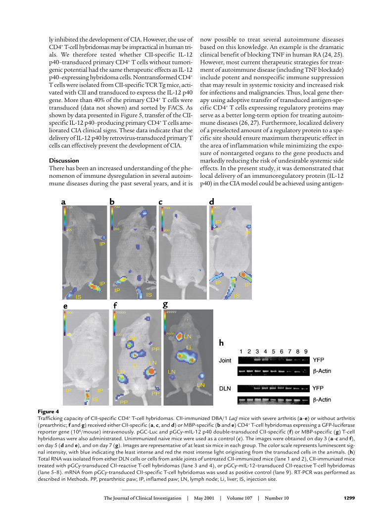
Antigen-specific T-cell hybridomas accumulate in the site ofinflammation. To examine directly whether CII-specific T-cell hybridomas home to the site of inflammation, wetransduced CII-specific T-cell hybridomas with a geneencoding a GFP-luciferase fusion protein, CII-GFP-Luc,and tested the patterns of cell trafficking using whole-body bioluminescence imaging of the labeled cells in liv-ing animals (13–16). This technique has been used tomonitor tumor-cell growth in vivo; it has demonstratedexcellent sensitivity (15, 16) and should allow visualiza-tion of the trafficking of antigen-specific CD4+ T cells invivo. Initially, we injected CII-GFP-Luc(derived from theA1 cell line) into mice that had severe arthritis and fol-lowed the mice with serial images for 2 weeks. Three daysafter the cell transfer, photons emitted from the cells weredetected in arthritic joints from all mice tested (Figure4a). Interestingly, GFP-Luc–transduced MBP-specific T-cell hybridomas (MBP-GFP-Luc) initially homed toinflamed joints as efficiently as the CII-GFP-Luc cells(Figure 4b). However, accumulation of CII-GFP-Luc cellsinto normal, noninflamed joints was not observed innaive mice (n = 6), suggesting that the MBP-GFP-Luc cellshomed nonspecifically to the inflammation (Figure 4c).The trafficking of MBP-GFP-Luc cells to the inflamedjoints was transient, whereas the CII-GFP-Luc cells weredetected in the arthritic joints for more than 7 days after
the injection (Figure 4, d and e). The other CII-reactive IL-12 p40–producing T-cell hybridomas, A2 and B6, migrat-ed into and remained in the inflamed joints in a mannersimilar to the A1 cells (data not shown). Asymptomaticimmunized mice were then given CII-specific or MBP-specific mIL-12 p40 and GFP-Luc double-transduced T-cell hybridomas (termed CII-p40-Luc or MBP-p40-Luc).Again, the CII-p40-Luc cells preferentially and rapidlyhomed to joints (five of six mice) that were not overtlyinflamed and to DLN (six of six mice) within 3 days andremained for more than 7 days, whereas MBP-p40-Luccells preferentially accumulated in the DLN and liver 7days after the injection, but not in the noninflamed joints(Figure 4, f and g). These results indicate that both CII-and MBP-specific transduced T cells efficiently traffickedinto the inflamed joints, but that TCR specificity seemedto be required for detection in preinflamed joints and,ultimately, for effective therapy in the inflamed joints.Taken together, these data suggest that CII-specific IL-12p40–producing hybridomas inhibited CIA inflammationlocally by homing to and remaining in the inflamedjoints. We performed RT-PCR analyses, which confirmedthe imaging data: we detected the YFP gene by RT-PCR inDLN and joints from CII-immunized mice treated withCII-YFP or A1p40 within 4 days after the cell transfer, butnot in nontreated mice (Figure 4h).
CIA treatment using CII-specific IL-12 p40–transduced pri-mary CD4+ T cells. We demonstrated that the transfer ofIL-12 p40–transduced CD4+ T-cell hybridomas effective-
1298 The Journal of Clinical Investigation | May 2001 | Volume 107 | Number 10
Figure 3T-cell and B-cell responses in mice treated with mIL-12 p40–transduced T-cell hybridomas. (a) DBA/1 LacJ mice were immunized with CIIand treated as described in Figure 2. DLN (inguinal and popliteal) cells were isolated 7 days after the booster immunization and cultured inthe presence of indicated amount of CII for 96 hours. Cultures were pulsed with [3H]-thymidine for the last 16 hours. Data are shown as themean nuclide incorporation ± SEM of triplicate cultures and are representative of three separate experiments with similar results. (b) Serafrom individual mice were collected 7 days after the booster immunization. CII-specific Ab levels were measured by subclass-specific ELISA.Data are representative of at least seven mice in each group. Similar results were obtained in two other experiments. (c) DLN cells were pre-pared from the mice treated with YFP mIL-12 p40 gene–transduced CII-specific or MBP-specific CD4+ T-cell hybridomas 4 days after thebooster immunization and cultured in the presence or absence of CII (40 µg/ml) for 48 hours. Culture supernatants were assayed for cytokinelevels of IFN-γ (left) and IL-4 (right). Data are shown as the mean cytokine level ± SEM of triplicate cultures. Similar results were obtained inthree independent experiments. AP < 0.05 and BP < 0.01 as compared with the control group.
ly inhibited the development of CIA. However, the use ofCD4+ T-cell hybridomas may be impractical in human tri-als. We therefore tested whether CII-specific IL-12p40–transduced primary CD4+ T cells without tumori-genic potential had the same therapeutic effects as IL-12p40–expressing hybridoma cells. Nontransformed CD4+
T cells were isolated from CII-specific TCR Tg mice, acti-vated with CII and transduced to express the IL-12 p40gene. More than 40% of the primary CD4+ T cells weretransduced (data not shown) and sorted by FACS. Asshown by data presented in Figure 5, transfer of the CII-specific IL-12 p40–producing primary CD4+ T cells ame-liorated CIA clinical signs. These data indicate that thedelivery of IL-12 p40 by retrovirus-transduced primary Tcells can effectively prevent the development of CIA.
DiscussionThere has been an increased understanding of the phe-nomenon of immune dysregulation in several autoim-mune diseases during the past several years, and it is
now possible to treat several autoimmune diseasesbased on this knowledge. An example is the dramaticclinical benefit of blocking TNF in human RA (24, 25).However, most current therapeutic strategies for treat-ment of autoimmune disease (including TNF blockade)include potent and nonspecific immune suppressionthat may result in systemic toxicity and increased riskfor infections and malignancies. Thus, local gene ther-apy using adoptive transfer of transduced antigen-spe-cific CD4+ T cells expressing regulatory proteins mayserve as a better long-term option for treating autoim-mune diseases (26, 27). Furthermore, localized deliveryof a preselected amount of a regulatory protein to a spe-cific site should ensure maximum therapeutic effect inthe area of inflammation while minimizing the expo-sure of nontargeted organs to the gene products andmarkedly reducing the risk of undesirable systemic sideeffects. In the present study, it was demonstrated thatlocal delivery of an immunoregulatory protein (IL-12p40) in the CIA model could be achieved using antigen-
The Journal of Clinical Investigation | May 2001 | Volume 107 | Number 10 1299
Figure 4Trafficking capacity of CII-specific CD4+ T-cell hybridomas. CII-immunized DBA/1 LacJ mice with severe arthritis (a–e) or without arthritis(prearthritic; f and g) received either CII-specific (a, c, and d) or MBP-specific (b and e) CD4+ T-cell hybridomas expressing a GFP-luciferasereporter gene (106/mouse) intravenously. pGC-Luc and pGCy-mIL-12 p40 double-transduced CII-specific (f) or MBP-specific (g) T-cellhybridomas were also administrated. Unimmunized naive mice were used as a control (c). The images were obtained on day 3 (a–c and f),on day 5 (d and e), and on day 7 (g). Images are representative of at least six mice in each group. The color scale represents luminescent sig-nal intensity, with blue indicating the least intense and red the most intense light originating from the transduced cells in the animals. (h)Total RNA was isolated from either DLN cells or cells from ankle joints of untreated CII-immunized mice (lane 1 and 2), CII-immunized micetreated with pGCy-transduced CII-reactive T-cell hybridomas (lane 3 and 4), or pGCy-mIL-12–transduced CII-reactive T-cell hybridomas(lane 5–8). mRNA from pGCy-transduced CII-specific T-cell hybridomas was used as positive control (lane 9). RT-PCR was performed asdescribed in Methods. PP, prearthritic paw; IP, inflamed paw; LN, lymph node; Li, liver; IS, injection site.
specific T cells as a gene-delivery vehicle. Ameliorationof CIA by CII-specific IL-12 p40–transduced CD4+ Tcells was due to local delivery and retention of IL-12 p40expression in the inflamed joint. This was demonstrat-ed by the following: First, antigen recognition wasrequired by the transduced T cells to ameliorate CIA,since MBP-specific IL-12 p40–expressing T cells thatproduced amounts of IL-12 p40 equivalent to CII-spe-cific IL-12 p40-expressing T cells and had proven effi-cacy in EAE (G.L. Costa et al., manuscript submitted forpublication) had no therapeutic effect in this model ofCIA despite transient migration to and localization inthe inflamed paws. The possibility that MBP-specific IL-12 p40–transduced T cells were not therapeutic due totheir B10.PL (H-2u) origin is unlikely, because both T-cell hybridomas had allogeneic properties similar to thehost derived from the BW5147 cell-fusion partner. Fur-thermore, both MBP-specific and CII-specific T cellscould be detected at least 2 weeks after transfer by bio-luminescence real-time imaging (data not shown).These data strongly suggest that CII-specific IL-12p40–expressing T cells have a site-specific effect medi-ated not only by homing to, but retention in, the site ofinflammation. Second, we demonstrated that CII-spe-cific CD4+ T cells preferentially accumulated in thejoints by bioluminescence real-time imaging (Figure 4).RT-PCR analysis confirmed the imaging data (Figure4h). Interestingly, MBP-specific T cells were also foundto migrate into the inflamed joints, but this phenome-non was transient. It is probable that antigen nonspe-cific T cells migrate nonspecifically into the site ofinflammation because of chemokines and adhesionmolecules expressed at the site of inflammation. This“nonspecific” migration was transient, the MBP-specif-ic T cells were not retained in the inflamed joints, andno therapeutic benefit was observed. The fact that MBP-specific T-cell hybridomas did not migrate into theprearthritic joints supports our hypothesis. Takentogether, these results strongly suggest that T cell–medi-ated adoptive gene therapy is site and antigen specificdue to local delivery and retention.
Additional support for the therapeutic benefit beingderived from the local effects of this form of adoptivegene therapy is that transfer of either CII-specific orMBP-specific IL-12 p40–transduced T cells did notinhibit T-cell proliferation, and both transiently sup-pressed Th1-type cytokine production and augmentedTh2-type cytokine production in the DLN cells.Despite these similarities, only CII-reactive T cells weretherapeutic in CIA. Thus, development of CIA seemedto be inhibited by local suppression of Th1-typeautoimmune responses in the joint.
The concept of local production of IL-12 p40 inhibit-ing CIA is supported by several reports that CIA is inhib-ited by systemic administration of anti–IL-12 mAb orIL-12 p40 (28, 29). From these published studies, how-ever, it was not clear whether anti–IL-12 mAb or IL-12p40 inhibited inflammatory responses locally, in thejoints, or systemically. Because the transfer of MBP-spe-
cific IL-12 p40–producing T-cell hybridomas modulat-ed cytokine production in DLN when examined 4 daysafter the cell transfer, but did not suppress CIA devel-opment, modulation of cytokine production in DLNdid not correlate with the therapeutic effects. In con-trast, CII-specific IL-12 p40 producing T cells homed toand remained in the joints, which correlated with sup-pressed joint inflammation. Modulation of cytokineproduction was IL-12 p40 specific, since transfer ofpGCy-transduced CII-specific T-cell hybridomas didnot affect cytokine production compared with untreat-ed CII-immunized mice (data not shown). Therefore,both CII-specific and MBP-specific T-cell hybridomasmigrated nonspecifically to DLN and modulatedcytokine production in the DLN of the immunizedmice. To exclude the possibility that allogenicity of theCII-specific IL-12 p40–producing T-cell hybridomasaffected immune response leading to amelioration ofCIA, we also used syngeneic CII-specific IL-12 p40–pro-ducing primary T cells from CII-specific TCR Tg DBA/1mice. As shown by data presented in Figure 5, transferof these transduced primary cells also efficiently sup-pressed the development of CIA. Taken together, anti-gen recognition by CII-specific TCR is necessary for theselective retention of T cells in the inflamed joints, andexpression of IL-12 p40 is necessary for the suppressionof joint inflammation, and both are required for thelocal gene therapy. Consistent with this observation arerecent reports suggesting that synovial cells from RApatients produce IL-12 and other proinflammatorycytokines in response to CD40 ligation (30). IL-12 has
1300 The Journal of Clinical Investigation | May 2001 | Volume 107 | Number 10
Figure 5The development of CIA is inhibited by the transfer of CII-spe-cific IL-12 p40–transduced primary CD4+ T cells. DBA/1 LacJmice (n = 10 for each group) were immunized as in Figure 2.These mice received YFP with or without IL-12 p40–transducedCII-specific primary CD4+ T cells (5 × 10⁶ cells/mice) intra-venously 1 day before the booster immunization. The develop-ment of CIA was evaluated as described in Figure 2. Data areshown as the mean ± SEM of clinical scores at the indicatedtime points after the booster immunization.
been shown to induce IFN-γ production and control theproduction of other proinflammatory cytokines (8).Thus, local production of IL-12 p40 by these transducedT cells likely suppressed joint inflammation locally byinhibiting the effect of bioactive IL-12.
In most autoimmune diseases, T cells play a criticalrole in the disease pathogenesis. The ability to transduceCD4+ T cells using retroviruses that carry genes encod-ing therapeutic proteins has lead to the possibility ofusing the homing of antigen-specific cells to deliver theimmunomodulatory cytokines locally (19). We demon-strated evidence for trafficking and retention ofautoantigen-specific CD4+ T cells in sites of autoim-mune inflammation. Previous barriers of retroviraltransduction for the application of gene therapy haveincluded low proviral integration frequency in immunecells, proviral promoter shutdown, and inadequate iso-lation and expansion of transduced cells. In fact, only3–5% transduction efficiency was obtained in previousstudies using retrovirus-mediated TGF-β or IL-10 genetransduction into T cells (31, 32). However, as we havereported, the system used in these studies has resultedin high transduction efficiency that has made retrovi-ral-mediated adoptive T cell–mediated gene therapy eas-ier in murine models of autoimmune disease (19).Adoptive T cell–mediated gene therapy may have greatadvantages over other gene delivery tools for treatingautoimmunity because autoantigen reactive T cells cantarget the inflamed autoantigen-expressing tissues.
In conclusion, we demonstrated directly thatautoantigen-specific CD4+ T cells trafficked intoinflamed autoantigen-expressing tissues. Using retro-viral-transduced CD4+ T cells as a vehicle for deliveryof IL-12 p40, we demonstrated that autoimmunearthritis could be efficiently controlled by CII-reactiveT cells transduced to express IL-12 p40 without sys-temic immune suppression. Therefore, targeting anti-gen-specific T cells by retroviral transduction is apromising strategy for controlling autoimmune dis-ease at the site of inflammation.
AcknowledgmentsWe thank Hirofumi Hamada for providing the mIL-12p40 cDNA and Robyn Kizer for assistance in the prepa-ration of this manuscript. This work was supported byNIH grants AI-39646 and AI-36535, the NIH contractNO1-AR-6-2227 (C.G. Fathman), unrestricted giftsfrom the Mary L. Johnson and Hess Research Funds(C.H. Contag), and an Arthritis Foundation PhysicianScientist Award (C.M. Seroogy). A. Nakajima was part-ly supported by the Japan Rheumatism Foundation.
1. Panayi, G.S. 1977. T cell-dependent pathways in rheumatoid arthritis.Curr. Opin. Rheumatol. 9:236–240.
2. O’Garra, A. 1998. Cytokines induce the development of functionally het-erogeneous T helper cell subsets. Immunity. 8:275–283.
3. Liblau, R.S., Singer, S.M, and McDevitt, H.O. 1995. Th1 and Th2 CD4+
T cells in the pathogenesis of organ-specific autoimmune diseases.Immunol. Today. 16:34–38.
4. Epstein, W.V. 1996. Expectation bias in rheumatoid arthritis clinical tri-als. The anti-CD4 monoclonal antibody experience. Arthritis Rheum.39:1773–1780.
5. Feldmann, M., Brennan, F.M., and Maini, R.N. 1996. Role of cytokinesin rheumatoid arthritis. Annu. Rev. Immunol. 14:397–440.
6. Shaw, M.K., et al. 1997. Local delivery of interleukin 4 by retrovirus-trans-duced T lymphocytes ameliorates experimental autoimmuneencephalomyelitis. J. Exp. Med. 185:1711–1714.
7. Mathisen, P.M., Yu, M., Johnson, J.M., Drazba, J.A., and Touhy, V.K. 1997.Treatment of experimental autoimmune encephalomyelitis with genet-ically modified memory T cells. J. Exp. Med. 186:159–164.
8. Gately, M.K., et al. 1998. The interleukin-12/interleukin-12-receptor sys-tem: role in normal and pathogenic immune responses. Annu. Rev.Immunol. 16:495–521.
9. Mattner, F., et al. 1993. The interleukin-12 subunit p40 specificallyinhibits effects of the interleukin-12 heterodimer. Eur. J. Immunol.23:2202–2208.
10. Kato, K., et al. 1996. Local production of the p40 subunit of interleukin12 suppresses T-helper 1-mediated immune responses and preventsallogeneic myoblast rejection. Proc. Natl. Acad. Sci. USA. 93:9085–9089.
11. Yasuda, H., et al. 1998. Local expression of immunoregulatory IL-12p40gene prolonged syngeneic islet graft survival in diabetic NOD mice. J.Clin. Invest. 102:1807–1814.
12. Trembleau, S., Germann, T., Gately, M.K., and Adorini, L. 1995. The roleof IL-12 in the induction of organ-specific autoimmune diseases.Immunol. Today. 16:383–386.
13. Contag, C.H., et al. 1995. Photonic detection of bacterial pathogens inliving hosts. Mol. Microbiol. 18:593–603.
14. Contag, P.R., Olomu, I.N., Stevenson, D.K., and Contag, C.H. 1999. Bio-luminescent indicator in living mammals. Nat. Med. 4:245–247.
15. Edinger, M., et al. 1999. Noninvasive assessment of tumor cell prolifer-ation in animal models. Neoplasia. 1:303–310.
16. Sweeney, T.J., et al. 1999. Visualizing the kinetics of tumor-cell clearancein living animals. Proc. Natl. Acad. Sci. USA. 96:12044–12049.
17. Osman, G.E., et al. 1998. Expression of a type II collagen-specific TCR trans-gene accelerates the onset of arthritis in mice. Int. Immunol. 10:1613–1622.
18. Lafaille, J.J., Nagashima, K., Katsuki, M., and Tonegawa, S. 1994. Highincidence of spontaneous autoimmune encephalomyelitis in immun-odeficient anti-myelin basic protein T cell receptor transgenic mice. Cell.78:399–408.
19. Costa, G.L., et al. 2000. Targeting rare populations of murine antigen-spe-cific T lymphocytes by retroviral transduction for potential applicationin gene therapy for autoimmune disease. J. Immunol. 164:3581–3590.
20. Cormack, B.P., Valdivia, R.H., and Falkow, S. 1996. FACS-optimizedmutants of the green fluorescent protein (GFP). Gene. 173:33–38.
21. McCafferty, J., Griffiths, A.D., Winter, G., and Chiswell, D.J. 1990. Phageantibodies: filamentous phage displaying antibody variable domains.Nature. 348:552–554.
22. Heim, R., and Tsien, R.Y. 1996. Engineering green fluorescent proteinfor improved brightness, longer wavelengths and fluorescence resonanceenergy transfer. Curr. Biol. 6:178–182.
23. Yoshioka, T., and Nakajima, A., et al. 2000. Contribution of OX40/OX40ligand interaction to the pathogenesis of rheumatoid arthritis. Eur. J.Immunol. 30:2815–2823.
24. Feldmann, M., Eliott, M.J., Woody, J.N., and Maini, R.N. 1997. Anti-tumor necrosis factor-α therapy of rheumatoid arthritis. Adv. Immunol.64:283–350.
25. Moreland, L.W., et al. 1997. Treatment of rheumatoid arthritis with arecombinant human tumor necrosis factor receptor (p75)-Fc fusion pro-tein. N. Engl. J. Med. 337:141–147.
26. Seroogy, C.M., and Fathman, C.G. 2000. The application of gene thera-py in autoimmune diseases. Gene Ther. 7:9–13.
27. Tsokos, G.C., and Nepom, G.T. 2000. Gene therapy in the treatment ofautoimmune diseases. J. Clin. Invest. 106:181–183.
28. Malfait, A.M., et al. 1998. Blockade of IL-12 during the induction of col-lagen-induced arthritis (CIA) markedly attenuates the severity of arthri-tis. Clin. Exp. Immunol. 111:377–383.
29. Germann, T., Hess, H., Szeliga, J., and Rude, E. 1996. Characterization ofthe adjuvant effect of IL-12 inhibitors in type II collagen-induced arthri-tis. Ann. NY Acad. Sci. 795:227–240.
30. Kitagawa, M., et al. 1999. Differential regulation of rheumatoid synovialcell interleukin-12 production by tumor necrosis factor alpha and CD40signals. Arthritis Rheum. 42:1917–1926.
31. Chen, L.Z., et al. 1998. Gene therapy in allergic encephalomyelitis usingmyelin basic protein-specific T cells engineered to express latent trans-forming growth factor-β1. Proc. Natl. Acad. Sci. USA. 95:12516–12521.
32. Setoguchi, K., et al. 2000. Antigen-specific T cells transduced with IL-10ameliorate experimentally induced arthritis without impairing the sys-temic immune response to the antigen. J. Immunol. 165:5980–5986.
The Journal of Clinical Investigation | May 2001 | Volume 107 | Number 10 1301


![A Survey of the 158 m [CII] Line at From Galaxies at z ~ 1 ...hosting.astro.cornell.edu/~spifiweb/zeus/publications/ZEUS_CII... · Accepted for publication in the Astrophysical Journal](https://static.fdocument.org/doc/165x107/5c69c0ac09d3f27a7e8b9aab/a-survey-of-the-158-m-cii-line-at-from-galaxies-at-z-1-spifiwebzeuspublicationszeuscii.jpg)

![1-2018 The [CII] 158 m Line Emission in High-Redshift Galaxies](https://static.fdocument.org/doc/165x107/622b52425b5d6f7f525b431f/1-2018-the-cii-158-m-line-emission-in-high-redshift-galaxies.jpg)