Anesthetic Management of a Child with β-Thalassemia Major ...
4
THIEME 217 Case Report Anesthetic Management of a Child with β-Thalassemia Major and Cortical Venous Thrombosis Mayank Tyagi 1 Surya K. Dube 1, Vanitha Rajagopalan 1, Gyaninder P. Singh 1, 1 Department of Neuroanaesthesiology and Critical Care, All India Institute of Medical Sciences, New Delhi, India published online November 19, 2020 Address for correspondence Surya K. Dube, MD, DM, Department of Neuroanaesthesiology and Critical Care, 7th Floor, Neurosciences Center, All India Institute of Medical Sciences, New Delhi 110029, India (e-mail: [email protected]). β-thalassemia are a group of inherited blood disorders with reduced hemoglobin lev- els. β-thalassemia major is the severe form of disease, and the patients often display an array of associated organ dysfunction which thus increase the risk associated with surgery and anesthesia. Patients with β-thalassemia major can have multiple patho- logical defects that may lead to thromboembolic events. Here, we report such a case who was complicated by occurrence of cerebral sinus thrombosis and presented for decompressive hemicraniectomy under general anesthesia. The anesthetic challenges during in such scenario have been discussed. Abstract Keywords ► beta-thalassemia major ► cortical venous thrombosis ► decompressive hemicraniectomy DOI https://doi.org/ 10.1055/s-0040-1715357 ISSN 2348-0548. © 2020. Indian Society of Neuroanaesthesiology and Critical Care. This is an open access article published by Thieme under the terms of the Creative Commons Attribution-NonDerivative-NonCommercial-License, permitting copying and reproduction so long as the original work is given appropriate credit. Contents may not be used for commercial purposes, or adapted, remixed, transformed or built upon. (https://creativecommons.org/licenses/by-nc-nd/4.0/) Thieme Medical and Scientific Publishers Pvt. Ltd., A-12, 2nd Floor, Sector 2, Noida-201301 UP, India Introduction Patients with β-thalassemia major have a chronic hyperco- agulable state with increased incidence of thromboembolic episodes. There are multiple pathological defects that lead to thromboembolic events, which may be life threatening. Cortical venous thrombosis with raised intracranial pres- sure presenting for emergency decompressive hemicra- niectomy is one such condition. We report here a case of β-thalassemia major complicated by cerebral sinus throm- bosis, who presented for decompressive hemicraniectomy under general anesthesia. We have discussed the anes- thetic concerns in management of this patient. In addition to highlighting the anesthetic challenges this report also emphasizes the importance of increased vigilance in the clinical management of this vulnerable group of patients. Case Report A 13-year-old (weight: 45 kg and height: 150 cm) female child presented to us with multiple episodes of seizures with altered consciousness (Glasgow Coma Scale [GCS] E 1 V 2 M 3 ). The seizure lasted more than 3 minutes without regaining consciousness in between and was managed initially by intra- venous midazolam 3 mg followed by levetiracetam 10 mg/kg given over a period of 30 minutes. Her heart rate, noninva- sive blood pressure, respiratory rate, and room air oxygen saturation were 121/min, 136/78 mm Hg, 22/min, and 88% respectively. She was pale, mildly icteric, and had hepato- splenomegaly. Ultrasonography revealed portal vein hyper- tension, cholelithiasis, and solitary right kidney. Her partial pressure of oxygen was 64 mm Hg, and the remarkable blood parameters were serum sodium of 125 mEq/L, hemoglobin of 7.9 g/dL, platelet count of 80,000/cm 3 , and prothrombin time (international normalized ratio [INR]) of 1.28. Her electrocar- diogram showed prolonged QT interval, and two-dimensional echocardiography revealed an ejection fraction of 55%, grade 2 diastolic dysfunction, and right ventricular enlargement. Her facial features included frontal bossing, malar prom- inence, and depressed nasal bridge. Airway assessment showed buck teeth and a normal range of neck movement. The patient was a case of β-thalassemia major (BTM) and was receiving two units of washed red blood cell units every third week along with deferoxamine mesylate. Computed J Neuroanaesthesiol Crit Care 2021;8:217–220. Published online: 2020-11-19
Transcript of Anesthetic Management of a Child with β-Thalassemia Major ...
THIEME
217Case Report
Anesthetic Management of a Child with β-Thalassemia Major and Cortical Venous ThrombosisMayank Tyagi1 Surya K. Dube1, Vanitha Rajagopalan1, Gyaninder P. Singh1,
1Department of Neuroanaesthesiology and Critical Care, All India Institute of Medical Sciences, New Delhi, India
published onlineNovember 19, 2020
Address for correspondence Surya K. Dube, MD, DM, Department of Neuroanaesthesiology and Critical Care, 7th Floor, Neurosciences Center, All India Institute of Medical Sciences, New Delhi 110029, India (e-mail: [email protected]).
β-thalassemia are a group of inherited blood disorders with reduced hemoglobin lev-els. β-thalassemia major is the severe form of disease, and the patients often display an array of associated organ dysfunction which thus increase the risk associated with surgery and anesthesia. Patients with β-thalassemia major can have multiple patho-logical defects that may lead to thromboembolic events. Here, we report such a case who was complicated by occurrence of cerebral sinus thrombosis and presented for decompressive hemicraniectomy under general anesthesia. The anesthetic challenges during in such scenario have been discussed.
Abstract
Keywords ► beta-thalassemia major ► cortical venous thrombosis ► decompressive hemicraniectomy
DOI https://doi.org/ 10.1055/s-0040-1715357 ISSN 2348-0548.
© 2020. Indian Society of Neuroanaesthesiology and Critical Care.This is an open access article published by Thieme under the terms of the Creative Commons Attribution-NonDerivative-NonCommercial-License, permitting copying and reproduction so long as the original work is given appropriate credit. Contents may not be used for commercial purposes, or adapted, remixed, transformed or built upon. (https://creativecommons.org/licenses/by-nc-nd/4.0/)Thieme Medical and Scientific Publishers Pvt. Ltd., A-12, 2nd Floor, Sector 2, Noida-201301 UP, India
IntroductionPatients with β-thalassemia major have a chronic hyperco-agulable state with increased incidence of thromboembolic episodes. There are multiple pathological defects that lead to thromboembolic events, which may be life threatening. Cortical venous thrombosis with raised intracranial pres-sure presenting for emergency decompressive hemicra-niectomy is one such condition. We report here a case of β-thalassemia major complicated by cerebral sinus throm-bosis, who presented for decompressive hemicraniectomy under general anesthesia. We have discussed the anes-thetic concerns in management of this patient. In addition to highlighting the anesthetic challenges this report also emphasizes the importance of increased vigilance in the clinical management of this vulnerable group of patients.
Case ReportA 13-year-old (weight: 45 kg and height: 150 cm) female child presented to us with multiple episodes of seizures with altered consciousness (Glasgow Coma Scale [GCS] E1V2M3).
The seizure lasted more than 3 minutes without regaining consciousness in between and was managed initially by intra-venous midazolam 3 mg followed by levetiracetam 10 mg/kg given over a period of 30 minutes. Her heart rate, noninva-sive blood pressure, respiratory rate, and room air oxygen saturation were 121/min, 136/78 mm Hg, 22/min, and 88% respectively. She was pale, mildly icteric, and had hepato-splenomegaly. Ultrasonography revealed portal vein hyper-tension, cholelithiasis, and solitary right kidney. Her partial pressure of oxygen was 64 mm Hg, and the remarkable blood parameters were serum sodium of 125 mEq/L, hemoglobin of 7.9 g/dL, platelet count of 80,000/cm3, and prothrombin time (international normalized ratio [INR]) of 1.28. Her electrocar-diogram showed prolonged QT interval, and two-dimensional echocardiography revealed an ejection fraction of 55%, grade 2 diastolic dysfunction, and right ventricular enlargement. Her facial features included frontal bossing, malar prom-inence, and depressed nasal bridge. Airway assessment showed buck teeth and a normal range of neck movement. The patient was a case of β-thalassemia major (BTM) and was receiving two units of washed red blood cell units every third week along with deferoxamine mesylate. Computed
J Neuroanaesthesiol Crit Care 2021;8:217–220.
Published online: 2020-11-19
218
Journal of Neuroanaesthesiology and Critical Care Vol. 8 No. 3/2021 © 2020. Indian Society of Neuroanaesthesiology and Critical Care.
Beta-Thalassemia with Cortical Venous Thrombosis Tyagi et al.
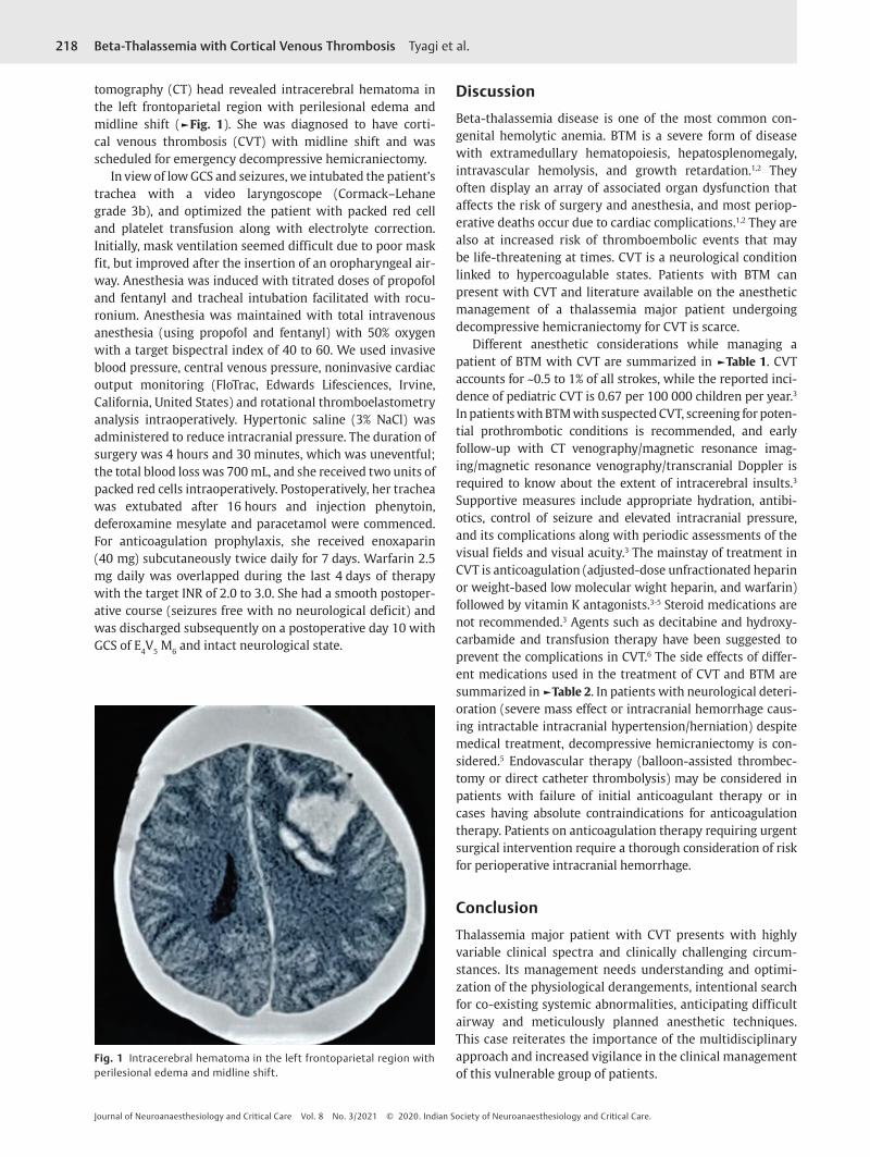
tomography (CT) head revealed intracerebral hematoma in the left frontoparietal region with perilesional edema and midline shift (►Fig. 1). She was diagnosed to have corti-cal venous thrombosis (CVT) with midline shift and was scheduled for emergency decompressive hemicraniectomy.
In view of low GCS and seizures, we intubated the patient’s trachea with a video laryngoscope (Cormack–Lehane grade 3b), and optimized the patient with packed red cell and platelet transfusion along with electrolyte correction. Initially, mask ventilation seemed difficult due to poor mask fit, but improved after the insertion of an oropharyngeal air-way. Anesthesia was induced with titrated doses of propofol and fentanyl and tracheal intubation facilitated with rocu-ronium. Anesthesia was maintained with total intravenous anesthesia (using propofol and fentanyl) with 50% oxygen with a target bispectral index of 40 to 60. We used invasive blood pressure, central venous pressure, noninvasive cardiac output monitoring (FloTrac, Edwards Lifesciences, Irvine, California, United States) and rotational thromboelastometry analysis intraoperatively. Hypertonic saline (3% NaCl) was administered to reduce intracranial pressure. The duration of surgery was 4 hours and 30 minutes, which was uneventful; the total blood loss was 700 mL, and she received two units of packed red cells intraoperatively. Postoperatively, her trachea was extubated after 16 hours and injection phenytoin, deferoxamine mesylate and paracetamol were commenced. For anticoagulation prophylaxis, she received enoxaparin (40 mg) subcutaneously twice daily for 7 days. Warfarin 2.5 mg daily was overlapped during the last 4 days of therapy with the target INR of 2.0 to 3.0. She had a smooth postoper-ative course (seizures free with no neurological deficit) and was discharged subsequently on a postoperative day 10 with GCS of E4V5 M6 and intact neurological state.
DiscussionBeta-thalassemia disease is one of the most common con-genital hemolytic anemia. BTM is a severe form of disease with extramedullary hematopoiesis, hepatosplenomegaly, intravascular hemolysis, and growth retardation.1,2 They often display an array of associated organ dysfunction that affects the risk of surgery and anesthesia, and most periop-erative deaths occur due to cardiac complications.1,2 They are also at increased risk of thromboembolic events that may be life-threatening at times. CVT is a neurological condition linked to hypercoagulable states. Patients with BTM can present with CVT and literature available on the anesthetic management of a thalassemia major patient undergoing decompressive hemicraniectomy for CVT is scarce.
Different anesthetic considerations while managing a patient of BTM with CVT are summarized in ►Table 1. CVT accounts for ~0.5 to 1% of all strokes, while the reported inci-dence of pediatric CVT is 0.67 per 100 000 children per year.3 In patients with BTM with suspected CVT, screening for poten-tial prothrombotic conditions is recommended, and early follow-up with CT venography/magnetic resonance imag-ing/magnetic resonance venography/transcranial Doppler is required to know about the extent of intracerebral insults.3 Supportive measures include appropriate hydration, antibi-otics, control of seizure and elevated intracranial pressure, and its complications along with periodic assessments of the visual fields and visual acuity.3 The mainstay of treatment in CVT is anticoagulation (adjusted-dose unfractionated heparin or weight-based low molecular wight heparin, and warfarin) followed by vitamin K antagonists.3-5 Steroid medications are not recommended.3 Agents such as decitabine and hydroxy-carbamide and transfusion therapy have been suggested to prevent the complications in CVT.6 The side effects of differ-ent medications used in the treatment of CVT and BTM are summarized in ►Table 2. In patients with neurological deteri-oration (severe mass effect or intracranial hemorrhage caus-ing intractable intracranial hypertension/herniation) despite medical treatment, decompressive hemicraniectomy is con-sidered.5 Endovascular therapy (balloon-assisted thrombec-tomy or direct catheter thrombolysis) may be considered in patients with failure of initial anticoagulant therapy or in cases having absolute contraindications for anticoagulation therapy. Patients on anticoagulation therapy requiring urgent surgical intervention require a thorough consideration of risk for perioperative intracranial hemorrhage.
ConclusionThalassemia major patient with CVT presents with highly variable clinical spectra and clinically challenging circum-stances. Its management needs understanding and optimi-zation of the physiological derangements, intentional search for co-existing systemic abnormalities, anticipating difficult airway and meticulously planned anesthetic techniques. This case reiterates the importance of the multidisciplinary approach and increased vigilance in the clinical management of this vulnerable group of patients.
Fig. 1 Intracerebral hematoma in the left frontoparietal region with perilesional edema and midline shift.
219
Journal of Neuroanaesthesiology and Critical Care Vol. 8 No. 3/2021 © 2020. Indian Society of Neuroanaesthesiology and Critical Care.
Beta-Thalassemia with Cortical Venous Thrombosis Tyagi et al.
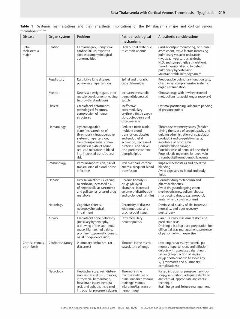
Table 1 Systemic manifestations and their anesthetic implications of the β-thalassemia major and cortical venous thrombosis1-3,5,7-9
Disease Organ system Problem Pathophysiological mechanisms
Anesthetic considerations
Beta-thalassemia major
Cardiac Cardiomegaly, Congestive cardiac failure, hyperten-sion, electrophysiological abnormalities
High output state due to chronic anemia
Cardiac output monitoring, acid-base assessment, avoid factors increasing pulmonary vascular resistance (hypoxia, hypercarbia, acidosis, N2O, and sympathetic stimulation), two-dimensional echo to detect pulmonary hypertensionMaintain stable hemodynamics
Respiratory Restrictive lung disease, pulmonary hypertension
Spinal and thoracic cage deformities
Preoperative pulmonary function test, chest X-ray, comprehensive systemic organs examination
Muscle Decreased weight gain, poor muscle development (leading to growth retardation)
Increased metabolic demand/decreased supply
Choose drugs with low hepatorenal metabolism (to avoid longer recovery)
Skeletal Craniofacial deformities, pathological fractures, compression of neural structures
Ineffective extramedullary erythroid tissue expan-sion, osteopenia and osteomalacia
Optimal positioning, adequate padding of pressure points
Hematology Hypercoagulable state (increased risk of thrombosis), intraoperative systemic hypertension, Hemolysis/anemia, abnor-malities in platelet count, reduced tolerance to bleed-ing, increased transfusional risk
Reduced nitric oxide, multiple blood transfusion, platelet and endothelial activation, decreased protein C and S level, disrupted membrane phospholipids
Thromboelastometry study (for iden-tifying the cause of coagulopathy and guiding administration of coagulation products) and coagulation tests, avoidance of hypoxemiaConsider blood salvageConsider risks of neuraxial anesthesiaProphylactic measures for deep vein thrombosis/thromboembolic events
Immunology Immunosuppression, risk of transmission of blood-borne infections
Iron overload, chronic anemia, frequent blood transfusion
Impaired hemostasis and operative bleedingAvoid exposure to blood and body fluids
Hepatic Liver failure/fibrosis leading to cirrhosis, increased risk of hepatocellular carcinoma and gall stones, altered drug metabolism
Chronic hemolysis, drugs (delayed clearance, increased volume of distribution and prolonged half-life)
Consider drug metabolism and pharmacokineticsAvoid drugs undergoing exten-sive hepatic metabolism (choose short-acting drugs, e.g., propofol, fentanyl, and cis-atracurium)
Neurology Cognitive defects, neuropsychological impairment
Chronicity of disease with emotional and psychosocial issues
Diminished quality of life, increased mortality, and poor recovery postsurgery
Airway Craniofacial bone deformity (maxillary hypertrophy, narrowing of the submental space, high-arched palate, prominent zygomatic bones, nasal bridge depression)
Extramedullary hematopoiesis
Careful airway assessment (bedside predictive tests)Drafting a backup plan, preparation for difficult airway management, presence of personnel with expertise
Cortical venous thrombosis
Cardiorespiratory Pulmonary embolism, car-diac arrest
Thrombi in the micro-vasculature of lungs
Low lung capacity, hypoxemia, pul-monary hypertension, and diffusion defects with associated right heart failure (Keep fraction of inspired oxygen 50% or above to avoid any V/Q mismatch and pulmonary complications)
Neurology Headache, scalp vein disten-sion, and visual disturbances, intracranial hemorrhage, focal brain injury, hemipa-resis and aphasia, increased intracranial pressure, seizures
Thrombi in the microvasculature of brain, impaired venous drainage, venous infarction/ischemia or hemorrhage
Raised intracranial pressure (laryngo-scopy/ intubation/ adequate depth of anesthesia), appropriate anesthetic techniqueBrain bulge and Seizure management
220 Beta-Thalassemia with Cortical Venous Thrombosis Tyagi et al.
3 Saposnik G, Barinagarrementeria F, Brown RD Jr, et al; American Heart Association Stroke Council and the Council on Epidemiology and Prevention. Diagnosis and management of cerebral venous thrombosis: a statement for healthcare pro-fessionals from the American Heart Association/American Stroke Association. Stroke 2011;42(4):1158–1192
4 Yasmin A, O’Keefe YA, Kranz PG, Dombrowski KE, Kolls BJ, James ML. Cerebral venous thrombosis: reviewing pathophysi-ology, diagnosis, and treatment strategies. J Neuroanaesth Crit Care 2019;06:140–144
5 Keller E, Pangalu A, Fandino J, Könü D, Yonekawa Y. Decompressive craniectomy in severe cerebral venous and dural sinus thrombosis. Acta Neurochir Suppl (Wien) 2005;94(94):177–183
6 Kalantri SA, Ray R, Chattopadhyay A, Bhattacharjee S, Biswas A, Bhattacharyya M. Efficacy of decitabine as hemo-globin F inducer in HbE/β-thalassemia. Ann Hematol 2018;97(9):1689–1694
7 Succar J, Musallam KM, Taher AT. Thalassemia and venous thromboembolism. Mediterr J Hematol Infect Dis 2011;3(1):e2011025
8 Bharti N, Devrajan J. Difficult airway management in a case of thalassaemia major. Indian J Anaesth 2008;52(1):87–89
9 Eldor A, Rachmilewitz EA. The hypercoagulable state in thalas-semia. Blood 2002;99(1):36–43
10 Mobarra N, Shanaki M, Ehteram H, et al. A review on iron che-lators in treatment of iron overload syndromes. Int J Hematol Oncol Stem Cell Res 2016;10(4):239–247
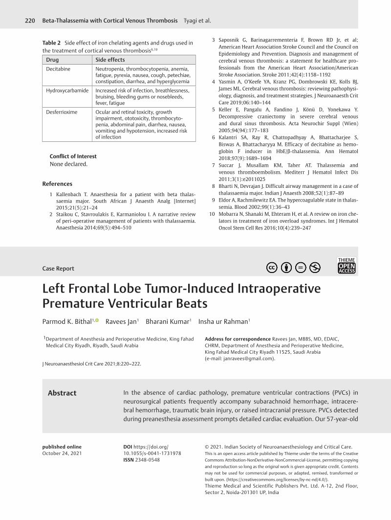
Table 2 Side effect of iron chelating agents and drugs used in the treatment of cortical venous thrombosis6,10
Drug Side effects
Decitabine Neutropenia, thrombocytopenia, anemia, fatigue, pyrexia, nausea, cough, petechiae, constipation, diarrhea, and hyperglycemia
Hydroxycarbamide Increased risk of infection, breathlessness, bruising, bleeding gums or nosebleeds, fever, fatigue
Desferrioxime Ocular and retinal toxicity, growth impairment, ototoxicity, thrombocyto-penia, abdominal pain, diarrhea, nausea, vomiting and hypotension, increased risk of infection
Conflict of InterestNone declared.
References
1 Kallenbach T. Anaesthesia for a patient with beta thalas-saemia major. South African J Anaesth Analg [Internet] 2015;21(5):21–24
2 Staikou C, Stavroulakis E, Karmaniolou I. A narrative review of peri-operative management of patients with thalassaemia. Anaesthesia 2014;69(5):494–510
Left Frontal Lobe Tumor-Induced Intraoperative Premature Ventricular BeatsParmod K. Bithal1, Ravees Jan1 Bharani Kumar1 Insha ur Rahman1
1Department of Anesthesia and Perioperative Medicine, King Fahad Medical City Riyadh, Riyadh, Saudi Arabia
Address for correspondence Ravees Jan, MBBS, MD, EDAIC, CHRM, Department of Anesthesia and Perioperative Medicine, King Fahad Medical City Riyadh 11525, Saudi Arabia (e-mail: [email protected]).
In the absence of cardiac pathology, premature ventricular contractions (PVCs) in neurosurgical patients frequently accompany subarachnoid hemorrhage, intracere-bral hemorrhage, traumatic brain injury, or raised intracranial pressure. PVCs detected during preanesthesia assessment prompts detailed cardiac evaluation. Our 57-year-old
Abstract
J Neuroanaesthesiol Crit Care 2021;8:220–222.
published onlineOctober 24, 2021
DOI https://doi.org/ 10.1055/s-0041-1731978 ISSN 2348-0548
© 2021. Indian Society of Neuroanaesthesiology and Critical Care.This is an open access article published by Thieme under the terms of the Creative Commons Attribution-NonDerivative-NonCommercial-License, permitting copying and reproduction so long as the original work is given appropriate credit. Contents may not be used for commercial purposes, or adapted, remixed, transformed or built upon. (https://creativecommons.org/licenses/by-nc-nd/4.0/).Thieme Medical and Scientific Publishers Pvt. Ltd. A-12, 2nd Floor, Sector 2, Noida-201301 UP, India
THIEME
Case Report