Analysis of β-Galactosidase During Fruit Development and ...
13
ORIGINAL RESEARCH published: 24 April 2018 doi: 10.3389/fpls.2018.00539 Edited by: Antonio Ferrante, Università degli Studi di Milano, Italy Reviewed by: Cristina Sgherri, Università degli Studi di Pisa, Italy Alessandra Francini, Scuola Sant’Anna di Studi Avanzati, Italy *Correspondence: Zhengyang Zhao [email protected] Specialty section: This article was submitted to Crop and Product Physiology, a section of the journal Frontiers in Plant Science Received: 18 November 2017 Accepted: 06 April 2018 Published: 24 April 2018 Citation: Yang H, Liu J, Dang M, Zhang B, Li H, Meng R, Qu D, Yang Y and Zhao Z (2018) Analysis of β-Galactosidase During Fruit Development and Ripening in Two Different Texture Types of Apple Cultivars. Front. Plant Sci. 9:539. doi: 10.3389/fpls.2018.00539 Analysis of β-Galactosidase During Fruit Development and Ripening in Two Different Texture Types of Apple Cultivars Huijuan Yang 1 , Junling Liu 1 , Meile Dang 1 , Bo Zhang 1 , Hongguang Li 1 , Rui Meng 1 , Dong Qu 2 , Yazhou Yang 1 and Zhengyang Zhao 1,3 * 1 State Key Laboratory of Crop Stress Biology in Arid Areas, College of Horticulture, Northwest A&F University, Yangling, China, 2 Shaanxi Province Key Laboratory of Bio-Resources, School of Biological Science and Engineering, Shaanxi University of Technology, Hanzhong, China, 3 Apple Engineering and Technology Research Center of Shaanxi Province, Northwest A&F University, Yangling, China β-galactosidase (β-Gal), one of the cell wall modifying enzymes, plays an important role in fruit ripening and softening. However, its role in apple fruit texture remains unclear. In this study, the role of β-Gal was analyzed in two apple cultivars, ‘Fuji’ and ‘Qinguan,’ which are characterized by different fruit texture types, during fruit development and ripening. The firmness and pectin content of the fruits rapidly decreased and were much lower in ‘Fuji’ than in ‘Qinguan’ from 105 days after full bloom (DAFB). Transmission electron microscopy showed that the pectin-rich middle lamella was substantially degraded from 105 to 180 DAFB in the two apple cultivars. However, the degradation was more severe in ‘Fuji’ than in ‘Qinguan.’ Subcellular localization analysis showed that the Mdβ-Gal1, Mdβ-Gal2, and Mdβ-Gal5 proteins were located in the cell wall. β-Gal activity continuously increased during all fruit developmental stages and was much higher in the mature fruits of ‘Fuji’ than in those of ‘Qinguan,’ indicating that pectin was degraded by β-Gal. Consistent with the enzyme activities, expression levels of β -Gal genes (Mdβ -Gal1, Mdβ -Gal2, and Mdβ -Gal5) showed only slight changes from 60 to 105 DAFB but then dramatically increased until fruit ripening, with higher values in ‘Fuji’ than in ‘Qinguan.’ Furthermore, we found that activities of deletion derivatives in the Mdβ -Gal2 promoter and transcript level of Mdβ -Gal2 were induced by the treatment with methyl jasmonate (MeJA) and ethylene (ETH) hormones. Two ETH and one MeJA hormone-responsive elements were identified by analyzing the promoter sequence. These results suggest that β-Gals, induced by ETH and MeJA, are involved in different fruit texture types of apple cultivars by influencing the degradation of pectin during the mature fruit stage. Keywords: apple, β-galactosidase, texture, fruit ripening, firmness INTRODUCTION Fruit ripening is a complex and coordinated developmental process that involves a range of physiological, biochemical, and genetic events affecting the qualitative traits, such as the color, texture, and taste (Prasanna et al., 2007). Apple fruit texture, described by firmness, crispness, and juiciness, has been extensively studied because of consumer preferences (Peneau et al., 2006). Fruit Frontiers in Plant Science | www.frontiersin.org 1 April 2018 | Volume 9 | Article 539
Transcript of Analysis of β-Galactosidase During Fruit Development and ...
fpls-09-00539 April 20, 2018 Time: 16:11 # 1
ORIGINAL RESEARCHpublished: 24 April 2018
doi: 10.3389/fpls.2018.00539
Edited by:Antonio Ferrante,
Università degli Studi di Milano, Italy
Reviewed by:Cristina Sgherri,
Università degli Studi di Pisa, ItalyAlessandra Francini,
Scuola Sant’Anna di Studi Avanzati,Italy
*Correspondence:Zhengyang Zhao
Specialty section:This article was submitted to
Crop and Product Physiology,a section of the journal
Frontiers in Plant Science
Received: 18 November 2017Accepted: 06 April 2018Published: 24 April 2018
Citation:Yang H, Liu J, Dang M, Zhang B,Li H, Meng R, Qu D, Yang Y and
Zhao Z (2018) Analysisof β-Galactosidase During Fruit
Development and Ripening in TwoDifferent Texture Types of Apple
Cultivars. Front. Plant Sci. 9:539.doi: 10.3389/fpls.2018.00539
Analysis of β-Galactosidase DuringFruit Development and Ripening inTwo Different Texture Types of AppleCultivarsHuijuan Yang1, Junling Liu1, Meile Dang1, Bo Zhang1, Hongguang Li1, Rui Meng1,Dong Qu2, Yazhou Yang1 and Zhengyang Zhao1,3*
1 State Key Laboratory of Crop Stress Biology in Arid Areas, College of Horticulture, Northwest A&F University, Yangling,China, 2 Shaanxi Province Key Laboratory of Bio-Resources, School of Biological Science and Engineering, ShaanxiUniversity of Technology, Hanzhong, China, 3 Apple Engineering and Technology Research Center of Shaanxi Province,Northwest A&F University, Yangling, China
β-galactosidase (β-Gal), one of the cell wall modifying enzymes, plays an important rolein fruit ripening and softening. However, its role in apple fruit texture remains unclear. Inthis study, the role of β-Gal was analyzed in two apple cultivars, ‘Fuji’ and ‘Qinguan,’which are characterized by different fruit texture types, during fruit development andripening. The firmness and pectin content of the fruits rapidly decreased and were muchlower in ‘Fuji’ than in ‘Qinguan’ from 105 days after full bloom (DAFB). Transmissionelectron microscopy showed that the pectin-rich middle lamella was substantiallydegraded from 105 to 180 DAFB in the two apple cultivars. However, the degradationwas more severe in ‘Fuji’ than in ‘Qinguan.’ Subcellular localization analysis showedthat the Mdβ-Gal1, Mdβ-Gal2, and Mdβ-Gal5 proteins were located in the cell wall.β-Gal activity continuously increased during all fruit developmental stages and was muchhigher in the mature fruits of ‘Fuji’ than in those of ‘Qinguan,’ indicating that pectin wasdegraded by β-Gal. Consistent with the enzyme activities, expression levels of β-Galgenes (Mdβ-Gal1, Mdβ-Gal2, and Mdβ-Gal5) showed only slight changes from 60 to105 DAFB but then dramatically increased until fruit ripening, with higher values in ‘Fuji’than in ‘Qinguan.’ Furthermore, we found that activities of deletion derivatives in theMdβ-Gal2 promoter and transcript level of Mdβ-Gal2 were induced by the treatmentwith methyl jasmonate (MeJA) and ethylene (ETH) hormones. Two ETH and one MeJAhormone-responsive elements were identified by analyzing the promoter sequence.These results suggest that β-Gals, induced by ETH and MeJA, are involved in differentfruit texture types of apple cultivars by influencing the degradation of pectin during themature fruit stage.
Keywords: apple, β-galactosidase, texture, fruit ripening, firmness
INTRODUCTION
Fruit ripening is a complex and coordinated developmental process that involves a range ofphysiological, biochemical, and genetic events affecting the qualitative traits, such as the color,texture, and taste (Prasanna et al., 2007). Apple fruit texture, described by firmness, crispness, andjuiciness, has been extensively studied because of consumer preferences (Peneau et al., 2006). Fruit
Frontiers in Plant Science | www.frontiersin.org 1 April 2018 | Volume 9 | Article 539
fpls-09-00539 April 20, 2018 Time: 16:11 # 2
Yang et al. β-Galactosidase in Apple Fruit Texture
texture is mainly affected by disassembly of the cell wallstructure and depolymerization of cell wall components(Brummell and Harpster, 2001), which involve the coordinatedand interdependent actions of cell wall hydrolytic enzymes,including polygalacturonase (PG), pectin methylesterase(PME), pectate lyase (PL), endo-1,4-β-D-glucanase (EGase),xyloglucan-endotransglycosylase (XET), β-galactosidase (β-Gal),α-L-arabinofuranosidase (α-AF), and expansion (EXP) (Goulaoet al., 2007). Among these enzymes, PG has been extensivelystudied and was believed to be the most potent enzymeregulating fruit softening (Grierson and Tucker, 1983; Li et al.,2013). However, this has not been proven in transgenic tomatoeswith suppressed PG expression (Giovannoni et al., 1989). Theloss of galactan residues from the cell wall seems to play a roleduring apple fruit development and ripening (Ross et al., 1994;Pena and Carpita, 2004; Ng et al., 2015). In our previous study,we have found that the β-Gal activity showed a prominentdifference among apple cultivars with different texture typesduring fruit ripening (Gao et al., 2016).
β-galactan is mainly present on side chains of thepolysaccharide rhamnogalacturonan-I (Schols et al., 1995). Thesechains are entangled with glucan chains of cellulose (Zykwinskaet al., 2007), forming a dense network that contributes to theextensibility, strength, and porosity of the cell wall (Ulvskovet al., 2005; Larsen et al., 2011). β-Gal (EC 3.2.1.23), a glycosidase,contains a consensus sequence of the putative active site, G–G–P–[LIVM]–x–Q–x–E–N–E–[FY], of glycosyl hydrolase family 35(GH35) proteins (Henrissat, 1998). The role of β-Gal is to removeterminal, non-reducing β-D-galactosyl residues of hemicelluloseand pectin from the cell wall (Smith and Gross, 2000). The β-Galenzyme is believed to accelerate fruit softening by increasing theporosity of the cell wall and enhancing the access of other cellwall-degrading enzymes (Pena and Carpita, 2004; Ng et al., 2013,2015).
β-Gals are typically encoded by members of large gene familiesand have been isolated from various plants, including Arabidopsis(Ahn et al., 2007), tomato (Smith and Gross, 2000), strawberry(Trainotti et al., 2001), pear (Tateishi et al., 2001), and papaya(do Prado et al., 2016). In mature tomatoes, the gene thatis most abundantly transcribed is Slβ-Gal4, which belongs toone of the seven β-Gal gene families (Smith and Gross, 2000).The increase in β-Gal activity and the decrease in the cell wallgalactosyl content in antisense Slβ-Gal4 tomato lines suggestedthat this gene may be involved in cell wall modification, thuspreventing fruit softening (Smith et al., 2002). Paniagua et al.(2016) have reached a similar conclusion regarding strawberrieswhen they used antisense-mediated downregulation of Faβ-Gal4.A recent study has identified some selective sweeps underlyingquantitative trait loci/genes of important fruit quality traits,including the fruit texture and flavor, and provided evidencesupporting the contribution of β-Gal to the constant selectionof cultivars with firm fruits in the history of apple domestication(Duan et al., 2017).
Methyl jasmonate (MeJA) is an important plant hormone inbiotic and abiotic stress tolerance as well as in flowering and seedand fruit maturation (Saniewski et al., 1987; Lalel et al., 2003).In addition, application of MeJA has been shown to increase
the activities of cell wall modifying enzymes (Concha et al.,2013; Wei et al., 2017). In apples, MeJA treatment was shown tomarkedly increase the ethylene (ETH) release and to acceleratefruit softening (Li et al., 2017). ETH also plays a major role inripening and softening of climacteric fruits (Bapat et al., 2010).Previous studies have shown that ETH induces the expressionof PG, β-Gal, PL, PME, XET, and EGase genes, resulting in therapid softening of fruits (Nishiyama et al., 2007; Shen et al.,2017). Moreover, a recent study has shown that β-Gal activity ishighly correlated with the ETH production in apples (Gwanpuaet al., 2014), thereby emphasizing the role of hormones inβ-Gal regulation during apple fruit softening. However, most ofprevious studies have focused on fruit softening, and the functionof β-Gal during fruit development and ripening in apple cultivarswith different texture types remains unclear.
Among two late cultivars selected for this study, ‘Fuji,’ amajor apple cultivar, has a soft and crisp texture type, whereas‘Qinguan’ is characterized by firmness and toughness of maturedfruits. We hypothesized that β-Gal, induced by ETH and MeJA,played important roles in the two different texture types ofapple cultivars by degrading pectin. In the present study, wemeasured the contents of cell wall components and observedthe ultrastructure of the cell wall in fruits of these two applecultivars. Additionally, we investigated the expression patternsof Mdβ-Gal family members and attempted to elucidate theroles of these genes in apple fruit development and ripening.We also isolated the Mdβ-Gal2 promoter and then used it toexpress a β-glucuronidase (GUS) reporter gene in transgenictobacco plants under hormonal treatment to study the role ofthe hormones in Mdβ-Gal2 transcription. Our findings may helpelucidate the potential role of β-Gal in apple texture, which is acritical fruit quality in the series of fruit evaluation indicators.
MATERIALS AND METHODS
Plant Materials‘Qinguan’ and ‘Fuji’ apple trees were planted in 2000 atExperimental Station of Northwest A&F University, BaishuiCounty, Shaanxi Province, China. Fruits of ‘Fuji’ and ‘Qinguan’were harvested at 180 days after full bloom (DAFB) determinedby taste, ground color and degree of starch clearance. Uniformsize, appearance and without external damage fruits, youngleaves, function leaves, young stems and full flowers were selectedfrom three trees at the same block. Fruits of ‘Qinguan’ and ‘Fuji’were collected every 15 days from 60 DAFB until harvest. Inaddition, ‘Qinguan’ fruits were sprayed with 0.5 mM MeJA or0.5 g·L−1 ETH at 165 DAFB, and these fruits were collectedat harvest. Untreated fruits were used as control. Fruits wereimmediately transported to the laboratory at Northwest A&FUniversity. Whole fruits were used for firmness tests, and theflesh tissues were cut into small pieces, pooled, immediatelyfrozen in liquid nitrogen, and stored at −80◦C for biochemicaland molecular analyses.
Tobacco plants (Nicotiana tabacum cv. NC89) were culturedin an artificial climate incubator under a 16/8 h photoperiod,with a 25/20◦C (day/night) temperature cycle and 70% relative
Frontiers in Plant Science | www.frontiersin.org 2 April 2018 | Volume 9 | Article 539
fpls-09-00539 April 20, 2018 Time: 16:11 # 3
Yang et al. β-Galactosidase in Apple Fruit Texture
humidity. Six- to eight-week-old, growth consistent plants wereused for Agrobacterium-mediated transient assays.
Firmness MeasureFruit firmness was measured at three equidistant sites near thefruit equatorial axis of 15 peeled fruits using a texture analyzer(FTA GS-15, Germany; test depth, 8 mm) equipped with a 10 mmdiameter flat probe. The maximum force formed during the testwas recorded. Firmness was calculated as the average force.
Cell Wall Material and β-Gal ActivityAnalysisThe cell wall extraction procedure used was a modified methodfrom Melton and Smith (2001). Briefly, cell wall material(CWM) was extracted using Tris-phenol buffer and dimethylsulfoxide (DMSO) from the frozen flesh (3.0 g). Pectin andhemicellulose polysaccharides were sequentially extracted using(1,2-cyclohhexylenedinitrilo)-tetraacetic acid (CDTA, 0.05 M),Na2CO3 (0.05 M), and KOH (1 and 4 M) from the obtainedCWM. The final cell wall residue was mainly cellulose.The carbazole-ethanol method and anthrone-sulfuric acidmethod were applied to determine the contents of pectin andhemicellulose, which were expressed as mg·g−1 fresh weight(FW). Cell wall enzymes were extracted used the methodsdescribed by Brummell et al. (2004). The β-Gal enzyme activitywas expressed as µmol PNP (p-nitrophenol) min−1
·g−1FW byusing p-nitrophenyl-β-D-galactopyranoside as substrate.
Ultrastructural Analysis of Cell WallThe ultrastructural analysis of the apple flesh cell wall bytransmission electron microscopy (TEM) was performed asdescribed previously (Tong et al., 1999) with modification. Smallpieces of apple flesh (10 mm3) were cut, fixed in fixative solution(4% glutaraldehyde) at 4◦C for 6 h, washed in phosphate-buffered saline (PBS, pH 6.8) at least three times, and postfixedin 1% osmium tetroxide for 2 h. After dehydration througha graded ethanol-water series, the samples were infiltrated,embedded, and polymerized in LR White resin (London ResinCompany, Reading, United Kingtom) at 55◦C for 48 h. Ultra-thin sections (approximately 70 nm thick) were cut with anultramicrotome (Ultracut-R, Leica, Germany), and then analyzedwith a transmission electron microscope (JEM 1230, JEOL,Japan).
Bioinformatic AnalysisThe nucleotide sequences of Mdβ-Gal genes were searched andconfirmed from National Center for Biotechnology Information(NCBI), designated as Mdβ-Gal1-13. A phylogenetic tree wasgenerated with MEGA 7.0 using the neighbor-joining methodand 1000 bootstrap replicates with default parameters fordeduced amino acid sequences of 13 apple β-Gal genes and 50β-Gal homologs from other species. The alignment of the aminoacid sequence was performed by DNAMAN, and conservedmotifs within the apple β-Gal proteins were identified using theConserved Domain Database1.1https://www.ncbi.nlm.nih.gov/cdd
RT-qPCRRNA was extracted and purified using a Quick RNA isolationkit (Huayueyang, Beijing, China). Reverse transcription wasperformed to synthesize cDNA from 1 µg total RNA byrandom primers using the PrimeScriptTM RT reagent Kitwith gDNA Eraser (TaKaRa, Kyoto, Japan). RT-qPCR wascarried out with an iQ5 Multicolor Real-Time PCR DetectionSystem (Bio-Rad, Hercules, CA, United States) using SYBR R©
Green Master Mix (TaKaRa, Kyoto, Japan). The RT-qPCRprogram was as follows: 94◦C for 30 s and then 40 cyclesof 95◦C for 5 s, 60◦C for 30 s. The mRNA data wasquantified by the 2−11Ct method (Livak and Schmittgen,2001), using the Actin gene served as an internal control andthe gene expression level in the stem tissue as a nominalvalue of 1. The specific primers used for RT-qPCR weredesigned and synthesized, and are shown in SupplementaryTable S1.
Subcellular LocalizationThe full Mdβ-Gal1/2/16 coding sequences without thestop codons were amplified by PCR (primers are listed inSupplementary Table S1). The amplified fragments were digestedby the appropriate restriction enzymes, which are underlinedin the primers list (Supplementary Table S1), and were thenligated into the pCAMBIA1302-GFP vector, digested by thecorresponding enzymes, resulting in Mdβ-Gal1/2/16-GFPvectors. The three fusion constructs and the control GFP vectorwere bombarded into onion epidermal cells by a Biolistic R©
PDS-1000/He particle delivery system (Bio-Rad, Hercules, CA,United States). All bombarded onion epidermal cells wereincubated on Murashige-Skoog medium for 24 h at 22◦C in thedark and were then plasmolyzed in 30% sucrose for 10 min.GFP fluorescence was observed with a fluorescence microscopy(BX51; OLYMPUS, Japan). All bombard tests were repeated atleast three times.
Promoter Cloning and BioinformaticAnalysisGenomic DNA was extracted from apple leaves using thePlant Genomic DNA Kit (Tiangen, Beijing, China). In orderto obtain the promoter region of the Mdβ-Gal2 gene, theprimers, Mdβ-Gal2p1-F and Mdβ-Gal2p1-R, were used toamplify about 2 kb from ‘Qinguan’ genomic DNA betweenan upstream DNA sequence and a portion of the Mdβ-Gal2gene downstream of the transcriptional start site. Then, thesecond round of PCR was performed using Mdβ-Gal2p2-F and Mdβ-Gal2p2-R as primers (all primers are listed inSupplementary Table S1) and the first PCR product as a template.The final purified product was cloned into the pGEMT R©-T EasyVector (Promega, Madison, WI, United States) and sequenced atAoke (Beijing, China). Sequence analyses were carried out usingDNASTAR software. The putative cis-element of the Mdβ-Gal2promoter was identified using PLANTCARE2 (Lescot et al.,2002).
2http://bioinformatics.psb.ugent.be/webtools/plantcare/html/
Frontiers in Plant Science | www.frontiersin.org 3 April 2018 | Volume 9 | Article 539
fpls-09-00539 April 20, 2018 Time: 16:11 # 4
Yang et al. β-Galactosidase in Apple Fruit Texture
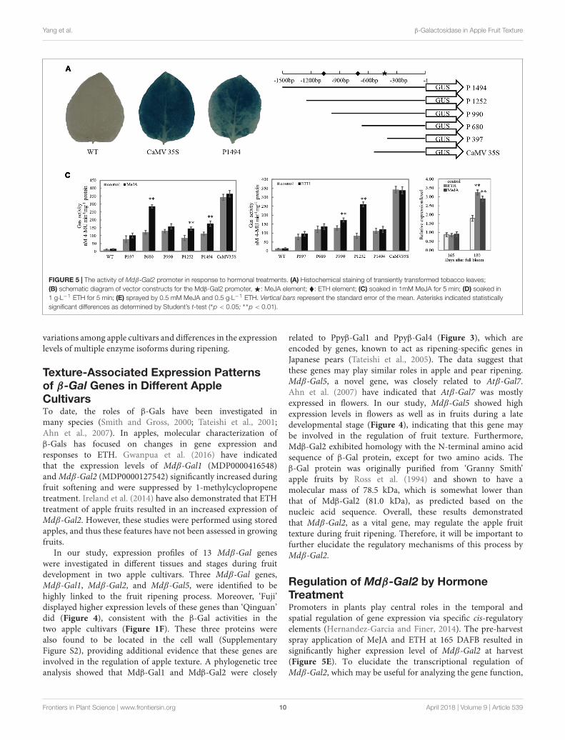
Construction of Promoter-GUS(β-Glucuronidase) Fusion and DeletionVectorsThe full length of Mdβ-Gal2 promoter (1494 bp) and a seriesof deletion fragment of the Mdβ-Gal2 promoter (−1252 bp,−990 bp, −680 bp, and −397 bp from the translational startsite of Mdβ-Gal2) were, respectively, amplified by PCR, addinga PstI restriction site in all forward primers and a BamHIsite in all reverse primers. Further, the Cauliflower mosaicvirus (CaMV) 35S promoter was amplified from pCAMBIA1302by using primers containing the same restriction enzymesites as mentioned above. All amplified regions were clonedupstream of the ATG in the GUS gene of the pCAMBIA1391zbinary vector, following double-digestion with PstI/BamHI andligation. These recombinants, which were positively identifiedby sequencing and were named as P1494, P1252, P990, P680,P397, and CaMV35S, were transformed into Agrobacteriumtumefaciens (strain GV3101) via the freeze-thaw method.A schematic diagram of promoter deletion is illustrated inFigure 5A. The primers that were used are presented inSupplementary Table S1.
Agrobacterium-Based Transient Assayand Hormone TreatmentThe Agrobacterium-based transient assay was performed asdescribed previously (Rothstein et al., 1987) with modification.The Agrobacterium strain, GV3101 (transformed with theappropriate construct) was streaked on LB agar with rifampicin(50 mg·L−1), kanamycin (50 mg·L−1) and gentamicin(50 mg·L−1) and grown at 28◦C for 2 days. Colonies wereselected and grown overnight in LB broth with the abovementioned antibiotics. Agrobacterium cultures were centrifuged,and then resuspended in infiltration solution (27.8 mMglucose, 100 µM acetosyringone, 50 mM 2-(N-morpholino)ethanesulfonic acid). The cultures were diluted until theirOD600 reached 0.5 and were then infiltrated into tobaccoleaves using a vacuum pump. The infiltrated leaves weremaintained on a wet filter in a controlled-environment growthchamber under normal growth conditions. After 24 h, theleaves were, respectively, soaked in MeJA (1 mM) and ETH(1 g·L−1) solutions for 5 min, and then incubation continued for24 h.
GUS Histochemical and FluorometricAssaysFor the histochemical analysis of GUS, the infiltrated tobaccoleaves were immersed in GUS staining solution (100 mMsodium phosphate buffer, pH 7.0, 0.5 mM K3Fe(CN)6,0.5 mM K4Fe(CN)6, 10 mM Na2EDTA, 0.5 mg·mL−1 5-bromo-4-chloro-3-indolyl β-D-glucuronide (X-GluC), 0.1%Triton X-100) for 12 h at 37◦C in darkness and were thendetained in multiple changes of 75% ethanol. Further, GUSactivity was expressed as nmol 4-methylumbelliferone (4-MU, Sigma-Aldrich) generated from the correspondingglucuronide (4-methylumbelliferyl β-D-glucuronide, MUG)per minute per milligram of soluble protein (Jefferson, 1987).
To measure this activity, the 4-MU product was quantifiedby fluorescence intensity with a Hitachi 850 FluorescenceSpectrophotometer (Hitachi, Tokyo, Japan). The proteinconcentration was determined by the method of Bradford(Bradford, 1976).
Statistical AnalysisAll data were obtained from at least three independentexperiments. Values were expressed as mean ± standard error ofthe mean. The data were tested through analysis of variance usingSPSS statistics v19.0 software, and the means were comparedwith student’s t-test. Difference between groups was consideredsignificantly different if p< 0.05.
RESULTS
Physiological Characterization of AppleFruits During Development and RipeningFruit firmness, CWMs, and β-Gal activity were measuredin ‘Fuji’ and ‘Qinguan’ fruits at 15-day intervals from 60DAFB. The flesh firmness rapidly decreased during the fruitletstage (before 90 DAFB) and then slowly decreased duringthe expanding fruit stage (from 90 to 150 DAFB) andmature fruit stage (from 150 to 180 DAFB). The degreeof decline was different between the cultivars, resulting ina difference in the final fruit firmness. Compared with thatat the fruitlet stage, the firmness of ‘Fuji’ at the maturefruit stage decreased by 67%, whereas that of ‘Qinguan’ wasslower to decrease, resulting in a 56% reduction. During thefruitlet and early expanding fruit stages, the fruit firmnesswas higher in ‘Fuji’ than in ‘Qinguan,’ but during the lateexpanding and mature fruit stages, the trend was reversed(Figure 1A).
The contents of CWMs, cellulose, and hemicellulose in thetwo apple cultivars showed similar decreasing trends through alldevelopmental stages. The decrease was rapid from the fruitletstage to the expanding fruit stage, with a slight change during themature fruit stage (Figures 1B–D). The contents were higher in‘Qinguan’ than in ‘Fuji,’ which was consistent with the firmnessobserved during the expanding and mature fruit stages butcontrasted with that in the fruitlet stage.
The pectin content in the two apple cultivars was only slightlydifferent at the fruitlet stage but showed distinct differencesduring the expanding and mature fruit stages (Figure 1E), alongwith corresponding firmness changes in the two apple cultivars.
The activities of β-Gal in the two apple cultivarsshowed almost no changes in the fruitlet stage but weresignificantly different in the expanding and mature fruitstages. Overall, the activity of β-Gal in ‘Qinguan’ remainedstable. However, in ‘Fuji,’ the activity of β-Gal progressivelyincreased, and the rate of the increase was significantlyaccelerated at the mature fruit stage. Furthermore, in maturefruits of ‘Fuji,’ the activity of β-Gal was 7.75-fold higherthan that in ‘Qinguan’ (Figure 1F). In summary, pectindegradation by β-Gal may affect the fruit texture of applecultivars.
Frontiers in Plant Science | www.frontiersin.org 4 April 2018 | Volume 9 | Article 539
fpls-09-00539 April 20, 2018 Time: 16:11 # 5
Yang et al. β-Galactosidase in Apple Fruit Texture
FIGURE 1 | The variation of physiological characters in ‘Fuji’ and ‘Qinguan’ apple during fruit development and ripening. (A) Fruit firmness, (B) the content of cell wallmaterial, (C) cellulose, (D) hemicellulose, (E) pectin and (F) β-galactosidase activity. Data are presented as means from three independent experiments, the verticalbars indicate the standard error of the mean.
Frontiers in Plant Science | www.frontiersin.org 5 April 2018 | Volume 9 | Article 539
fpls-09-00539 April 20, 2018 Time: 16:11 # 6
Yang et al. β-Galactosidase in Apple Fruit Texture
FIGURE 2 | Transmission electron microscope of two apple cultivars during fruit development and ripening. (A,C,E) Fuji; (B,D,F) Qinguan; (A,B) 105 days after fullbloom; (C,D) 180 days after full bloom. CW: cell wall, ML: middle lamella. (A,B): 20000 × , size bars, 500 nm; (C,D): 6000 × , size bars, 2 µm.
Ultrastructural Analysis of Apple Fruits atDifferent StagesPectin, the main component of the middle lamella, affects the cellwall loosening and cell adhesion and shows difference betweenthe two apple cultivars. Ultrastructural changes in cell wallswere determined by TEM during the fruitlet and mature fruitstages in ‘Qinguan’ and ‘Fuji’ (Figure 2). In the fruitlet stage,adjacent regions of cell walls in the two cultivars appeared asbroad light–dark–light bands. The cell wall structure showed anorderly arrangement, with tightly packed fibrous material anda conspicuous middle lamella (Figures 2A,B). Ripening fruitsshowed a severely reduced dark region and damaged cell wallintegrity. The broad light and dark bands nearly disappearedand were replaced by thin lines or cumulate laminates. Theelectron-dense cell wall became electron-lucent, particularly atcell corners. Differences were observed between the two cultivarsin that the region previously occupied by the middle lamella wasmore obscure, particularly at tricellular junctions, and the fibrousmaterial from the cell wall was more degraded and appeared tobe dispersed in ‘Fuji’ (Figures 2C,D). These results indicated thatthe differences in the pectin content were the primary reason forthe different texture types of apple cultivars.
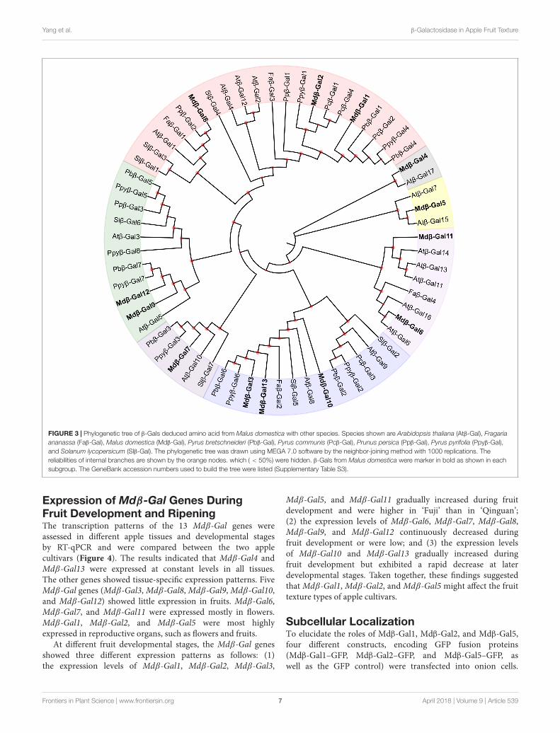
Phylogenetic Analysis of Mdβ-GalProteinsThe β-Gal enzymes are encoded by a family of Mdβ-Galgenes. To determine the evolutionary relationship of the β-Gals
among plant species, a phylogenetic tree was created using full-length β-Gal protein sequences from apple, tomato, and otherselected fruit species (Figure 3). Thirteen Mdβ-Gal proteins wereclustered into seven subgroups (A–G). Subgroup A includedMdβ-Gal1, Mdβ-Gal2, and Mdβ-Gal8, together with Slβ-Gal4,which has previously been confirmed to affect fruit softening,and Faβ-Gal1, which has shown increased expression duringfruit ripening, reaching a maximum in red fruits. Mdβ-Gal4,which lacked the signal peptide, was classified in subgroup B,together with Atβ-Gal17. Mdβ-Gal5, Mdβ-Gal6, and Mdβ-Gal11were classified in subgroup C/D, together with Atβ-Gal8,which was expressed mostly in flowers. Additionally, Mdβ-Gal3,Mdβ-Gal7, Mdβ-Gal10, and Mdβ-Gal13 were classified insubgroup E/F, together with Pcβ-Gal3, which was highlyexpressed in early stages of fruit development and decreasedtoward fruit maturity. Mdβ-Gal9 and Mdβ-Gal12 belonged tosubgroup G.
All the Mdβ-Gal proteins contained a putativeconsensus sequence, G–G–P–[LIVM](2)–x(2)–Q–x–E–N–E–[FY], belonging to GH35 proteins, and all, exceptionMdβ-Gal4, contained a predicted signal peptide. Ten Mdβ-Galproteins also possessed a Gal lectin domain at their C-terminus,which contributes to the substrate specificity of β-Gals.Mdβ-Gal5 carried an additional β-Gal jelly roll domain betweenthe GH35 and Gal_lectin domains, whereas Mdβ-Gal11 andMdβ-Gal12 carried two GH2N domains between the GH35 andGal_lectin domains (Supplementary Figure S1). The functions ofthese extra domains remain unclear.
Frontiers in Plant Science | www.frontiersin.org 6 April 2018 | Volume 9 | Article 539
fpls-09-00539 April 20, 2018 Time: 16:11 # 7
Yang et al. β-Galactosidase in Apple Fruit Texture
FIGURE 3 | Phylogenetic tree of β-Gals deduced amino acid from Malus domestica with other species. Species shown are Arabidopsis thaliana (Atβ-Gal), Fragariaananassa (Faβ-Gal), Malus domestica (Mdβ-Gal), Pyrus bretschneideri (Pbβ-Gal), Pyrus communis (Pcβ-Gal), Prunus persica (Ppβ-Gal), Pyrus pyrifolia (Ppyβ-Gal),and Solanum lycopersicum (Slβ-Gal). The phylogenetic tree was drawn using MEGA 7.0 software by the neighbor-joining method with 1000 replications. Thereliabilities of internal branches are shown by the orange nodes. which ( < 50%) were hidden. β-Gals from Malus domestica were marker in bold as shown in eachsubgroup. The GeneBank accession numbers used to build the tree were listed (Supplementary Table S3).
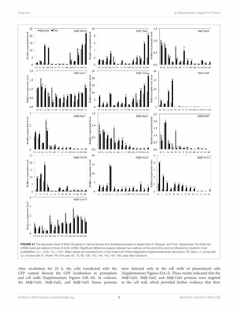
Expression of Mdβ-Gal Genes DuringFruit Development and RipeningThe transcription patterns of the 13 Mdβ-Gal genes wereassessed in different apple tissues and developmental stagesby RT-qPCR and were compared between the two applecultivars (Figure 4). The results indicated that Mdβ-Gal4 andMdβ-Gal13 were expressed at constant levels in all tissues.The other genes showed tissue-specific expression patterns. FiveMdβ-Gal genes (Mdβ-Gal3, Mdβ-Gal8, Mdβ-Gal9, Mdβ-Gal10,and Mdβ-Gal12) showed little expression in fruits. Mdβ-Gal6,Mdβ-Gal7, and Mdβ-Gal11 were expressed mostly in flowers.Mdβ-Gal1, Mdβ-Gal2, and Mdβ-Gal5 were most highlyexpressed in reproductive organs, such as flowers and fruits.
At different fruit developmental stages, the Mdβ-Gal genesshowed three different expression patterns as follows: (1)the expression levels of Mdβ-Gal1, Mdβ-Gal2, Mdβ-Gal3,
Mdβ-Gal5, and Mdβ-Gal11 gradually increased during fruitdevelopment and were higher in ‘Fuji’ than in ‘Qinguan’;(2) the expression levels of Mdβ-Gal6, Mdβ-Gal7, Mdβ-Gal8,Mdβ-Gal9, and Mdβ-Gal12 continuously decreased duringfruit development or were low; and (3) the expression levelsof Mdβ-Gal10 and Mdβ-Gal13 gradually increased duringfruit development but exhibited a rapid decrease at laterdevelopmental stages. Taken together, these findings suggestedthat Mdβ-Gal1, Mdβ-Gal2, and Mdβ-Gal5 might affect the fruittexture types of apple cultivars.
Subcellular LocalizationTo elucidate the roles of Mdβ-Gal1, Mdβ-Gal2, and Mdβ-Gal5,four different constructs, encoding GFP fusion proteins(Mdβ-Gal1–GFP, Mdβ-Gal2–GFP, and Mdβ-Gal5–GFP, aswell as the GFP control) were transfected into onion cells.
Frontiers in Plant Science | www.frontiersin.org 7 April 2018 | Volume 9 | Article 539
fpls-09-00539 April 20, 2018 Time: 16:11 # 8
Yang et al. β-Galactosidase in Apple Fruit Texture
FIGURE 4 | The expression level of Mdβ-Gal genes in various tissues and developing phases on apple fruits of ‘Qinguan’ and ‘Fuji,’ respectively. The Mdβ-GalmRNA levels are relative to those of Actin mRNA. Significant difference analysis between two cultivars at the same time-point is indicated by student’s t-testprobabilities: ∗p < 0.05; ∗∗p < 0.01. Mean values and standard error of the mean from three independent experimental series are shown. ST: stem; L1: young leaf;L2: function leaf; FL: flower; PE: fruit peel; 60, 75, 90, 105, 120, 135, 150, 165, 180: days after full bloom.
After incubation for 24 h, the cells transfected with theGFP control showed the GFP localization in protoplastsand cell walls (Supplementary Figures S2K–M). In contrast,the Mdβ-Gal1, Mdβ-Gal2, and Mdβ-Gal5 fusion proteins
were detected only in the cell walls of plasmolyzed cells(Supplementary Figures S2A–J). These results indicated that theMdβ-Gal1, Mdβ-Gal2, and Mdβ-Gal5 proteins were targetedto the cell wall, which provided further evidence that their
Frontiers in Plant Science | www.frontiersin.org 8 April 2018 | Volume 9 | Article 539
fpls-09-00539 April 20, 2018 Time: 16:11 # 9
Yang et al. β-Galactosidase in Apple Fruit Texture
encoding genes were involved in the regulation of appletexture.
Isolation and Sequence Analysis of theMdβ-Gal2 PromoterTo explore the regulation of Mdβ-Gal2, a 1494 bp 5′ flankingregion, designated pMdβ-Gal2, was isolated from ‘Qinguan’leaves and analyzed for putative cis-regulatory elements. ThepMdβ-Gal2 sequence and putative plant regulatory elementsare shown in Supplementary Figure S3. The cis-regulatoryelements in the promoter were classified into the followingfour functional groups (Supplementary Table S2): abiotic stress-, biotic stress-, light response-, and hormone response-relatedelements. Heat stress-responsive elements are an importanttype of abiotic stress-responsive elements. The biotic stress-responsive elements consisted of anaerobic-responsive elements,which are involved in the regulation of zein metabolism (O2site), and a fungal elicitor-responsive element (Box-W1). Thelight-responsive elements consisted of a G-box and other typicalelements, including an ABRE box, Box I, a GAG motif, andan Sp1 element. The hormone-responsive elements includeda MeJA-responsive element (TGACG motif) and two ETH-responsive elements (EREs). The presence of these putative cis-regulatory elements indicated that Mdβ-Gal2 may be partiallyinvolved in responses to hormones.
Responsiveness of the Mdβ-Gal2 toHormonal StressTo test the activity of the Mdβ-Gal2 promoter, the promoter–GUS reporter construct (P1494) was analyzed in anAgrobacterium-mediated transient expression system. TheCaMV35S–GUS (CaMV35S) construct was used as the positivecontrol. No GUS activity was observed in the wild-type strain.A histochemical assay confirmed that the 1494 bp Mdβ-Gal2promoter was able to drive the expression of the GUS reportergene (Figure 5A), although the promoter activity of Mdβ-Gal2was much lower than that of the positive control.
To elucidate whether the differential gene expression patternsof Mdβ-Gal2 in the two apple cultivars were correlatedwith the regulatory elements in its promoter, we prepareda series of pMdβ-Gal2 deletions and fused the clones withthe GUS reporter gene (Figure 5B). Compared with thatof the control, the GUS activity of pMdβ-Gal2–GUS wassubstantially increased by MeJA and ETH treatments, byapproximately 2.02- and 1.62-fold, respectively. SignificantMeJA-inducible promoter activity was detected in tobacco leavesharboring the P1494, P680, and P397 constructs (Figure 5C).Additionally, significant ETH-inducible promoter activity wasdetected in the transformants with the P1252 and P990constructs (Figure 5D). In all of the treatments, wild-typeleaves and those transformed with the positive control constructshowed no obvious inducible GUS activity. In addition, thetranscript level of Mdβ-Gal2 increased by the pre-harvestapplication of MeJA and ETH at harvest (Figure 5E). Theseresults indicated that the Mdβ-Gal2 was induced by hormonalstress.
DISCUSSION
The apple cultivars ‘Fuji’ and ‘Qinguan’ show different fruittexture during development and ripening. The firmness of ‘Fuji’apples decreased rapidly from an initial value of 16.32 N in thefruitlet stage to 6.86 N in the mature stage, whereas that of‘Qinguan’ showed a slower decrease during fruit developmentand ripening. β-Gal, a pectin enzyme, plays an important rolein fruit ripening and softening (Ng et al., 2015; Dheilly et al.,2016). In our study, we explored the regulatory mechanisms offruit texture by β-Gal in two different apple cultivars.
Cell Wall Composition and Ultrastructureand β-Gal Enzyme in Different AppleCultivarsTexture changes in fruits are a consequence of the degradationof cell wall polysaccharides, including cellulose, hemicellulose,and pectin (Forster et al., 2002). Cybulska et al. (2013) and Chenet al. (2009) have reported that thicker cellulose and hemicelluloselayers were associated with desirable texture properties, whereasZhang et al. (2012) have demonstrated that long and linearsingle pectin chains were detached during peach storage. In ourstudy, we found that the contents of cellulose and hemicellulosewere higher in ‘Qinguan’ than in ‘Fuji’ (Figures 1C,D), whichwas consistent with the observed firmness in the expandingand mature fruit stages but not with that in the fruitlet stage.However, the pectin content was markedly different between thetwo cultivars, consistent with the apple firmness at all stages(Figures 1A,E). Thus, we concluded that the pectin content isthe main contributor to the differences in fruit texture betweenthe two apple cultivars. This hypothesis was confirmed by TEMobservations during the fruitlet and mature stages (Figure 2). Inthe fruitlet stage, the middle lamella was clear, and the cell wallnetwork was dense. In contrast, in the mature fruit stage, themiddle lamella was almost invisible, and the cell wall structurebecame loose. Moreover, the region of the middle lamella wasmore dispersed in ‘Fuji’.
Pectin, the main component of the middle lamella, is degradedby a series of enzymes, including PG, PL, PME, β-Gal, and α-AF(Jarvis et al., 2003; Ng et al., 2013). β-Gal and α-AF affect thestorability of apples more than PG and PME do (Wei et al.,2010). Pena and Carpita (2004) have suggested that the lossof galactan occurs during the growth and maturation phase,whereas the loss of highly branched arabinans occurs in storage.In our study, β-Gal levels were the lowest in the fruitlet stage andthen increased until the mature fruit stage. The activity in ‘Fuji’was higher than that in ‘Qinguan’ at all developmental stages,and the difference became increasingly significant from the fruitexpanding stage (Figure 1F). These changes were consistent withthe results of Dheilly et al. (2016) and Gwanpua et al. (2016),implying that alterations in the β-Gal activity lead to differencesin pectin solubilization and the cell wall structure, and result indifferent texture types. Other investigators have shown that β-Galactivity decreases from the fruitlet to harvest or peaks at theexpanding fruit stage and then decreases until harvest (Goulaoet al., 2007; Ng et al., 2015). This discrepancy may be due to
Frontiers in Plant Science | www.frontiersin.org 9 April 2018 | Volume 9 | Article 539
fpls-09-00539 April 20, 2018 Time: 16:11 # 10
Yang et al. β-Galactosidase in Apple Fruit Texture
FIGURE 5 | The activity of Mdβ-Gal2 promoter in response to hormonal treatments. (A) Histochemical staining of transiently transformed tobacco leaves;(B) schematic diagram of vector constructs for the Mdβ-Gal2 promoter,F: MeJA element; �: ETH element; (C) soaked in 1mM MeJA for 5 min; (D) soaked in1 g·L−1 ETH for 5 min; (E) sprayed by 0.5 mM MeJA and 0.5 g·L−1 ETH. Vertical bars represent the standard error of the mean. Asterisks indicated statisticallysignificant differences as determined by Student’s t-test (∗p < 0.05; ∗∗p < 0.01).
variations among apple cultivars and differences in the expressionlevels of multiple enzyme isoforms during ripening.
Texture-Associated Expression Patternsof β-Gal Genes in Different AppleCultivarsTo date, the roles of β-Gals have been investigated inmany species (Smith and Gross, 2000; Tateishi et al., 2001;Ahn et al., 2007). In apples, molecular characterization ofβ-Gals has focused on changes in gene expression andresponses to ETH. Gwanpua et al. (2016) have indicatedthat the expression levels of Mdβ-Gal1 (MDP0000416548)and Mdβ-Gal2 (MDP0000127542) significantly increased duringfruit softening and were suppressed by 1-methylcyclopropenetreatment. Ireland et al. (2014) have also demonstrated that ETHtreatment of apple fruits resulted in an increased expression ofMdβ-Gal2. However, these studies were performed using storedapples, and thus these features have not been assessed in growingfruits.
In our study, expression profiles of 13 Mdβ-Gal geneswere investigated in different tissues and stages during fruitdevelopment in two apple cultivars. Three Mdβ-Gal genes,Mdβ-Gal1, Mdβ-Gal2, and Mdβ-Gal5, were identified to behighly linked to the fruit ripening process. Moreover, ‘Fuji’displayed higher expression levels of these genes than ‘Qinguan’did (Figure 4), consistent with the β-Gal activities in thetwo apple cultivars (Figure 1F). These three proteins werealso found to be located in the cell wall (SupplementaryFigure S2), providing additional evidence that these genes areinvolved in the regulation of apple texture. A phylogenetic treeanalysis showed that Mdβ-Gal1 and Mdβ-Gal2 were closely
related to Ppyβ-Gal1 and Ppyβ-Gal4 (Figure 3), which areencoded by genes, known to act as ripening-specific genes inJapanese pears (Tateishi et al., 2005). The data suggest thatthese genes may play similar roles in apple and pear ripening.Mdβ-Gal5, a novel gene, was closely related to Atβ-Gal7.Ahn et al. (2007) have indicated that Atβ-Gal7 was mostlyexpressed in flowers. In our study, Mdβ-Gal5 showed highexpression levels in flowers as well as in fruits during a latedevelopmental stage (Figure 4), indicating that this gene maybe involved in the regulation of fruit texture. Furthermore,Mdβ-Gal2 exhibited homology with the N-terminal amino acidsequence of β-Gal protein, except for two amino acids. Theβ-Gal protein was originally purified from ‘Granny Smith’apple fruits by Ross et al. (1994) and shown to have amolecular mass of 78.5 kDa, which is somewhat lower thanthat of Mdβ-Gal2 (81.0 kDa), as predicted based on thenucleic acid sequence. Overall, these results demonstratedthat Mdβ-Gal2, as a vital gene, may regulate the apple fruittexture during fruit ripening. Therefore, it will be important tofurther elucidate the regulatory mechanisms of this process byMdβ-Gal2.
Regulation of Mdβ-Gal2 by HormoneTreatmentPromoters in plants play central roles in the temporal andspatial regulation of gene expression via specific cis-regulatoryelements (Hernandez-Garcia and Finer, 2014). The pre-harvestspray application of MeJA and ETH at 165 DAFB resulted insignificantly higher expression level of Mdβ-Gal2 at harvest(Figure 5E). To elucidate the transcriptional regulation ofMdβ-Gal2, which may be useful for analyzing the gene function,
Frontiers in Plant Science | www.frontiersin.org 10 April 2018 | Volume 9 | Article 539
fpls-09-00539 April 20, 2018 Time: 16:11 # 11
Yang et al. β-Galactosidase in Apple Fruit Texture
the promoter of Mdβ-Gal2 was isolated and functionallycharacterized. Previous studies have demonstrated that MeJAalters the expression levels of the EG1 and XTH1 genesin Fragaria chiloensis via MeJA-responsive elements in theirpromoter regions (Concha et al., 2013; Opazo et al., 2013).Similarly, the banana EXP gene is also regulated by MeJA (Ghoshet al., 2012). A histochemical GUS assay using transgenic tobaccoplants suggested that the pMdβ-Gal2 contained all the cis-acting regulatory elements required for the regulation of β-Gal(Figure 5A). For regulatory elements analysis, we identified aTGACG motif in the Mdβ-Gal2 promoter sequence betweenpositions -680 and -397 (Figure 5B), which corresponds toa cis-acting element involved in MeJA-responsiveness (Finket al., 1988). We found that the activities of derivatives withdeletions in the Mdβ-Gal2 promoter, containing the TGACGmotif, were strongly induced in tobacco leaves treated withMeJA (Figure 5C). Furthermore, we identified two EREs inthe Mdβ-Gal2 promoter region (Figure 5B). ETH, as the mostimportant fruit ripening-related hormone, also regulated theactivity of the Mdβ-Gal2 promoter (Figure 5D). Taken together,our results demonstrated that the transcription of the β-Gal genemay be induced by MeJA and ETH via promoter activity, in whichthe TGACG motif and ERE act as important recognition sites.
CONCLUSION
A broad analysis of β-Gal was performed to reveal enzymeactivity, gene expression patterns, and hormone response duringfruit development and ripening in two apple cultivars. Our resultssuggest that β-Gals, induced by ETH and MeJA, are involved indifferent fruit texture types of apple cultivars by influencing thedegradation of pectin during the mature fruit stage.
AUTHOR CONTRIBUTIONS
HY performed the experiments, analyzed the data, and preparedthe manuscript. JL, MD, BZ, and HL contributed to preparationof the materials, sample collection, and data analysis. RM, DQ,and YY contributed to analysis and discussion of the results andpreparation of the manuscript. ZZ designed the experiments,discussed the data, and drafted the manuscript. All authorsreviewed and approved the final manuscript.
FUNDING
This work was financially supported by the fund through TheNational Natural Science Foundation of China (No. 31471845),Modern Agro-industry Technology Research System of China(CARS-27), and Chinese Universities Scientific (2014YB086).
ACKNOWLEDGMENTS
We thank Prof. Yuhong Li and Prof. Haijun Gong (NorthwestA&F University, China) for providing Agrobacterium strainGV3101 and pCAMBIA1391z-GUS vector. We also thank Prof.Qiaochun Wang and Dr. Yu Wang (Northwest A&F University,China) for their critically reading the manuscript.
SUPLEMENTARY MATERIAL
The Supplementary Material for this article can be found onlineat: https://www.frontiersin.org/articles/10.3389/fpls.2018.00539/full#supplementary-material
REFERENCESAhn, Y. O., Zheng, M., Bevan, D. R., Esen, A., Shiu, S. H., Benson, J., et al.
(2007). Functional genomic analysis of Arabidopsis thaliana glycoside hydrolasefamily 35. Phytochemistry 68, 1510–1520. doi: 10.1016/j.phytochem.2007.03.021
Bapat, V. A., Trivedi, P. K., Ghosh, A., Sane, V. A., Ganapathi, T. R., and Nath, P.(2010). Ripening of fleshy fruit: molecular insight and the role of ethylene.Biotechnol. Adv. 28, 94–107. doi: 10.1016/j.biotechadv.2009.10.002
Bradford, M. M. (1976). A rapid and sensitive method for the quantitation ofmicrogram quantities of protein utilizing the principle of protein-dye binding.Anal. Biochem. 72, 248–254. doi: 10.1016/0003-2697(76)90527-3
Brummell, D. A., Dal Cin, V., Crisosto, C. H., and Labavitch, J. M. (2004). Cell wallmetabolism during maturation, ripening and senescence of peach fruit. J. Exp.Bot. 55, 2029–2039. doi: 10.1093/jxb/erh227
Brummell, D. A., and Harpster, M. H. (2001). Cell wall metabolism in fruitsoftening and quality and its manipulation in transgenic plants. Plant Mol. Biol.47, 311–340. doi: 10.1023/A:1010656104304
Chen, F., Zhang, L., An, H., Yang, H., Sun, X., Liu, H., et al. (2009). Thenanostructure of hemicellulose of crisp and soft Chinese cherry (Prunuspseudocerasus L.) cultivars at different stages of ripeness. LWT Food Sci.Technol. 42, 125–130. doi: 10.1016/j.lwt.2008.03.016
Concha, C. M., Figueroa, N. E., Poblete, L. A., Onate, F. A., Schwab, W., andFigueroa, C. R. (2013). Methyl jasmonate treatment induces changes in fruitripening by modifying the expression of several ripening genes in Fragaria
chiloensis fruit. Plant Physiol. Bioch. 70, 433–444. doi: 10.1016/j.plaphy.2013.06.008
Cybulska, J., Zdunek, A., Psonka-Antonczyk, K. M., and Stokke, B. T. (2013). Therelation of apple texture with cell wall nanostructure studied using an atomicforce microscope. Carbohyd. Polym. 92, 128–137. doi: 10.1016/j.carbpol.2012.08.103
Dheilly, E., Gall, S. L., Guillou, M. C., Renou, J. P., Bonnin, E., Orsel, M., et al.(2016). Cell wall dynamics during apple development and storage involveshemicellulose modifications and related expressed genes. BMC Plant Biol.16:201. doi: 10.1186/s12870-016-0887-0
do Prado, S. B., Melfi, P. R., Castro-Alves, V. C., Broetto, S. G., Araujo, E. S., doNascimento, J. R., et al. (2016). Physiological degradation of pectin in papayacell walls: release of long chains galacturonans derived from insoluble fractionsduring postharvest fruit ripening. Front. Plant Sci. 7:1120. doi: 10.3389/fpls.2016.01120
Duan, N., Bai, Y., Sun, H., Wang, N., Ma, Y., Li, M., et al. (2017). Genomere-sequencing reveals the history of apple and supports a two-stage modelfor fruit enlargement. Nat. Commun. 8:249. doi: 10.1038/s41467-017-00336-7
Fink, J. S., Verhave, M., Kasper, S., Tsukada, T., Mandel, G., and Goodman, R. H.(1988). The CGTCA sequence motif is essential for biological activity of thevasoactive intestinal peptide gene cAMP-regulated enhancer. Proc. Natl. Acad.Sci. U.S.A. 85, 6662–6666. doi: 10.1073/pnas.85.18.6662
Forster, S., Dongowski, G., and Kunzek, H. (2002). Structure, physicochemicalproperties and in vitro fermentation of enzymatically degraded cell
Frontiers in Plant Science | www.frontiersin.org 11 April 2018 | Volume 9 | Article 539

fpls-09-00539 April 20, 2018 Time: 16:11 # 12
Yang et al. β-Galactosidase in Apple Fruit Texture
wall materials from apples. Nahrung 46, 158–166. doi: 10.1002/1521-3803(20020501)46:3<158::AID-FOOD158>3.0.CO;2-D
Gao, Z., Fan, X., Yang, H., Jiang, X., Yang, Y., Zhao, Z., et al. (2016). Correlationamong cell wall components, related enzyme acitivities and texture ofdeveloping fruits of different apple (Malus × domestica) cultivars. Food Sci. 37,70–75. doi: 10.7506/spkx1002-6630-201619012
Ghosh, A., Shekhawat, U. K. S., Ganapathi, T. R., and Bapat, V. A. (2012). Analysisof banana fruit-specific promoters using transient expression in embryogeniccells of banana cultivar Robusta (AAA Group). J. Plant Biochem. Biot. 21,189–197. doi: 10.1007/s13562-011-0070-5
Giovannoni, J. J., DellaPenna, D., Bennett, A. B., and Fischer, R. L. (1989).Expression of a chimeric polygalacturonase gene in transgenic rin (ripeninginhibitor) tomato fruit results in polyuronide degradation but not fruitsoftening. Plant Cell 1, 53–63. doi: 10.1105/tpc.1.1.53
Goulao, L. F., Santos, J., de Sousa, I., and Oliveira, C. A. (2007). Patterns ofenzymatic activity of cell wall-modifying enzymes during growth and ripeningof apples. Postharvest Biol. Tec. 43, 307–318. doi: 10.1016/j.postharvbio.2006.10.002
Grierson, D., and Tucker, G. A. (1983). Timing of ethylene and polygalacturonasesynthesis in relation to the control of tomato fruit ripening. Planta 157,174–179. doi: 10.1007/BF00393652
Gwanpua, S. G., Van Buggenhout, S., Verlinden, B. E., Christiaens, S.,Shpigelman, A., Vicent, V., et al. (2014). Pectin modifications andthe role of pectin-degrading enzymes during postharvest softening ofJonagold apples. Food Chem. 158, 283–291. doi: 10.1016/j.foodchem.2014.02.138
Gwanpua, S. G., Verlinden, B. E., Hertog, M. L., Nicolai, B. M., Hendrickx, M.,and Geeraerd, A. (2016). Slow softening of Kanzi apples (Malus domestica L.) isassociated with preservation of pectin integrity in middle lamella. Food Chem.211, 883–891. doi: 10.1016/j.foodchem.2016.05.138
Henrissat, B. (1998). Glycosidase families. Biochem. Soc. T. 26, 153–156.doi: 10.1042/bst0260153
Hernandez-Garcia, C. M., and Finer, J. J. (2014). Identification and validationof promoters and cis-acting regulatory elements. Plant Sci. 21, 109–119. doi:10.1016/j.plantsci.2013.12.007
Ireland, H. S., Gunaseelan, K., Muddumage, R., Tacken, E. J., Putterill, J.,Johnston, J. W., et al. (2014). Ethylene regulates apple (Malus domestica) fruitsoftening through a dose time-dependent mechanism and through differentialsensitivities and dependencies of cell wall-modifying genes. Plant Cell Physiol.55, 1005–1016. doi: 10.1093/pcp/pcu034
Jarvis, M. C., Briggs, S. P. H., and Knox, J. P. (2003). Intercellular adhesion and cellseparation in plants. Plant Cell Environ. 26, 977–989. doi: 10.1046/j.1365-3040.2003.01034.x
Jefferson, R. A. (1987). Assaying chimeric genes in plants: the GUS genefusion system. Plant Mol. Biol. Report. 5, 387–405. doi: 10.1007/BF02667740
Lalel, H., Singh, Z., and Tan, S. C. (2003). The role of methyl jasmonate in mangoripening and biosynthesis of aroma volatile compounds. J. Hortic. Sci. Biotech.78, 470–484. doi: 10.1080/14620316.2003.11511652
Larsen, F. H., Byg, I., Damager, I., Diaz, J., Engelsen, S. B., and Ulvskov, P.(2011). Residue specific hydration of primary cell wall potato pectin identifiedby solid-state 13C single-pulse MAS and CP/MAS NMR spectroscopy.Biomacromolecules 12, 1844–1850. doi: 10.1021/bm2001928
Lescot, M., Dehais, P., Thijs, G., Marchal, K., Moreau, Y., Van de Peer, Y., et al.(2002). PlantCARE, a database of plant cis-acting regulatory elements and aportal to tools for in silico analysis of promoter sequences. Nucleic Acids Res.30, 325–327. doi: 10.1093/nar/30.1.325
Li, M., Zhang, Y., Zhang, Z., Ji, X., Zhang, R., and Liu, D. (2013). Hypersensitiveethylene signaling and ZMdPG1 expression lead to fruit softening anddehiscence. PLoS One 8:e58745. doi: 10.1371/journal.pone.0058745
Li, T., Xu, Y., Zhang, L., Ji, Y., Tan, D., Yuan, H., et al. (2017). The jasmonate-activated transcription factor MdMYC2 regulates ETHYLENE RESPONSEFACTOR and ethylene biosynthetic genes to promote ethylene biosynthesisduring apple fruit ripening. Plant Cell 29, 1316–1334. doi: 10.1105/tpc.17.00349
Livak, K. J., and Schmittgen, T. D. (2001). Analysis of relative gene expressiondata using real-time quantitative PCR and the 2−1 1 CT Method. Methods 25,402–408. doi: 10.1006/meth.2001.1262
Melton, L. D., and Smith, B. G. (2001). Isolation of Plant Cell Walls andFractionation of Cell Wall Polysaccharides. Current Protocols in Food AnalyticalChemistry. Hoboken, NJ: John Wiley & Sons, Inc.
Ng, J. K., Schroder, R., Brummell, D. A., Sutherland, P. W., Hallett, I. C., Smith,B. G., et al. (2015). Lower cell wall pectin solubilisation and galactose lossduring early fruit development in apple (Malus domestica) cultivar ‘Scifresh’are associated with slower softening rate. J. Plant Physiol. 176, 129–137. doi:10.1016/j.jplph.2014.12.012
Ng, J. K., Schroder, R., Sutherland, P. W., Hallett, I. C., Hall, M. I., Prakash, R.,et al. (2013). Cell wall structures leading to cultivar differences in softeningrates develop early during apple (Malus domestica) fruit growth. BMC PlantBiol. 13:183. doi: 10.1186/1471-2229-13-183
Nishiyama, K., Guis, M., Rose, J. K., Kubo, Y., Bennett, K. A., Wangjin, L., et al.(2007). Ethylene regulation of fruit softening and cell wall disassembly inCharentais melon. J. Exp. Bot. 58, 1281–1290. doi: 10.1093/jxb/erl283
Opazo, M. C., Lizana, R., Pimentel, P., Herrera, R., and Moya-Leon, M. A.(2013). Changes in the mRNA abundance of FcXTH1 and FcXTH2 promotedby hormonal treatments of Fragaria chiloensis fruit. Postharvest Biol. Tec. 77,28–34. doi: 10.1016/j.postharvbio.2012.11.007
Paniagua, C., Blancoportales, R., Barcelómuñoz, M., Garcíagago, J. A., Waldron,K. W., Quesada, M. A., et al. (2016). Antisense down-regulation of thestrawberry β-galactosidase gene Faβgal4 increases cell wall galactose levelsand reduces fruit softening. J. Exp. Bot. 67, 619–631. doi: 10.1093/jxb/erv462
Pena, M. J., and Carpita, N. C. (2004). Loss of highly branched arabinans anddebranching of rhamnogalacturonan I accompany loss of firm texture and cellseparation during prolonged storage of apple. Plant Physiol. 135, 1305–1313.doi: 10.1104/pp.104.043679
Peneau, S., Hoehn, E., Roth, H. R., Escher, F., and Nuessli, J. (2006). Importanceand consumer perception of freshness of apples. Food Qual. Prefer. 17, 9–19.doi: 10.1016/j.foodqual.2005.05.002
Prasanna, V., Prabha, T. N., and Tharanathan, R. N. (2007). Fruit ripeningphenomena–an overview. Crit. Rev. Food Sci. Nutr. 47, 1–19. doi: 10.1080/10408390600976841
Ross, G. S., Wegrzyn, T., MacRae, E. A., and Redgwell, R. J. (1994). Appleβ-galactosidase. Activity against cell wall polysaccharides and characterizationof a related cDNA clone. Plant Physiol. 106, 521–528. doi: 10.1104/pp.106.2.521
Rothstein, S. J., Lahners, K. N., Lotstein, R. J., Carozzi, N. B., Jayne, S. M., and Rice,D. A. (1987). Promoter cassettes, antibiotic-resistance genes, and vectors forplant transformation. Gene 53, 153–161. doi: 10.1016/0378-1119(87)90003-5
Saniewski, M., Nowacki, J., and Czapski, J. (1987). The effect of methyl jasmonateon ethylene production and ethylene-forming enzyme activity in tomatoes.J. Plant Physiol. 129, 175–180. doi: 10.1016/S0176-1617(87)80114-1
Schols, H. A., Bakx, E. J., Schipper, D., and Voragen, A. G. J. (1995).A xylogalacturonan subunit present in the modified hairy regions of applepectin. Carbohyd. Res. 279, 265–279. doi: 10.1016/0008-6215(95)00287-1
Shen, Y. H., Lu, B. G., Feng, L., Yang, F. Y., Geng, J. J., Ming, R., et al.(2017). Isolation of ripening-related genes from ethylene/1-MCP treated papayathrough RNA-seq. BMC Genomics 18:671. doi: 10.1186/s12864-017-4072-0
Smith, D. L., Abbott, J. A., and Gross, K. C. (2002). Down-regulation of tomatoβ-galactosidase 4 results in decreased fruit softening. Plant Physiol. 129, 1755–1762. doi: 10.1104/pp.011025
Smith, D. L., and Gross, K. C. (2000). A family of at least seven β-galactosidasegenes is expressed during tomato fruit development. Plant Physiol. 123, 1173–1183. doi: 10.1104/pp.123.3.1173
Tateishi, A., Inoue, H., Shiba, H., and Yamaki, S. (2001). Molecular cloning ofβ-galactosidase from Japanese pear (Pyrus pyrifolia) and its gene expressionwith fruit ripening. Plant Cell Physiol. 42, 492–498. doi: 10.1093/pcp/pce059
Tateishi, A., Nagashima, K., Mathooka, F. M., Mwaniki, M. W., Kubo, Y., andInaba, A. (2005). Differential expression of members of the β-Galactosidasegene family during Japanese pear (Pyrus pyrifolia L.) fruit growth and on-treeripening. J. Am. Soc. Hortic. Sci. 130, 819–829.
Tong, C., Krueger, D., Vickers, Z., Bedford, D., Luby, J., El-Shiekh, A., et al. (1999).Comparison of softening-related changes during storage of ‘Honeycrisp’ apple,its parents, and ‘Delicious’. J. Am. Soc. Hortic. Sci. 124, 407.
Trainotti, L., Spinello, R., Piovan, A., Spolaore, S., and Casadoro, G. (2001).β-galactosidases with a lectin-like domain are expressed in strawberry. J. Exp.Bot. 52, 1635–1645. doi: 10.1093/jexbot/52.361.1635
Frontiers in Plant Science | www.frontiersin.org 12 April 2018 | Volume 9 | Article 539
fpls-09-00539 April 20, 2018 Time: 16:11 # 13
Yang et al. β-Galactosidase in Apple Fruit Texture
Ulvskov, P., Wium, H., Bruce, D., Jorgensen, B., Qvist, K. B., Skjot, M., et al.(2005). Biophysical consequences of remodeling the neutral side chains ofrhamnogalacturonan I in tubers of transgenic potatoes. Planta 220, 609–620.doi: 10.1007/s00425-004-1373-8
Wei, J., Ma, F., Shi, S., Qi, X., Zhu, X., and Yuan, J. (2010). Changesand postharvest regulation of activity and gene expression ofenzymes related to cell wall degradation in ripening apple fruit.Postharvest Biol. Tec. 56, 147–154. doi: 10.1016/j.postharvbio.2009.12.003
Wei, J., Wen, X., and Tang, L. (2017). Effect of methyl jasmonic acid on peachfruit ripening progress. Sci. Hortic. (Amsterdam) 220, 206–213. doi: 10.1016/j.scienta.2017.03.004
Zhang, L., Chen, F., Yang, H., Ye, X., Sun, X., Liu, D., et al. (2012). Effects oftemperature and cultivar on nanostructural changes of water-soluble pectin andchelate-soluble pectin in peaches. Carbohyd. Polym. 87, 816–821. doi: 10.1016/j.carbpol.2011.08.074
Zykwinska, A., Thibault, J. F., and Ralet, M. C. (2007). Organization of pecticarabinan and galactan side chains in association with cellulose microfibrils inprimary cell walls and related models envisaged. J. Exp. Bot. 58, 1795–1802.doi: 10.1093/jxb/erm037
Conflict of Interest Statement: The authors declare that the research wasconducted in the absence of any commercial or financial relationships that couldbe construed as a potential conflict of interest.
Copyright © 2018 Yang, Liu, Dang, Zhang, Li, Meng, Qu, Yang and Zhao. This is anopen-access article distributed under the terms of the Creative Commons AttributionLicense (CC BY). The use, distribution or reproduction in other forums is permitted,provided the original author(s) and the copyright owner are credited and that theoriginal publication in this journal is cited, in accordance with accepted academicpractice. No use, distribution or reproduction is permitted which does not complywith these terms.
Frontiers in Plant Science | www.frontiersin.org 13 April 2018 | Volume 9 | Article 539