
Analyses of the Interactions among Plasmid- and Host-Encoded Components
12
MOLECULAR AND CELLULAR BIOLOGY, 0270-7306/98/$04.0010 Dec. 1998, p. 7466–7477 Vol. 18, No. 12 Copyright © 1998, American Society for Microbiology. All Rights Reserved. The 2mm Plasmid Stability System: Analyses of the Interactions among Plasmid- and Host-Encoded Components SOUNDARAPANDIAN VELMURUGAN, YONG-TAE AHN, XIAN-MEI YANG, XU-LI WU, AND MAKKUNI JAYARAM* Department of Microbiology and Institute of Cell and Molecular Biology, University of Texas at Austin, Austin, Texas 78712 Received 9 June 1998/Returned for modification 27 July 1998/Accepted 24 August 1998 The stable inheritance of the 2mm plasmid in a growing population of Saccharomyces cerevisiae is dependent on two plasmid-encoded proteins (Rep1p and Rep2p), together with the cis-acting locus REP3 (STB). In this study we demonstrate that short carboxy-terminal deletions of Rep1p and Rep2p severely diminish their nor- mal capacity to localize to the yeast nucleus. The nuclear targeting, as well as their functional role in plasmid partitioning, can be restored by the addition of a nuclear localization sequence to the amino or the carboxy terminus of the shortened Rep proteins. Analyses of deletion derivatives of the Rep proteins by using the in vivo dihybrid genetic test in yeast, as well as by glutathione S-transferase fusion trapping assays in vitro demon- strate that the amino-terminal portion of Rep1p (ca. 150 amino acids long) is responsible for its interactions with Rep2p. In a monohybrid in vivo assay, we have identified Rep1p, Rep2p, and a host-encoded protein, Shf1p, as being capable of interacting with the STB locus. The Shf1 protein expressed in Escherichia coli can bind with high specificity to the STB sequence in vitro. In a yeast strain deleted for the SHF1 locus, a 2mm circle-derived plasmid shows relatively poor stability. The 2mm circle, a relatively small circular plasmid (6,318 bp) present in most common strains of Saccharomyces cerevisiae, has optimized a partitioning system and an amplification sys- tem that allow it to be propagated stably in a cell population at a copy number of approximately 60 to 100 per cell (reviewed in reference 2). Genetic analyses suggest that two plasmid-coded proteins, Rep1p and Rep2p, in conjunction with a cis-acting locus STB (also called REP3) contribute to the stability func- tion (16, 17, 19, 23). One plausible mechanism for plasmid sta- bility is that the interaction of Rep1p and Rep2p with the STB element serves to overcome the normal bias in plasmid segre- gation that tends to favor the mother cell over the daughter cell (22). The evidence for this suspected DNA-protein interaction is quite preliminary and rests almost entirely on the observa- tion that urea-solubilized yeast extracts expressing Rep1p and Rep2p or [cir 0 ] extracts supplemented exogenously with Rep1p and Rep2p can bind STB (14). The need for plasmid amplification arises only if and when there is a decrease in copy number below the steady-state value. Normally, each plasmid molecule is replicated once, and only once, per cell cycle (35), and the daughter molecules are partitioned efficiently at cytokinesis (27). When there is a drop in copy number, the amplification system overrides the cell cycle restriction of a single round of plasmid replication during one S phase. Plasmid amplification is absolutely dependent on the 2mm circle Flp site-specific recombination system (33). A currently favored model for amplification proposes the recom- binational inversion of a bidirectional replication fork and the resultant double-rolling-circle replication mode as the means for obtaining multiple replicas of the plasmid from a single initiation event (9–11, 26). The cessation of amplification would require a second recombination event that can restore the normal direction of fork movement. The time interval between the two successive recombination events would determine the degree of amplification. How is the amplification system kept silent under normal steady-state growth conditions? And how is it rapidly commis- sioned into action at short notice? Biochemical answers to these fundamental question are sparse. On the basis primarily of genetic studies, it has been proposed that a regulatory com- plex containing Rep1p and Rep2p may provide an indirect readout of the copy number and, either at or above a critical concentration, may negatively control amplification by turning down expression of the FLP gene (24, 25, 28, 30). Recently, we have demonstrated self- and cross-interactions between Rep1p and Rep2p by immunoprecipitations of these proteins from mixed extracts of Escherichia coli cells that express them and by baiting assays with hybrid glutathione S-transferase (GST)-Rep proteins (1). Furthermore, these findings were corroborated by in vivo assays in yeast cells. We now present an analysis of the polypeptide regions in Rep1p and Rep2p responsible for their cellular localization as well as for their functional interactions. We also show here that an in vivo assay with the STB locus as a DNA bait fishes out three proteins: the Rep1 and Rep2 pro- teins, as well as a chromosomally encoded protein (the product of the YIL036W locus in the yeast genome bank). The chro- mosomal protein can bind to the STB element in vitro as well. Furthermore, its absence in vivo causes high instability of a 2mm circle-derived test plasmid. The potential implications of these findings in the benign parasitism of the 2mm circle plas- mid are discussed here. MATERIALS AND METHODS Yeast strains. The following [cir 0 ] and [cir 1 ] yeast strains were used in this study: FVY2-6B (MATa leu2-3,112 ura3-52 his3-d1 [cir 0 ]; Gal 1 ), FVY889 (MATa ade2 leu2-3,112 ura3-52 his5-2 [cir 0 ]; Gal 1 ), FVY93154 (MATa leu2- 3,112 ura3-52 trp1-289 ade2 [cir 1 ]; Gal 1 ), CCY666-1A (MATa ade2 his3-D200 leu2-3,112 ura3-52 [cir 1 ]; Gal 1 ), CCY666-7B (MATa ade2 his3-D200 leu2-3,112 ura3-52 [cir 1 ]; Gal 1 ), and MJY101 (CCY666-7B, shf1D::URA3). The cellular localizations of green fluorescent protein (GFP) hybrids were done in strains FVY2 [cir 0 ] and FVY93154 [cir 1 ], respectively. Results were unchanged when FVY889 and CCY666-1A were used as the [cir 0 ] host and the [cir 1 ] host, respectively. In the Results section, only the data from FVY2 [cir 0 ] are pre- * Corresponding author. Mailing address: Department of Microbi- ology, University of Texas at Austin, Austin, TX 78712. Phone: (512) 471-0966. Fax: (512) 471-5546. E-mail: [email protected]. 7466 Downloaded from https://journals.asm.org/journal/mcb on 19 November 2021 by 45.165.8.38.
Transcript of Analyses of the Interactions among Plasmid- and Host-Encoded Components
MOLECULAR AND CELLULAR BIOLOGY,0270-7306/98/$04.0010
Dec. 1998, p. 7466–7477 Vol. 18, No. 12
Copyright © 1998, American Society for Microbiology. All Rights Reserved.
The 2mm Plasmid Stability System: Analyses of the Interactionsamong Plasmid- and Host-Encoded ComponentsSOUNDARAPANDIAN VELMURUGAN, YONG-TAE AHN, XIAN-MEI YANG,
XU-LI WU, AND MAKKUNI JAYARAM*
Department of Microbiology and Institute of Cell and Molecular Biology,University of Texas at Austin, Austin, Texas 78712
Received 9 June 1998/Returned for modification 27 July 1998/Accepted 24 August 1998
The stable inheritance of the 2mm plasmid in a growing population of Saccharomyces cerevisiae is dependenton two plasmid-encoded proteins (Rep1p and Rep2p), together with the cis-acting locus REP3 (STB). In thisstudy we demonstrate that short carboxy-terminal deletions of Rep1p and Rep2p severely diminish their nor-mal capacity to localize to the yeast nucleus. The nuclear targeting, as well as their functional role in plasmidpartitioning, can be restored by the addition of a nuclear localization sequence to the amino or the carboxyterminus of the shortened Rep proteins. Analyses of deletion derivatives of the Rep proteins by using the in vivodihybrid genetic test in yeast, as well as by glutathione S-transferase fusion trapping assays in vitro demon-strate that the amino-terminal portion of Rep1p (ca. 150 amino acids long) is responsible for its interactionswith Rep2p. In a monohybrid in vivo assay, we have identified Rep1p, Rep2p, and a host-encoded protein,Shf1p, as being capable of interacting with the STB locus. The Shf1 protein expressed in Escherichia coli canbind with high specificity to the STB sequence in vitro. In a yeast strain deleted for the SHF1 locus, a 2mmcircle-derived plasmid shows relatively poor stability.
The 2mm circle, a relatively small circular plasmid (6,318 bp)present in most common strains of Saccharomyces cerevisiae,has optimized a partitioning system and an amplification sys-tem that allow it to be propagated stably in a cell population ata copy number of approximately 60 to 100 per cell (reviewed inreference 2). Genetic analyses suggest that two plasmid-codedproteins, Rep1p and Rep2p, in conjunction with a cis-actinglocus STB (also called REP3) contribute to the stability func-tion (16, 17, 19, 23). One plausible mechanism for plasmid sta-bility is that the interaction of Rep1p and Rep2p with the STBelement serves to overcome the normal bias in plasmid segre-gation that tends to favor the mother cell over the daughter cell(22). The evidence for this suspected DNA-protein interactionis quite preliminary and rests almost entirely on the observa-tion that urea-solubilized yeast extracts expressing Rep1p andRep2p or [cir0] extracts supplemented exogenously with Rep1pand Rep2p can bind STB (14).
The need for plasmid amplification arises only if and whenthere is a decrease in copy number below the steady-statevalue. Normally, each plasmid molecule is replicated once, andonly once, per cell cycle (35), and the daughter molecules arepartitioned efficiently at cytokinesis (27). When there is a dropin copy number, the amplification system overrides the cellcycle restriction of a single round of plasmid replication duringone S phase. Plasmid amplification is absolutely dependent onthe 2mm circle Flp site-specific recombination system (33). Acurrently favored model for amplification proposes the recom-binational inversion of a bidirectional replication fork and theresultant double-rolling-circle replication mode as the meansfor obtaining multiple replicas of the plasmid from a singleinitiation event (9–11, 26). The cessation of amplification wouldrequire a second recombination event that can restore thenormal direction of fork movement. The time interval between
the two successive recombination events would determine thedegree of amplification.
How is the amplification system kept silent under normalsteady-state growth conditions? And how is it rapidly commis-sioned into action at short notice? Biochemical answers tothese fundamental question are sparse. On the basis primarilyof genetic studies, it has been proposed that a regulatory com-plex containing Rep1p and Rep2p may provide an indirectreadout of the copy number and, either at or above a criticalconcentration, may negatively control amplification by turningdown expression of the FLP gene (24, 25, 28, 30). Recently, wehave demonstrated self- and cross-interactions between Rep1pand Rep2p by immunoprecipitations of these proteins frommixed extracts of Escherichia coli cells that express them and bybaiting assays with hybrid glutathione S-transferase (GST)-Repproteins (1). Furthermore, these findings were corroborated byin vivo assays in yeast cells. We now present an analysis of thepolypeptide regions in Rep1p and Rep2p responsible for theircellular localization as well as for their functional interactions.We also show here that an in vivo assay with the STB locus asa DNA bait fishes out three proteins: the Rep1 and Rep2 pro-teins, as well as a chromosomally encoded protein (the productof the YIL036W locus in the yeast genome bank). The chro-mosomal protein can bind to the STB element in vitro as well.Furthermore, its absence in vivo causes high instability of a2mm circle-derived test plasmid. The potential implications ofthese findings in the benign parasitism of the 2mm circle plas-mid are discussed here.
MATERIALS AND METHODS
Yeast strains. The following [cir0] and [cir1] yeast strains were used in thisstudy: FVY2-6B (MATa leu2-3,112 ura3-52 his3-d1 [cir0]; Gal1), FVY889(MATa ade2 leu2-3,112 ura3-52 his5-2 [cir0]; Gal1), FVY93154 (MATa leu2-3,112 ura3-52 trp1-289 ade2 [cir1]; Gal1), CCY666-1A (MATa ade2 his3-D200leu2-3,112 ura3-52 [cir1]; Gal1), CCY666-7B (MATa ade2 his3-D200 leu2-3,112ura3-52 [cir1]; Gal1), and MJY101 (CCY666-7B, shf1D::URA3). The cellularlocalizations of green fluorescent protein (GFP) hybrids were done in strainsFVY2 [cir0] and FVY93154 [cir1], respectively. Results were unchanged whenFVY889 and CCY666-1A were used as the [cir0] host and the [cir1] host,respectively. In the Results section, only the data from FVY2 [cir0] are pre-
* Corresponding author. Mailing address: Department of Microbi-ology, University of Texas at Austin, Austin, TX 78712. Phone: (512)471-0966. Fax: (512) 471-5546. E-mail: [email protected].
7466
Dow
nloa
ded
from
http
s://j
ourn
als.
asm
.org
/jour
nal/m
cb o
n 19
Nov
embe
r 20
21 b
y 45
.165
.8.3
8.
sented. The stabilities of the ADE2 containing test plasmids pSTB1 and pSTB2(see below) were assessed in yeast strains FVY889 and CCY666-1A. The strainEGY48 (his3 trp1 ura3-52 leu2::pLEU2-LexAop6 [cir1]) used for the dihybridassays was kindly provided by Roger Brent (8), and the strains used for themonohybrid assays were obtained from Clontech Laboratories (Palo Alto, Cal-if.). The dihybrid test was also performed in a [cir0] strain harboring the requisitemarkers. The outcomes were identical in the [cir0] and [cir1] strains. The host forthe monohybrid assay, supplied by Clontech Laboratories, was a [cir1] strain.This strain was first crossed with a [cir0] partner, and a haploid [cir0] strain withthe appropriate markers (ura3, his3, leu2, and trp1) was derived from the cross.The monohybrid analysis was done in this [cir0] derivative.
Plasmids. The pGFP-Rep plasmids, obtained by joining the 2mm circle REP1and REP2 coding regions to the 39 end of the GFP coding region via a shortin-frame adapter sequence, have been described previously (1). The cloningmanipulations were done in the yeast GFP expression vector pTS408 (an ARS-CEN-URA3 plasmid obtained as a gift from the Botstein laboratory throughClarence Chan). In this plasmid, the GFP gene was controlled by the yeastGAL1-10 promoter. The deletions in the Rep portions of these plasmids wereobtained by PCR with primers containing appropriate restriction enzyme sites tofacilitate cloning. The nuclear localization signal (NLS) derived from the simianvirus 40 (SV40) T antigen was inserted into some of these plasmids in the formof a synthetic DNA fragment encoding the heptapeptide (2HN-PKKKRKV-COOH). The DNA sequence corresponding to this heptapeptide was the sameas that in plasmid pGAD424 (from nucleotide positions 452 to 472), which wassupplied by Clontech Laboratories.
The plasmids used for assaying stability, pSTB, pSTB1, and pSTB2, wereconstructed as derivatives of pYES2 (Invitrogen, Carlsbad, Calif.). The pSTB1and pSTB2 plasmids contained the 2mm circle origin, the STB locus, the yeastLEU2 and ADE2 markers, and one of the two REP loci (REP1 in pSTB1 andREP2 in pSTB2). Neither one of the two REP genes was present in pSTB. Thedetails of their construction have been described earlier (1).
The plasmids pGST-Rep1 and pGST-Rep2 expressing the hybrid proteinsused for the GST-baiting assays were described by Ahn et al. (1), who alsooutlined the features of the plasmids pSRep1 and pSRep2 used to express theRep proteins containing the S-peptide tag. For the purpose of this study, the Repcoding regions in the pSRep plasmids were replaced by their deletion-harboringcounterparts by using PCR-based cloning strategies.
The plasmids for the dihybrid analysis were a gift from the Brent laboratory(8); those for the monohybrid analysis were purchased from Clontech Labora-tories.
Fluorescence microscopy of cells. The protocols for assaying the green fluo-rescence from GFP-Rep protein fusions and the blue fluorescence from DAPI(49,6-diamidino-2-phenylindole) complexed with DNA were as detailed by Ahnet al. (1).
Stability assays for plasmids pSTB1 and pSTB2. Stabilities of the pSTB1 andpSTB2 plasmids in a [cir0] host harboring pGFP-Rep or pGFP-Rep deletionplasmids were assayed as follows. Purified colonies of the plasmid-bearing strains(LEU2 and ADE2 markers on pSTB1 or pSTB2; URA3 on pGFP-Rep) weremaintained on SD plates without leucine or uracil-glucose or on SD plateswithout leucine or uracil-galactose. Single colonies from the glucose and galac-tose master plates were spread out on yeast extract-peptone-dextrose (YEPD)and YEP-galactose plates, respectively, and grown for 3 days at 30°C. The redand white colonies on each plate were counted. Sectored colonies were groupedwith the white colonies if the sector size was smaller than one-fourth the colonysize, and with the red colonies if the sector size was larger. The plasmid stabilityindex (SI) was then expressed as the ratio of the white colonies to the sum of thewhite plus red colonies multiplied by 100. The values of SI given here are fortransfer of colonies from selective galactose plates to YEP-galactose plates (seeTable 2).
Stability of the pSTB plasmid in an shf1D::URA3 yeast strain. The test plasmidwas pSTB, containing the 2mm plasmid origin, the STB locus, and the yeastADE2 and LEU2 markers (1). However, it did not contain either REP1 or REP2.Plasmid stability was assayed simultaneously in the parent strain CCY666-7B(wild type for the SHF1 locus) and in its shf1D::URA3 derivative (strainMJY101). Individual transformants purified on SD plates lacking leucine anduracil were grown in YEPD liquid medium for approximately 10 generations andplated on YEPD plates, and the colony color was screened after approximately60 h of growth at 30°C.
Baiting assays with hybrid GST-Rep proteins. The expression of the GST-Rephybrid proteins or the S-tagged Rep proteins in E. coli and the preparation of cellsupernatants for the baiting assays followed the protocols detailed by Ahn et al.(1). Each baiting mixture contained 1.0 ml of the GST-Rep1p (or GST-Rep2p)plus 1.0 ml of the S-Rep2p (or S-Rep1p) supernatant.
Dihybrid and monohybrid assays in yeast cells. The dihybrid assays werecarried out according to procedures described by Finley and Brent (8). Theb-galactosidase activities were assayed according to the method of Guarente(12). The monohybrid assays were done according to the protocols provided byClontech Laboratories. An approximately 375-bp fragment from the 2mm plas-mid spanning the STB locus was amplified by PCR and cloned upstream of thebasal promoter of the HIS3 reporter gene. This transcriptional cassette wasintegrated into the chromosomal HIS3 locus. Titrations with 3-AT (3-amino-1,2,4-triazole) indicated that complete a cessation of growth of the host strain
harboring the integration occurred with a 15 mM concentration of the inhibitor.Selection assays with a cDNA-activation domain fusion library from [cir1] yeastwere done at an inhibitor concentration of 45 mM. The positive candidates weresubjected to two additional rounds of 3-AT selection before they were subclonedinto E. coli and characterized.
Western blot analysis of yeast extracts. Next, 5-ml yeast cultures (opticaldensity at 600 nm of ca. 0.4 to 0.6) were centrifuged, and the pelleted cells werewashed twice at 4°C in 20 mM Tris-HCl (pH 7.5), 100 mM NaCl, 1 mM EDTA(pH 8.0), 1 mM phenylmethylsulfonyl fluoride (added from a 2003 stock, freshlyprepared in isopropanol), and 13 protease inhibitor cocktail from BoehringerMannheim. The cells were resuspended in 250 ml of the washing buffer and thendisrupted by vortexing (six 30-s cycles, with a 30-s intermission on ice betweencycles) with acid-washed glass beads (425 to 600 mm in diameter). The superna-tants were collected by centrifugation at 4°C in an Eppendorf centrifuge.
Aliquots were fractionated in sodium dodecyl sulfate (SDS)–12% polyacryl-amide gels; proteins were then electrophoretically transferred to polyvinylidenedifluoride membranes and probed with either a monoclonal antibody against theLexA protein or a polyclonal antiserum against the HA1 (hemagglutinin) epi-tope tag.
In vitro DNA binding assays. A 65-bp synthetic DNA, representing one STBrepeat element, was obtained by hybridization of two complementary deoxyoli-gonucleotides and used as the substrate for the in vitro binding reactions. TheShf1 protein tagged with His-6 was expressed in E. coli from the pTrcHis vector(Invitrogen) and was purified by nickel-affinity chromatography. The proteinused in the assays was approximately 50% pure. 32P-end-labeled DNA (ca. 0.02pmol) was incubated with roughly 0.4 pmol of the Shf1 protein in 50 mM HEPES(pH 7.5), 50 mM KCl, 5 mM EDTA, and 10% glycerol (14) in the presence orabsence of competitor DNA at 30°C for 30 min. Binding reactions were analyzedby electrophoresis in 12% polyacrylamide gels (acrylamide to bisacrylamide,29:1) under nondenaturing conditions. The DNA-protein complexes were visu-alized by autoradiography or phosphorimaging.
RESULTS
Cellular localization and functional competence of deletionderivatives of Rep1 and Rep2 proteins. Given that the 2mmplasmid exists primarily in the yeast nucleus (with perhaps onlyoccasional excursions outside of it), it is reasonable to suspectthat the logical site of action of the proposed Rep1p-Rep2pcomplex would be the nucleus. Our recent results that thegreen fluorescence form of GFP-Rep fusions was primarilylocalized in the nucleus is consistent with this hypothesis (1).The pattern of fluorescence, though, was distinct for Rep1pand Rep2p. Whereas the GFP-Rep1p fluorescence spilled overweakly into the cytoplasm (Fig. 1A), the GFP-Rep2p fluores-cence was much more localized and was confined virtuallyexclusively to the nucleus in the majority of the cells (Fig. 1D).Relatively large amino-terminal deletions of 125 amino acidresidues in Rep1p or 150 amino acids in Rep2p did not affectthe compartmentation of the corresponding GFP fusions (Fig.1B and E, respectively). In sharp contrast, very short carboxy-terminal deletions of 25 amino acids in Rep1p (Fig. 1C) or 20amino acids in Rep2p (Fig. 1F) abolished the nuclear associ-ation of fluorescence from these GFP fusion derivatives. Thepatterns shown in Fig. 1C and F were indistinguishable fromthe fluorescence pattern seen with GFP alone. The resultsobtained with all of the Rep deletions tested are summarizedin Table 1.
We have previously shown that GFP-Rep1p and GFP-Rep2p can substitute for the corresponding native proteins inplasmid partitioning with reasonable efficiency. These stabilityassays were done with pSTB1 and pSTB2, two related plasmidsthat contain the 2mm circle replication origin, the STB locus,the yeast ADE2 gene, and a second yeast marker (LEU2).Whereas pSTB1 harbored the 2mm circle REP1 gene, pSTB2harbored the REP2 gene. In these plasmids, expression fromthe REP1 and REP2 genes was controlled by their native pro-moters. In a [cir0] ade2 strain, pSTB1 or pSTB2 was lost rapidlyunder nonselective growth conditions, giving rise to ade2 cellsat a very high frequency (easily identified as red colonies orwhite colonies with large red sectors). In the [cir1] strain, theloss rate was reduced significantly as a result of complemen-
VOL. 18, 1998 2mm PLASMID STABILITY IN S. CEREVISIAE 7467
Dow
nloa
ded
from
http
s://j
ourn
als.
asm
.org
/jour
nal/m
cb o
n 19
Nov
embe
r 20
21 b
y 45
.165
.8.3
8.
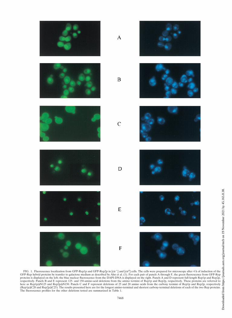
FIG. 1. Fluorescence localization from GFP-Rep1p and GFP-Rep2p in [cir1] and [cir0] cells. The cells were prepared for microscopy after 4 h of induction of theGFP-Rep hybrid proteins by transfer to galactose medium as described by Ahn et al. (1). For each pair of panels A through F, the green fluorescence from GFP-Repproteins is displayed on the left; the blue nuclear fluorescence from the DAPI-DNA is displayed on the right. Panels A and D represent full-length Rep1p and Rep2p,respectively. Panels B and E represent 125- and 150-amino-acid deletions from the amino termini of Rep1p and Rep2p, respectively. These proteins are referred tohere as Rep1pDN125 and Rep2pDN150. Panels C and F represent deletions of 25 and 20 amino acids from the carboxy termini of Rep1p and Rep2p, respectively(Rep1pDC20 and Rep2pDC25). The results presented here are for the longest amino-terminal and shortest carboxy-terminal deletions of each of the two Rep proteins.The fluorescence profiles for the other deletions tested are summarized in Table 1.
7468
Dow
nloa
ded
from
http
s://j
ourn
als.
asm
.org
/jour
nal/m
cb o
n 19
Nov
embe
r 20
21 b
y 45
.165
.8.3
8.
tation by the REP1 or REP2 gene function provided by theresident 2mm circle (Table 2).
To test the functional competence of the Rep deletions, weassayed the stabilities of pSTB1 in a [cir0] host that harboredpGFP-Rep2p or its deletion derivatives. Similarly, the stabili-ties of pSTB2 were assayed in the same host in the presence ofpGFP-Rep1p and the deletion constructs obtained from it.The results are summarized in Table 2. Note that, since theGFP plasmids contained a centromere, they were not lost atany appreciable rate even under nonselective growth condi-tions. A marked drop in plasmid stability was observed with allof the amino-terminal deletions of Rep1p and Rep2p tested.This result suggests that the amino-terminal domains of theRep proteins are critical for their activity, even though thesepeptide regions are apparently dispensable in localizing themto the nucleus, the presumed cellular site at which they func-tion. None of the carboxy-terminal deletions of Rep1p orRep2p assayed were able to sustain the stable propagation ofthe test plasmids. Either the extreme carboxy-terminal regionsof the Rep proteins are directly required for expressing thestability function, or they are required indirectly for steeringthe Rep proteins to their proper cellular destination.
Restoration of localization and of function in carboxy-ter-minal Rep deletions by the addition of an NLS sequence. Wetested whether the carboxy-terminal deletions of Rep1p andRep2p can be targeted to the nucleus by the addition of asynthetic heptapeptide derived from the SV40 T antigen that isknown to act as an NLS in yeast cells. The results shown in Fig.2 demonstrate that the two shortest deletions (Rep1DC25 andRep2DC20), when provided with the NLS at their carboxyterminus, were efficiently transported to the nucleus. Identicalresults were obtained when the NLS was placed at the aminoterminus of each of these two proteins (data not shown). Fur-thermore, once they were escorted to their normal cellular lo-
cale, both Rep1DC25-NLS and Rep2DC20-NLS were able toelevate the stability of pSTB2 and pSTB1, respectively, in a[cir0] background (Table 3).
Thus, we conclude that the peptide segments comprising thecarboxy-terminal extremities of Rep1p and Rep2p are not di-rectly involved in mediating plasmid partitioning. They affectRep protein function indirectly by their role in targeting theseproteins to the proper subcellular location.
Interactions among Rep1p and Rep2p and their deletion de-rivatives assayed in vivo in yeast cells. By using the two-hybridtranscriptional activation assay (8), we tested the interactionsamong pairwise combinations of full-length Rep proteins and asubset of their deletion derivatives. The “bait” protein, a hy-brid between LexA and Rep (or a Rep protein containing adeletion), was expressed constitutively from the yeast ADHpromoter harbored by an HIS3 plasmid. The prey, a fusionbetween Rep (or a Rep protein containing a deletion) and atranscriptional activation domain, was expressed from the in-ducible GAL1 promoter contained within a TRP1 plasmid. Thereporter cassette was a chromosomally integrated copy of theyeast LEU2 gene placed under the control of the LexA oper-ator DNA serving as the UAS (upstream activating sequence).The simultaneous presence of the plasmids expressing the baitand the prey was verified for all of the pairwise combinations,as indicated by the growth of the tester strain in SD mediumlacking histidine and tryptophan (Fig. 3, row A). The labelsabove each column in Fig. 3 represent the two proteins underscrutiny (indicated as the bait [LexA fusion] “1” the prey [theactivation domain fusion] above each column). A growingpatch in row C (LEU2 reporter; SD medium lacking histidine,tryptophan, and leucine-galactose) indicated a positive inter-action between a pair of proteins being tested. Note that, where-as the LexA hybrid would be constitutively expressed (from theADH promoter), the fusion protein containing the transcrip-
TABLE 1. Cellular localization of hybrid proteins formed betweenGFP and Rep deletionsa
a The deleted portions of Rep1p or Rep2p are indicated by the blanks in their schematic represen-tations. All of the deletion derivatives were expressed as hybrid proteins fused to the carboxy terminusof GFP. The numbers indicate the extent of the deletions; for example, DN125 refers to an amino-terminal deletion that includes the amino acid residue at position 125. The patterns of localization werecharacterized as A through F, with reference to the profiles shown in Fig. 1.
VOL. 18, 1998 2mm PLASMID STABILITY IN S. CEREVISIAE 7469
Dow
nloa
ded
from
http
s://j
ourn
als.
asm
.org
/jour
nal/m
cb o
n 19
Nov
embe
r 20
21 b
y 45
.165
.8.3
8.
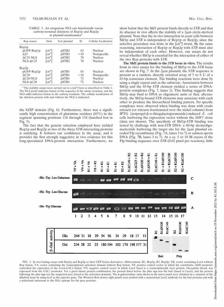
tional activator would be induced only in the presence of ga-lactose (from the GAL1 promoter). As glucose would repressexpression of the latter, row B should not support growth.Thus, growth in row C and no growth in row B provided thecritical criterion for interaction. The positive control used here,a direct fusion of the activation domain to LexA (i.e., column1), was an exception to this rule. Since this protein was con-stitutively made from the ADH promoter, the growth of thehost strain in rows C and B (column 1) was expected.
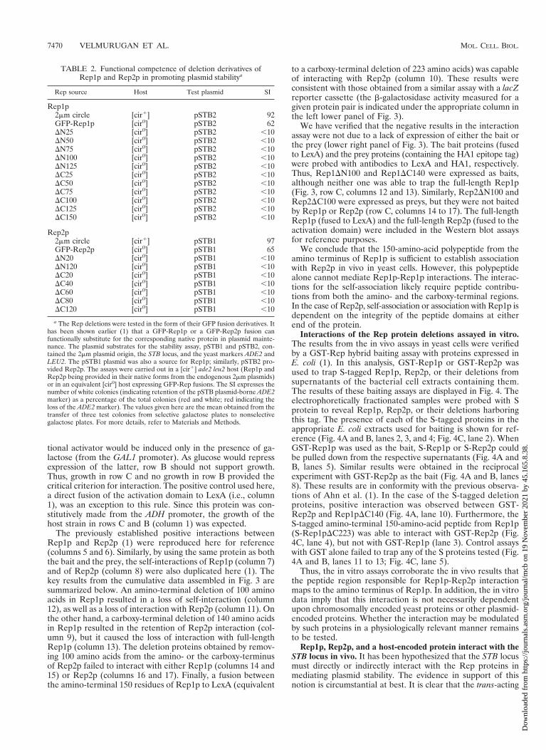
The previously established positive interactions betweenRep1p and Rep2p (1) were reproduced here for reference(columns 5 and 6). Similarly, by using the same protein as boththe bait and the prey, the self-interactions of Rep1p (column 7)and of Rep2p (column 8) were also duplicated here (1). Thekey results from the cumulative data assembled in Fig. 3 aresummarized below. An amino-terminal deletion of 100 aminoacids in Rep1p resulted in a loss of self-interaction (column12), as well as a loss of interaction with Rep2p (column 11). Onthe other hand, a carboxy-terminal deletion of 140 amino acidsin Rep1p resulted in the retention of Rep2p interaction (col-umn 9), but it caused the loss of interaction with full-lengthRep1p (column 13). The deletion proteins obtained by remov-ing 100 amino acids from the amino- or the carboxy-terminusof Rep2p failed to interact with either Rep1p (columns 14 and15) or Rep2p (columns 16 and 17). Finally, a fusion betweenthe amino-terminal 150 residues of Rep1p to LexA (equivalent
to a carboxy-terminal deletion of 223 amino acids) was capableof interacting with Rep2p (column 10). These results wereconsistent with those obtained from a similar assay with a lacZreporter cassette (the b-galactosidase activity measured for agiven protein pair is indicated under the appropriate column inthe left lower panel of Fig. 3).
We have verified that the negative results in the interactionassay were not due to a lack of expression of either the bait orthe prey (lower right panel of Fig. 3). The bait proteins (fusedto LexA) and the prey proteins (containing the HA1 epitope tag)were probed with antibodies to LexA and HA1, respectively.Thus, Rep1DN100 and Rep1DC140 were expressed as baits,although neither one was able to trap the full-length Rep1p(Fig. 3, row C, columns 12 and 13). Similarly, Rep2DN100 andRep2DC100 were expressed as preys, but they were not baitedby Rep1p or Rep2p (row C, columns 14 to 17). The full-lengthRep1p (fused to LexA) and the full-length Rep2p (fused to theactivation domain) were included in the Western blot assaysfor reference purposes.
We conclude that the 150-amino-acid polypeptide from theamino terminus of Rep1p is sufficient to establish associationwith Rep2p in vivo in yeast cells. However, this polypeptidealone cannot mediate Rep1p-Rep1p interactions. The interac-tions for the self-association likely require peptide contribu-tions from both the amino- and the carboxy-terminal regions.In the case of Rep2p, self-association or association with Rep1p isdependent on the integrity of the peptide domains at eitherend of the protein.
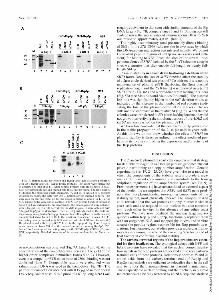
Interactions of the Rep protein deletions assayed in vitro.The results from the in vivo assays in yeast cells were verifiedby a GST-Rep hybrid baiting assay with proteins expressed inE. coli (1). In this analysis, GST-Rep1p or GST-Rep2p wasused to trap S-tagged Rep1p, Rep2p, or their deletions fromsupernatants of the bacterial cell extracts containing them.The results of these baiting assays are displayed in Fig. 4. Theelectrophoretically fractionated samples were probed with Sprotein to reveal Rep1p, Rep2p, or their deletions harboringthis tag. The presence of each of the S-tagged proteins in theappropriate E. coli extracts used for baiting is shown for ref-erence (Fig. 4A and B, lanes 2, 3, and 4; Fig. 4C, lane 2). WhenGST-Rep1p was used as the bait, S-Rep1p or S-Rep2p couldbe pulled down from the respective supernatants (Fig. 4A andB, lanes 5). Similar results were obtained in the reciprocalexperiment with GST-Rep2p as the bait (Fig. 4A and B, lanes8). These results are in conformity with the previous observa-tions of Ahn et al. (1). In the case of the S-tagged deletionproteins, positive interaction was observed between GST-Rep2p and Rep1pDC140 (Fig. 4A, lane 10). Furthermore, theS-tagged amino-terminal 150-amino-acid peptide from Rep1p(S-Rep1pDC223) was able to interact with GST-Rep2p (Fig.4C, lane 4), but not with GST-Rep1p (lane 3). Control assayswith GST alone failed to trap any of the S proteins tested (Fig.4A and B, lanes 11 to 13; Fig. 4C, lane 5).
Thus, the in vitro assays corroborate the in vivo results thatthe peptide region responsible for Rep1p-Rep2p interactionmaps to the amino terminus of Rep1p. In addition, the in vitrodata imply that this interaction is not necessarily dependentupon chromosomally encoded yeast proteins or other plasmid-encoded proteins. Whether the interaction may be modulatedby such proteins in a physiologically relevant manner remainsto be tested.
Rep1p, Rep2p, and a host-encoded protein interact with theSTB locus in vivo. It has been hypothesized that the STB locusmust directly or indirectly interact with the Rep proteins inmediating plasmid stability. The evidence in support of thisnotion is circumstantial at best. It is clear that the trans-acting
TABLE 2. Functional competence of deletion derivatives ofRep1p and Rep2p in promoting plasmid stabilitya
Rep source Host Test plasmid SI
Rep1p2mm circle [cir1] pSTB2 92GFP-Rep1p [cir0] pSTB2 62DN25 [cir0] pSTB2 ,10DN50 [cir0] pSTB2 ,10DN75 [cir0] pSTB2 ,10DN100 [cir0] pSTB2 ,10DN125 [cir0] pSTB2 ,10DC25 [cir0] pSTB2 ,10DC50 [cir0] pSTB2 ,10DC75 [cir0] pSTB2 ,10DC100 [cir0] pSTB2 ,10DC125 [cir0] pSTB2 ,10DC150 [cir0] pSTB2 ,10
Rep2p2mm circle [cir1] pSTB1 97GFP-Rep2p [cir0] pSTB1 65DN20 [cir0] pSTB1 ,10DN120 [cir0] pSTB1 ,10DC20 [cir0] pSTB1 ,10DC40 [cir0] pSTB1 ,10DC60 [cir0] pSTB1 ,10DC80 [cir0] pSTB1 ,10DC120 [cir0] pSTB1 ,10
a The Rep deletions were tested in the form of their GFP fusion derivatives. Ithas been shown earlier (1) that a GFP-Rep1p or a GFP-Rep2p fusion canfunctionally substitute for the corresponding native protein in plasmid mainte-nance. The plasmid substrates for the stability assay, pSTB1 and pSTB2, con-tained the 2mm plasmid origin, the STB locus, and the yeast markers ADE2 andLEU2. The pSTB1 plasmid was also a source for Rep1p; similarly, pSTB2 pro-vided Rep2p. The assays were carried out in a [cir1] ade2 leu2 host (Rep1p andRep2p being provided in their native forms from the endogenous 2mm plasmids)or in an equivalent [cir0] host expressing GFP-Rep fusions. The SI expresses thenumber of white colonies (indicating retention of the pSTB plasmid-borne ADE2marker) as a percentage of the total colonies (red and white; red indicating theloss of the ADE2 marker). The values given here are the mean obtained from thetransfer of three test colonies from selective galactose plates to nonselectivegalactose plates. For more details, refer to Materials and Methods.
7470 VELMURUGAN ET AL. MOL. CELL. BIOL.
Dow
nloa
ded
from
http
s://j
ourn
als.
asm
.org
/jour
nal/m
cb o
n 19
Nov
embe
r 20
21 b
y 45
.165
.8.3
8.

Rep proteins and the cis-acting STB locus are functionallytightly interrelated, since mutations in any one of the three loci(REP1, REP2, or STB) lead to the same phenotype, namely,plasmid instability. Preliminary in vitro binding studies (14)have suggested that Rep1p and Rep2p can bind to STB butonly in association with a host factor (or factors).
We have therefore searched for STB-binding protein factorsin yeast cells by using the monohybrid positive-selection meth-od. The assay was standardized (see Materials and Methods)to establish conditions under which the growth of a yeast col-ony in the presence of a titrated amount of 3-AT was contin-gent upon the enhanced transcriptional activation of the HIS3reporter gene via STB-protein interaction. Under the strin-gency conditions applied here, only three types of cloneswere obtained by this procedure. Two of them were the REP1and REP2 genes, and the third was the as-yet-uncharacterized
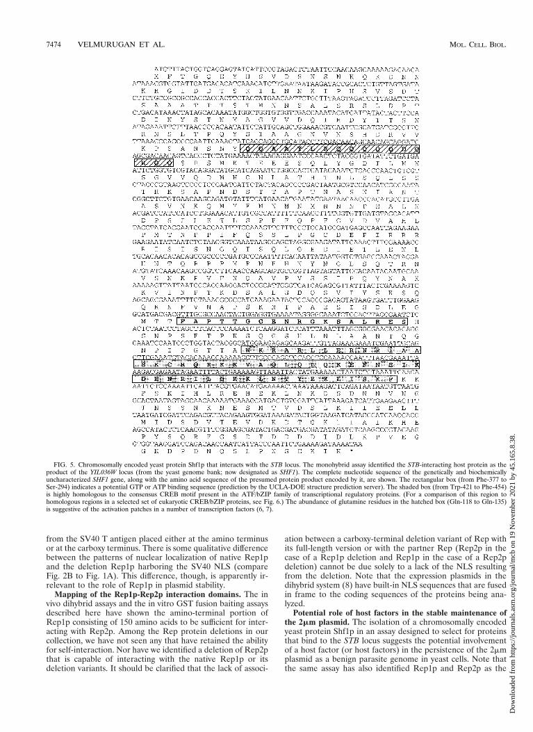
chromosomal gene YIL036W (designation in the yeast genomebank). The frequencies of REP1, REP2, and YIL036W amongthe more than 80 positive clones obtained after three rounds of3-AT selection were approximately 12, 32, and 56%, respec-tively. The nucleotide sequence of YIL036W, which we desig-nate as SHF1 (STB binding host factor), and the derived aminoacid sequence are shown in Fig. 5. Certain interesting struc-tural motifs could be gleaned in the protein (Shf1p) sequence.For example, the peptide region from amino acids 377 to 394(the open rectangular box in Fig. 5) was suggestive of GTP orATP binding (as predicted by the UCLA-DOE Structure Pre-diction Server). Similarly, the peptide stretch from amino acids421 to 454 (shaded box in Fig. 5; see also Fig. 6) showed stronghomology to the cyclic AMP responsive element binding(CREB) motif found in the activating transcription factor(ATF)/CREB family of transcriptional regulators containing
FIG. 2. Localization of short carboxy-terminal deletions of Rep1p and Rep2p when provided with the NLS from SV40 T antigen. The fluorescence patterns ofRep1pDC25 and Rep2pDC20 are shown in panels A and C, respectively, for reference. Panels B and D illustrate the localization of the same deletions when aheptapeptide NLS from an SV40 T antigen was fused to their carboxy termini (B, Rep1pDC25-NLS; D, Rep1pDC20-NLS). When the NLS was fused to the aminotermini of these deletions, the fluorescence localization was indistinguishable from that shown in panels B and D (data not shown).
VOL. 18, 1998 2mm PLASMID STABILITY IN S. CEREVISIAE 7471
Dow
nloa
ded
from
http
s://j
ourn
als.
asm
.org
/jour
nal/m
cb o
n 19
Nov
embe
r 20
21 b
y 45
.165
.8.3
8.
the bZIP domain (Fig. 6). Furthermore, there was a signifi-cantly high concentration of glutamine residues (65%) in thesegment spanning positions 118 through 134 (hatched box inFig. 5).
The fact that the genetic selection employed here yieldedRep1p and Rep2p as two of the three STB-interacting proteinsis satisfying. It bolsters our confidence in the assay, and itprovides the first strongly suggestive in vivo evidence for thislong-speculated DNA-protein interaction. Furthermore, we
show below that the Shf1 protein binds directly to STB and thatits absence in vivo affects the stability of a 2mm circle-derivedplasmid. Note that the in vivo interaction in yeast cells betweenShf1p and STB is independent of Rep1p or Rep2p, since theselective procedure was done in the [cir0] strain. By the samereasoning, interaction of Rep1p or Rep2p with STB must alsobe independent of each other. However, our assays do notreveal whether Shf1p is essential for the interaction of either ofthe two Rep proteins with STB.
The Shf1 protein binds to the STB locus in vitro. The resultsfrom in vitro assays for the binding of Shf1p to the STB locusare shown in Fig. 7. In the 2mm plasmid, the STB sequence ispresent as a tandem, directly oriented array of 5 to 6 U of a65-bp consensus element. The binding reactions were done byusing a single repeat unit as the substrate. Association betweenShf1p and the 65-bp STB element yielded a series of DNA-protein complexes (Fig. 7, lanes 2). This finding suggests thatShf1p may bind to DNA as oligomeric units or that, alterna-tively, the Shf1p-bound STB elements may associate with eachother to produce the hierarchical binding pattern. No specificcomplexes were observed when binding was done with crudeextracts (or extracts fractionated over the nickel column) fromIPTG (isopropyl-b-D-thiogalactopyranoside)-induced E. colicells harboring the expression vector without the SHF1 clone(data not shown). The specificity of Shf1p-STB binding wastested by challenge with non-STB DNA: a 60-bp deoxyoligo-nucleotide harboring the target site for the 2mm plasmid en-coded Flp recombinase (Fig. 7A, lanes 3 to 7) or salmon spermDNA (Fig. 7B, lanes 3 to 7). At a ca. 5 or 10 M excess of theFlp binding sequence over STB (0.02 pmol per reaction), little
FIG. 3. In vivo baiting assays with Rep1p and Rep2p or their GFP fusion derivatives. Abbreviations: R1, Rep1p; R2, Rep2p; VB, vector containing LexA withoutRep fusion; VA, vector containing the transcriptional activation domain without Rep fusion; VP, positive control vector in which the constitutive ADH promotercontrolled the expression of the LexA-GAL4 fusion; VN, negative control vector in which LexA fused to a transcriptionally inert protein, Drosophila bicoid, wasexpressed from the GAL1 promoter. For a given binary protein combination, the protein listed before the plus sign was the bait (fused to LexA), and the proteinfollowing the plus sign was the suspected prey (fused to the activation domain). The b-galactosidase units shown in the lower panel were obtained in a variation of thedihybrid assay by using lacZ as the reporter gene. The Western blots (lower right panel) were probed with a monoclonal LexA antibody for the bait proteins and witha polyclonal antiserum to the HA1 epitope for the prey proteins.
TABLE 3. An exogenous NLS can functionally rescuecarboxy-terminal deletions of Rep1p and Rep2p
in plasmid maintenancea
Rep source Host Test plasmid SI Cellular localization
Rep1ppGFP-Rep1p [cir0] pSTB2 62 NuclearD25 [cir0] pSTB2 ,10 NonspecificDC25-NLS [cir0] pSTB2 70 NuclearNLS-DC25 [cir0] pSTB2 70 Nuclear
Rep2ppGFP-Rep2p [cir0] pSTB1 65 NuclearDC20 [cir0] pSTB1 ,10 NonspecificDC20-NLS [cir0] pSTB1 72 NuclearNLS-DC20 [cir0] pSTB1 72 Nuclear
a The stability assays were carried out in a [cir0] host as described in Table 2.The NLS prefix indicates fusion of this sequence at the amino terminus, and theNLS suffix indicates fusion to the carboxy terminus. The cellular localization ofthe deletion protein with and without the NLS is indicated.
7472 VELMURUGAN ET AL. MOL. CELL. BIOL.
Dow
nloa
ded
from
http
s://j
ourn
als.
asm
.org
/jour
nal/m
cb o
n 19
Nov
embe
r 20
21 b
y 45
.165
.8.3
8.
or no competition was observed (Fig. 7A, lanes 3 and 4). As theconcentration of the competitor was increased, the yield of thehigher-order complexes diminished (lanes 5 to 7). However,even at a competitor/STB molar ratio of 250:1, binding was notabolished (lane 7). Comparable results were obtained whensalmon sperm DNA was used as the competitor (Fig. 7B). Thepattern of competition obtained with 0.15 mg of salmon spermDNA (equivalent to ca. 5 to 6 pmol of a 60-bp-long DNA) was
roughly equivalent to that seen with similar amounts of the FlpDNA target (Fig. 7B, compare lanes 3 and 7). Binding was stillevident when the molar ratio of salmon sperm DNA to STBwas raised to approximately 4,000:1 (lane 7).
The highly discriminatory (and presumably direct) bindingof Shf1p to the STB DNA validates the in vivo assay by whichthis DNA-protein interaction was inferred initially. We do notknow what peptide regions of Shf1p are necessary (and suffi-cient) for binding to STB. From the sizes of the several inde-pendent clones of SHF1 isolated by the 3-AT selection assay invivo, we surmise that they encode full-length or nearly full-length Shf1p.
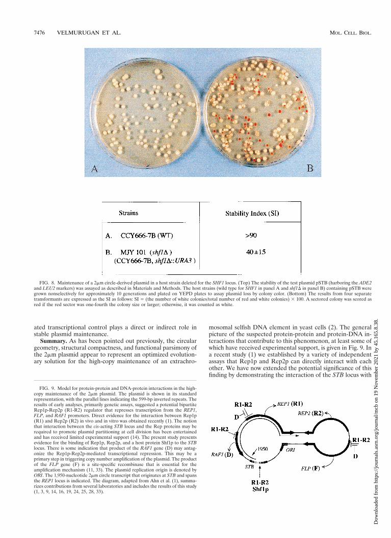
Plasmid stability in a host strain harboring a deletion of theSHF1 locus. Does the lack of SHF1 function affect the stabilityof a 2mm circle-derived test plasmid? To address this issue, themaintenance of plasmid pSTB (harboring the 2mm plasmidreplication origin and the STB locus) was followed in a [cir1]SHF1 strain (Fig. 8A) and a derivative strain lacking this locus(Fig. 8B) (see Materials and Methods for details). The plasmidloss rate was significantly higher in the shf1 deletion strain, asindicated by the increase in the number of red colonies (indi-cating the loss of the plasmid-borne ADE2 marker). The re-sults are also expressed as the relative SI (Fig. 8). When the redcolonies were transferred to SD plates lacking leucine, they didnot grow, thus verifying the simultaneous loss of the ADE2 andLEU2 markers carried on the plasmid pSTB.
We therefore conclude that the host factor Shf1p plays a rolein the stable propagation of the 2mm plasmid in yeast cells.At this time we do not know whether the effect of SHF1 onplasmid stability is direct or indirect, the effect mediated per-haps by its role in controlling the expression and/or activity ofthe Rep proteins.
DISCUSSION
The 2mm circle plasmid in yeast cells employs a dual strategyfor its stable propagation as a benign parasite genome: efficientplasmid partitioning and copy number amplification. Geneticexperiments (16, 19, 24, 25, 28) have given rise to a model inwhich the components of the stability system provide a mea-sure of the plasmid copy number and contribute to the tran-scriptional regulation of the amplification system (see Fig. 9).Previous experiments (1) have substantiated one central aspectof the model: the assumption that REP1 and REP2 gene prod-ucts, the two plasmid-coded trans-acting components of thestability system, must physically interact. The analyses of Ahnet al. revealed that the two proteins not only interact in vivo inyeast cells and are targeted to the nucleus but also associatewith each other in vitro in the absence of any other yeastproteins. We have now localized the nuclear targeting se-quences within Rep1p and Rep2p, functionally replaced themwith an exogenous NLS, and mapped (by in vivo and in vitroassays) the peptide regions responsible for Rep protein asso-ciations. Furthermore, our studies provide a molecular frame-work for examining the role of the cis-acting STB locus and ofhost factors in conferring plasmid stability.
The carboxy-terminal regions of the Rep proteins are essen-tial for their localization. The cytological assays with GFP andhybrid proteins have revealed that the nuclear compartmenta-tion signals in the Rep proteins are located at the very carboxy-terminal ends of these proteins. Deletions as short as 25 and 20amino acids from the carboxy-terminal end (of Rep1p andRep2p, respectively) are sufficient to delocalize them in the celland to render them nonfunctional in plasmid maintenance.Their capacity for nuclear homing and their activity in plasmidmaintenance can be fully restored by an NLS sequence derived
FIG. 4. Baiting assays for Rep1p and Rep2p and their deletions performedwith GST-Rep1p and GST-Rep2p hybrid proteins. The assays were carried outas described by Ahn et al. (1). After baiting, proteins were fractionated in SDS–12% polyacrylamide gels and probed with the S-protein probe. The lane markedM displays the molecular-weight standards. (A and B) In lanes 2 to 4, proteinsextracted by boiling the cells from 200-ml portions of the induced cultures (thatwere also the starting materials for the assays depicted in lanes 5 to 13) in theSDS sample buffer were run as controls. The S-Rep protein bands of interest inlanes 2 to 4 are indicated by the arrowheads. The data in panel A were obtainedwith S-tagged Rep1p or its derivatives; the data in panel B were obtained withS-tagged Rep2p or its derivatives. The GST-Rep hybrids used as the baits andthe corresponding baited S-Rep proteins (either full length or partially deleted)are indicated above lanes 5 to 10. In the reactions represented in lanes 11 to 13,the baiting was performed with GST and not with the GST-Rep hybrids. (C)Results obtained with Rep1pDC223 fused to the S-peptide tag. Lane 2 repre-sents the extract from 200 ml of the induced culture expressing Rep1pDC223;lanes 3 to 5 correspond to baiting assays with GST-Rep1p, GST-Rep2p, andGST, respectively. Detailed protocols of the assays are described in Ahn et al.(1).
VOL. 18, 1998 2mm PLASMID STABILITY IN S. CEREVISIAE 7473
Dow
nloa
ded
from
http
s://j
ourn
als.
asm
.org
/jour
nal/m
cb o
n 19
Nov
embe
r 20
21 b
y 45
.165
.8.3
8.
from the SV40 T antigen placed either at the amino terminusor at the carboxy terminus. There is some qualitative differencebetween the patterns of nuclear localization of native Rep1pand the deletion Rep1p harboring the SV40 NLS (compareFig. 2B to Fig. 1A). This difference, though, is apparently ir-relevant to the role of Rep1p in plasmid stability.
Mapping of the Rep1p-Rep2p interaction domains. The invivo dihybrid assays and the in vitro GST fusion baiting assaysdescribed here have shown the amino-terminal portion ofRep1p consisting of 150 amino acids to be sufficient for inter-acting with Rep2p. Among the Rep protein deletions in ourcollection, we have not seen any that have retained the abilityfor self-interaction. Nor have we identified a deletion of Rep2pthat is capable of interacting with the native Rep1p or itsdeletion variants. It should be clarified that the lack of associ-
ation between a carboxy-terminal deletion variant of Rep withits full-length version or with the partner Rep (Rep2p in thecase of a Rep1p deletion and Rep1p in the case of a Rep2pdeletion) cannot be due solely to a lack of the NLS resultingfrom the deletion. Note that the expression plasmids in thedihybrid system (8) have built-in NLS sequences that are fusedin frame to the coding sequences of the proteins being ana-lyzed.
Potential role of host factors in the stable maintenance ofthe 2mm plasmid. The isolation of a chromosomally encodedyeast protein Shf1p in an assay designed to select for proteinsthat bind to the STB locus suggests the potential involvementof a host factor (or host factors) in the persistence of the 2mmplasmid as a benign parasite genome in yeast cells. Note thatthe same assay has also identified Rep1p and Rep2p as the
FIG. 5. Chromosomally encoded yeast protein Shf1p that interacts with the STB locus. The monohybrid assay identified the STB-interacting host protein as theproduct of the YIL036W locus (from the yeast genome bank; now designated as SHF1). The complete nucleotide sequence of the genetically and biochemicallyuncharacterized SHF1 gene, along with the amino acid sequence of the presumed protein product encoded by it, are shown. The rectangular box (from Phe-377 toSer-294) indicates a potential GTP or ATP binding sequence (prediction by the UCLA-DOE structure prediction server). The shaded box (from Trp-421 to Phe-454)is highly homologous to the consensus CREB motif present in the ATF/bZIP family of transcriptional regulatory proteins. (For a comparison of this region tohomologous regions in a selected set of eukaryotic CREB/bZIP proteins, see Fig. 6.) The abundance of glutamine residues in the hatched box (Gln-118 to Gln-135)is suggestive of the activation patches in a number of transcription factors (6, 7).
7474 VELMURUGAN ET AL. MOL. CELL. BIOL.
Dow
nloa
ded
from
http
s://j
ourn
als.
asm
.org
/jour
nal/m
cb o
n 19
Nov
embe
r 20
21 b
y 45
.165
.8.3
8.
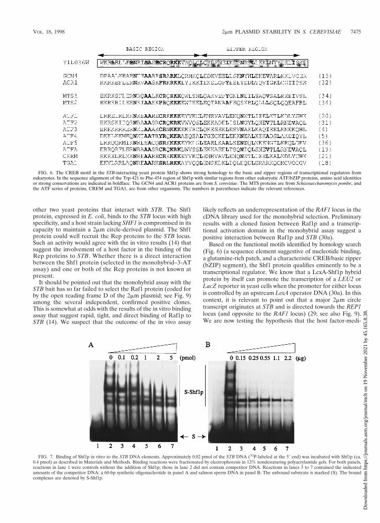
other two yeast proteins that interact with STB. The Shf1protein, expressed in E. coli, binds to the STB locus with highspecificity, and a host strain lacking SHF1 is compromised in itscapacity to maintain a 2mm circle-derived plasmid. The Shf1protein could well recruit the Rep proteins to the STB locus.Such an activity would agree with the in vitro results (14) thatsuggest the involvement of a host factor in the binding of theRep proteins to STB. Whether there is a direct interactionbetween the Shf1 protein (selected in the monohybrid–3-ATassay) and one or both of the Rep proteins is not known atpresent.
It should be pointed out that the monohybrid assay with theSTB bait has so far failed to select the Raf1 protein (coded forby the open reading frame D of the 2mm plasmid; see Fig. 9)among the several independent, confirmed positive clones.This is somewhat at odds with the results of the in vitro bindingassay that suggest rapid, tight, and direct binding of Raf1p toSTB (14). We suspect that the outcome of the in vivo assay
likely reflects an underrepresentation of the RAF1 locus in thecDNA library used for the monohybrid selection. Preliminaryresults with a cloned fusion between Raf1p and a transcrip-tional activation domain in the monohybrid assay suggest apositive interaction between Raf1p and STB (30a).
Based on the functional motifs identified by homology search(Fig. 6) (a sequence element suggestive of nucleotide binding,a glutamine-rich patch, and a characteristic CREB/basic zipper(bZIP) segment), the Shf1 protein qualifies eminently to be atranscriptional regulator. We know that a LexA-Shf1p hybridprotein by itself can promote the transcription of a LEU2 orLacZ reporter in yeast cells when the promoter for either locusis controlled by an upstream LexA operator DNA (30a). In thiscontext, it is relevant to point out that a major 2mm circletranscript originates at STB and is directed towards the REP1locus (and opposite to the RAF1 locus) (29; see also Fig. 9).We are now testing the hypothesis that the host factor-medi-
FIG. 6. The CREB motif in the STB-interacting yeast protein Shf1p shows strong homology to the basic and zipper regions of transcriptional regulators fromeukaryotes. In the sequence alignment of the Trp-421 to Phe-454 region of Shf1p with similar regions from other eukaryotic ATF/bZIP proteins, amino acid identitiesor strong conservations are indicated in boldface. The GCN4 and ACR1 proteins are from S. cerevisiae. The MTS proteins are from Schizosaccharomyces pombe, andthe ATF series of proteins, CREM and TGA1, are from other organisms. The numbers in parentheses indicate the relevant references.
FIG. 7. Binding of Shf1p in vitro to the STB DNA elements. Approximately 0.02 pmol of the STB DNA (32P-labeled at the 59 end) was incubated with Shf1p (ca.0.4 pmol) as described in Materials and Methods. Binding reactions were fractionated by electrophoresis in 12% nondenaturing polyacrylamide gels. For both panels,reactions in lane 1 were controls without the addition of Shf1p; those in lane 2 did not contain competitor DNA. Reactions in lanes 3 to 7 contained the indicatedamounts of the competitor DNA: a 60-bp synthetic oligonucleotide in panel A and salmon sperm DNA in panel B. The unbound substrate is marked (S). The boundcomplexes are denoted by S-Shf1p.
VOL. 18, 1998 2mm PLASMID STABILITY IN S. CEREVISIAE 7475
Dow
nloa
ded
from
http
s://j
ourn
als.
asm
.org
/jour
nal/m
cb o
n 19
Nov
embe
r 20
21 b
y 45
.165
.8.3
8.
ated transcriptional control plays a direct or indirect role instable plasmid maintenance.
Summary. As has been pointed out previously, the circulargeometry, structural compactness, and functional parsimony ofthe 2mm plasmid appear to represent an optimized evolution-ary solution for the high-copy maintenance of an extrachro-
mosomal selfish DNA element in yeast cells (2). The generalpicture of the suspected protein-protein and protein-DNA in-teractions that contribute to this phenomenon, at least some ofwhich have received experimental support, is given in Fig. 9. Ina recent study (1) we established by a variety of independentassays that Rep1p and Rep2p can directly interact with eachother. We have now extended the potential significance of thisfinding by demonstrating the interaction of the STB locus with
FIG. 8. Maintenance of a 2mm circle-derived plasmid in a host strain deleted for the SHF1 locus. (Top) The stability of the test plasmid pSTB (harboring the ADE2and LEU2 markers) was assayed as described in Materials and Methods. The host strains (wild type for SHF1 in panel A and shf1D in panel B) containing pSTB weregrown nonselectively for approximately 10 generations and plated on YEPD plates to assay plasmid loss by colony color. (Bottom) The results from four separatetransformants are expressed as the SI as follows: SI 5 (the number of white colonies/total number of red and white colonies) 3 100. A sectored colony was scored asred if the red sector was one-fourth the colony size or larger; otherwise, it was counted as white.
FIG. 9. Model for protein-protein and DNA-protein interactions in the high-copy maintenance of the 2mm plasmid. The plasmid is shown in its standardrepresentation, with the parallel lines indicating the 599-bp inverted repeats. Theresults of early analyses, primarily genetic assays, suggested a potential bipartiteRep1p-Rep2p (R1-R2) regulator that represses transcription from the REP1,FLP, and RAF1 promoters. Direct evidence for the interaction between Rep1p(R1) and Rep2p (R2) in vivo and in vitro was obtained recently (1). The notionthat interaction between the cis-acting STB locus and the Rep proteins may berequired to promote plasmid partitioning at cell division has been entertainedand has received limited experimental support (14). The present study presentsevidence for the binding of Rep1p, Rep2p, and a host protein Shf1p to the STBlocus. There is some indication that product of the RAF1 gene (D) may antag-onize the Rep1p-Rep2p-mediated transcriptional repression. This may be aprimary step in triggering copy number amplification of the plasmid. The productof the FLP gene (F) is a site-specific recombinase that is essential for theamplification mechanism (11, 33). The plasmid replication origin is denoted byORI. The 1,950-nucleotide 2mm circle transcript that originates at STB and spansthe REP1 locus is indicated. The diagram, adapted from Ahn et al. (1), summa-rizes contributions from several laboratories and includes the results of this study(1, 3, 9, 14, 16, 19, 24, 25, 28, 33).
7476 VELMURUGAN ET AL. MOL. CELL. BIOL.
Dow
nloa
ded
from
http
s://j
ourn
als.
asm
.org
/jour
nal/m
cb o
n 19
Nov
embe
r 20
21 b
y 45
.165
.8.3
8.
Rep1p, Rep2p, and the product of the chromosomal geneSHF1. The results of the present study therefore offer newinsight into the analysis of the molecular contributions madeby the host and the plasmid to this benign parasitism.
ACKNOWLEDGMENT
This work was supported by a grant from the Council for TobaccoResearch.
ADDENDUM IN PROOF
Scott-Drew and Murray (S. Scott-Drew and J. A. Murray, J.Cell Sci. 111:1779–1789, 1998) have recently demonstratedthat Rep1p and Rep2p form a complex. By confocal micros-copy they have further demonstrated that the proteins occupyspecific sites in the nucleus and that these sites are distributedto both mother and daughter cells during cell division.
REFERENCES
1. Ahn, Y. T., X. L. Wu, S. Biswal, S. Velmurugan, F. C. Volkert, and M.Jayaram. 1997. The 2mm-plasmid-encoded Rep1 and Rep2 proteins interactwith each other and colocalize to the Saccharomyces cerevisiae nucleus. J.Bacteriol. 179:7497–7506.
2. Broach, J. R., and F. C. Volkert. 1991. Circular DNA plasmids of yeasts:genome dynamics, protein synthesis and energetics, p. 297–331. In J. R.Broach, J. R. Pringle, and E. W. Jones (ed.), The molecular and cellularbiology of the yeast Saccharomyces. Cold Spring Harbor Press, Cold SpringHarbor, N.Y.
3. Cashmore, J. R., M. S. Albury, C. Hadfield, and P. A. Meacock. 1986.Genetic analysis of partitioning functions encoded by the 2mm circle ofSaccharomyces cerevisiae. Mol. Gen. Genet. 203:154–162.
4. Chen, B. P., G. Liang, J. Whelan, and T. Hai. 1994. ATF3 and ATF3DZip.Transcriptional repression versus activation by alternatively spliced isoforms.J. Biol. Chem. 269:15819–15826.
5. Chevray, P. M., and D. Nathans. 1992. Protein interaction cloning in yeast:identification of mammalian proteins that react with the leucine zipper ofJun. Proc. Natl. Acad. Sci. USA 89:5789–5793.
6. Courney, A. J., and R. Tjian. 1988. Analysis of Sp1 in vivo reveals multipletranscriptional domains, including a novel glutamine-rich activation motif.Cell 55:887–898.
7. Courney, A. J., D. A. Holtzman, S. P. Jackson, and R. Tjian. 1988. Synergisticactivation by glutamine-rich domains of human transcription factor Sp1. Cell59:827–836.
8. Finley, Jr., R. L., and R. Brent. 1996. Interaction trap cloning with yeast, p.169–203. In D. Glover and B. D. Hames (ed.), DNA cloning-expressionsystems: a practical approach. Oxford University Press, Oxford, United King-dom.
9. Futcher, A. B., and B. S. Cox. 1983. Maintenance of the 2mm circle plasmidin populations of Saccharomyces cerevisiae. J. Bacteriol. 154:612–622.
10. Futcher, A. B. 1986. Copy number amplification of the 2mm circle plasmid ofSaccharomyces cerevisiae. J. Theor. Biol. 119:197–204.
11. Futcher, A. B. 1988. The 2mm circle plasmid of Saccharomyces cerevisiae.Yeast 4:27–40.
12. Guarente, L. 1983. Yeast promoters and lacZ fusions designed to study theexpression of cloned genes in yeast. Methods Enzymol. 101:181–191.
13. Goetz, J., B. Chatton, M. G. Mattei, and C. Kedinger. 1996. Structure andexpression of the ATFa gene. J. Biol Chem. 271:29589–29598.
14. Hadfield, C., R. C. Mount, and A. M. Cashmore. 1995. Protein bindinginteractions at the STB locus of the yeast 2mm plasmid. Nucleic Acids Res.23:995–1002.
15. Hinnebusch, A. G. 1984. Evidence for translational regulation of the activa-tor of general amino acid control in yeast. Proc. Natl. Acad. Sci. USA 81:6442–6446.
16. Jayaram, M., A. Sutton, and J. R. Broach. 1985. Properties of REP3: acis-acting locus required for stable propagation of the Saccharomyces cerevi-siae plasmid 2mm circle. Mol. Cell. Biol. 5:2466–2475.
17. Jayaram, M., S. Sumida, and L.-J. Young. 1986. Inducible expression ofREP1 causes inducible expression of the 2 micron circle stability system.Curr. Genet. 11:85–91.
18. Katagiri, F., E. Lam, and N. H. Chua. 1989. Two tobacco DNA-bindingproteins with homology to the nuclear factor CREB. Nature 340:727–730.
19. Kikuchi, Y. 1983. Yeast plasmid requires a cis-acting locus and two plasmidproteins for its stable maintenance. Cell 35:487–493.
20. Lee, M.-R., C. S. Chung, M. L. Liou, M. Wu, W. Li, Y. P. Hsueh, and M. Z.Lai. 1992. Isolation and characterization of nuclear proteins that bind to Tcell receptor V-beta decamer motif. J. Immunol. 148:1906–1912.
21. Masquilier, D., N. S. Foulkes, M. G. Mattei, and P. Sassone-Corsi. 1993.Human CREM gene: evolutionary conservation, chromosomal localization,and inducibility of the transcript. Cell Growth Differ. 4:931–937.
22. Murray, A. W., and J. W. Szostak. 1983. Pedigree analysis of plasmid seg-regation in yeast. Cell 34:961–970.
23. Murray, J. A. H., and G. Cesareni. 1986. Functional analysis of the yeastplasmid partition locus STB. EMBO J. 5:3391–3399.
24. Murray, J. A. H., M. Scarpa, N. Rossi, and G. Cesareni. 1987. Antagonisticcontrols regulate copy number of the yeast 2mm plasmid. EMBO J. 6:4205–4212.
25. Reynolds, A. E., A. W. Murray, and J. W. Szostak. 1987. Roles of the 2mmgene products in stable maintenance of the 2 mm plasmid of Saccharomycescerevisiae. Mol. Cell. Biol. 7:3566–3573.
26. Russo, F. D., I. Scherson, and J. R. Broach. 1992. Direct simulations of yeast2 micron circle plasmid amplification J. Theor. Biol. 155:369–385.
27. Sigurdson, D. C., M. E. Garder, and D. M. Livingston. 1981. Characteriza-tion of the transmission during cytoductant formation of the 2 micron DNAplasmid from Saccharomyces. Mol. Gen. Genet. 183:59–65.
28. Som, T., K. A. Armstrong, F. C. Volkert, and J. R. Broach. 1988. Autoreg-ulation of 2mm circle gene expression provides a model for maintenance ofstable plasmid copy levels. Cell 52:27–37.
29. Sutton, A., and J. R. Broach. 1985. Signals for transcription initiation andtermination in the Saccharomyces cerevisiae plasmid 2mm circle. Mol. Cell.Biol. 5:2770–2780.
30. Veit, B. E., and W. L. Fangman. 1988. Copy number and partition of theSaccharomyces cerevisiae 2mm plasmid controlled by transcription regulators.Mol. Cell. Biol. 8:4949–4957.
30a.Velmurugan, S., and M. Jayaram. Unpublished data.31. Villarreal, X. C., and J. D. Richter. 1995. Analysis of ATF2 gene expression
during early Xenopus laevis development. Gene 153:225–229.32. Vincent, A. C., and K. Struhl. 1992. ACR1, a yeast ATF/CREB repressor.
Mol. Cell. Biol. 12:5394–5405.33. Volkert, F. C., and J. R. Broach. 1986. Site-specific recombination promotes
plasmid amplification in yeast. Cell 46:541–550.34. Wahls, W. P., and G. R. Smith. 1994. A heteromeric protein that binds to a
meiotic homologous recombination hot spot: correlation of binding and hotspot activity. Genes Dev. 14:1693–1702.
35. Zakian, V. A., B. J. Brewer, and W. L. Fangman. 1979. Replication of eachcopy of the yeast 2 micron DNA plasmid occurs during the S phase. Cell 17:923–934.
36. Zhu, C., F. E. Johansen, and R. Prywes. 1997. Interaction of ATF6 andserum response factor. Mol. Cell. Biol. 17:4957–4966.
VOL. 18, 1998 2mm PLASMID STABILITY IN S. CEREVISIAE 7477
Dow
nloa
ded
from
http
s://j
ourn
als.
asm
.org
/jour
nal/m
cb o
n 19
Nov
embe
r 20
21 b
y 45
.165
.8.3
8.


![Ph.D. Thesis Erika Lehoczkyné Birkás · 3.6. Ligand-stimulated [35 S] ... as in the case . 2 ... GABA B receptors have 2 subunits, which are encoded by 2 different genes.](https://static.fdocument.org/doc/165x107/5ad922b87f8b9ae1768b6f21/phd-thesis-erika-lehoczkyn-ligand-stimulated-35-s-as-in-the-case-2-.jpg)