ANÁLISE DO PERFIL GENÉTICO E FUNCIONAL DAS … · Garcia, Paula (orientadora) II. Silva,...
137
UNIVERSIDADE FEDERAL DE PERNAMBUCO CENTRO DE BIOCIÊNCIAS DEPARTAMENTO DE GENÉTICA PROGRAMA DE PÓS-GRADUAÇÃO EM GENÉTICA CAMILLA ALBERTINA DANTAS DE LIMA ANÁLISE DO PERFIL GENÉTICO E FUNCIONAL DAS CITOCINAS IL-23, IL-17, IL-12 E IFN-γ E SUAS RELAÇÕES COM A OSTEOPOROSE PRIMÁRIA PÓS-MENOPAUSA Recife 2017
-
Upload
truongminh -
Category
Documents
-
view
222 -
download
0
Transcript of ANÁLISE DO PERFIL GENÉTICO E FUNCIONAL DAS … · Garcia, Paula (orientadora) II. Silva,...
1i
UNIVERSIDADE FEDERAL DE PERNAMBUCO
CENTRO DE BIOCIÊNCIAS
DEPARTAMENTO DE GENÉTICA
PROGRAMA DE PÓS-GRADUAÇÃO EM GENÉTICA
CAMILLA ALBERTINA DANTAS DE LIMA
ANÁLISE DO PERFIL GENÉTICO E FUNCIONAL DAS
CITOCINAS IL-23, IL-17, IL-12 E IFN-γ E SUAS RELAÇÕES COM
A OSTEOPOROSE PRIMÁRIA
PÓS-MENOPAUSA
Recife
2017
2
CAMILLA ALBERTINA DANTAS DE LIMA
ANÁLISE DO PERFIL GENÉTICO E FUNCIONAL DAS
CITOCINAS IL-23, IL-17, IL-12 E IFN-γ E SUAS RELAÇÕES
COM A OSTEOPOROSE PRIMÁRIA
PÓS-MENOPAUSA
Tese apresentada ao Programa de Pós-Graduação
em Genética da Universidade Federal de
Pernambuco como parte dos requisitos exigidos
para obtenção do título de Doutora em Genética.
Orientadora: Paula Sandrin Garcia
Co-orientadores: Jaqueline de Azevêdo Silva
Paulo Roberto Eleutério de Souza
Recife
2017
3
Catalogação na fonte
Elaine Barroso
CRB 1728
Lima, Camilla Albertina Dantas de Análise do perfil genético e funcional das citocinas IL-23, IL-17,IL-12 e IFN-γ e suas relações com a osteoporose primária pós-menopausa / Camilla Albertina Dantas de Lima- Recife: O Autor, 2017. 137 folhas: il., fig., tab. Orientadora: Paula Sandrin-Garcia Coorientadores: Jaqueline de Azevedo Silva e Paulo Roberto Eleutério de Souza Tese (doutorado) – Universidade Federal de Pernambuco. Centro de Biociências. Genética, 2017. Inclui referências e anexo
1. Osteoporose em mulheres 2. Polimorfismos (genética) 3. Genes I. Sandrin-Garcia, Paula (orientadora) II. Silva, Jaqueline de Azevedo (coorientadora) III. Souza, Paulo Roberto Eleutério de (coorientador) IV. Título
616.716 CDD (22.ed.) UFPE/CB-2017-258
4
ANÁLISE DO PERFIL GENÉTICO E FUNCIONAL DAS
CITOCINAS IL-23, IL-17, IL-12 E IFN- γ E SUAS RELAÇÕES
COM A OSTEOPOROSE PRIMÁRIA PÓS-MENOPAUSA
Aprovado em 24/02/2017
Banca Examinadora
____________________________________________
Dra. Paula Sandrin Garcia
Universidade Federal de Pernambuco - UFPE
____________________________________________
Dr. Will Barros Pita
Universidade Federal de Pernambuco - UFPE
____________________________________________
Dra. Michelly Cristiny Pereira
Universidade Federal de Pernambuco - UFPE
____________________________________________
Dr. Ronaldo Celerino da Silva
Universidade Federal de Pernambuco - UFPE
____________________________________________
Dr. Fabrício Oliveira Souto
Universidade Federal de Pernambuco - UFPE
____________________________________________
Dr. Luydson Richardson Silva Vasconcelos
Centro de Pesquisas Aggeu Magalhães – CpqAM/Fiocruz

5
Aos pacientes que em meio a suas dores físicas se
doam na esperança da cura para todos.

6
AGRADECIMENTOS
A Deus que através da sua bondade e amor tem permitido todas as
descobertas que trazem alívio para as dores da humanidade. Porque é nEle que
todas as coisas encontram um sentido. Por todo amor que me fez viver nessa longa
jornada.
À minha orientadora Paula Sandrin pelos ensinamentos não só profissionais,
mas também pessoais. Por todo seu carinho, cuidado e dedicação. Você é uma
grade pesquisadora, mas acima de tudo uma grande pessoa com quem aprendi
muitas coisas que levarei para a vida inteira.
À minha co-orientadora Jaqueline de Azevêdo Silva por todos os
ensinamentos, carinho, dedicação e amizade. Toda essa pesquisa está cheia das
suas ideias e intervenções. Sua assinatura. Sua vida e inteligência me inspiram a
continuar nessa jornada.
Ao meu co-orientador Paulo Souza, por me acompanhar em toda essa
caminhada acadêmica. Sua dedicação, humildade, inteligência e humanidade me
fazem acreditar num futuro feliz para a ciência que vivemos. Mais uma etapa que se
encerra e posso contar com sua força e amizade. Quando crescer, quero ser, pelo
menos parecida com você.
Aos meus queridos companheiros de pesquisa em Osteoporose (a turma do
terceiro turno das sextas-feiras): Anninha, pelos desafios que aceitou enfrentar
comigo ao longo de todos esses dias. Por todas nossas placas, coletas, planilhas e
reuniões, que nos uniram e fizeram nascer nossa amizade. Pela sua sinceridade,
dedicação, inteligência e carinho. Você é única e agradeço a Deus ter sua
companhia nessa jornada. Nat, por ter abraçado nossas ideias e executar com tanto
cuidado e inteligência, vejo seu carinho em todos os passos desse trabalho e sei
que tudo isso não seria possível sem você. Guaraná, porque não é fácil encontrar
alguém para abraçar nossos projetos em horas difíceis, mas tivemos sorte por você
ter chegado. Vejo feliz em você um potencial promissor para ciência brasileira.
Continue!! Alexandre, por toda disponibilidade em nos atender diante dos grandes
desafios da Reumatologia e por cuidar com tanto carinho das pacientes atendidas.

7
Essa atitudes nos fazem acreditar num mundo melhor. A Jorginho, que por muito
pouco não é do grupo da sexta, mas que disponibiliza tanta inteligência, amizade e
cuidado. Não tenho palavras para você.
Ao grupo de Autoimunidade, que tem me ensinado tanto sobre pesquisa,
trabalho em equipe e dedicação. Vocês me inspiram. Em especial a Catarina pelo
acolhimento e Eduardo pelas parcerias e todo empenho em colocar nossas ideias
para frente.
A todo grupo PatGen por toda ajuda e aprofundamento de conhecimento. É
uma honra para mim poder conviver com todos. Em especial a Ronaldo, Heitor,
Antônio e Anselmo por toda paciência e ensinamentos compartilhados. Aprendi e
aprendo muito com vocês.
Ao Prof. Sergio Crovella por sempre apoiar nossas ideias e acrescentar
opiniões mais que relevantes a todas elas.
À minha querida amiga e parceira científica Ana Paula Valença, por ter
dedicado tanto do seu tempo a compartilhar seus conhecimentos comigo. Minha
amiga, agradecer é pouco lembrando de tudo que aprendi com você nesses dias.
Obrigada por todo seu carinho.
A Cida Seabra, pela sua dedicação e amor à pesquisa, pelos fins de semana
que abriu mão para abraçar nosso trabalho, pelas tardes de reunião e todos os
lanches de comemoração de experimento. Nossa parceria é para sempre!
À minha mãe e a minha irmã Carol em quem encontro meu suporte e lugar
seguro em todos os dias, nos fáceis, difíceis e até mesmo naqueles que parecem
insuportáveis. Sem dúvidas estar aqui é crédito de vocês. Palavras não podem
definir tudo o que vivemos juntas, mas nós bem sabemos e guardamos no coração.
Vocês são tudo que mais amo nessa vida.
Ao meu marido Welton Simões, amigo, confidente e companheiro, que
suportou pacientemente todo o meu estresse e inabilidade nas planilhas do
Excel.rsrsrsrs Minha admiração e respeito por você tem aumentado ainda mais

8
nessa jornada. Obrigada pelos conselhos e pelo colo, por todo seu amor e cuidado.
Tudo isso é seu também. Amo você para sempre.
Ao Prof Batista que me mostrou que um pesquisador é muito além de uma
publicação e um Lattes. Um pesquisador se faz com uma mente curiosa e disposta
somada a um coração que valoriza o que encontra pela frente. Obrigada por
oferecer coisas que não tem preço.
A todos meus amigos do Doutorado, por todo companheirismo, amizade,
cuidado e amor vividos nesses 4 anos. Pelas tarde de estudo, pelas apresentações,
pelos trabalhos... Netinho, Bruna, Rafa, Carol, Ludi e Wagner esse desfecho é nosso.
Ao Prof. Alex Costa, meu chefe no Dept. de Oceanografia por toda sua
compreensão e paciência, visando o avanço da ciência e meu crescimento pessoal
e profissional. Pessoas como você me fazem ter esperança em um mundo melhor.
Aos meus amigos e companheiros de trabalho no Docean, em especial a
minha companheira de sempre Luciana Dantas por toda sua amizade, paciência e
sabedoria compartilhadas. Você é especial e que bom poder aproveitar dos dias na
sua presença.
A todos os meus amigos e irmãos de fé, que sempre torceram, oraram, se
alegraram e algumas vezes choraram comigo. Nossos encontros de redes e
pequenos grupos foram essenciais para a continuidade dessa caminhada. Em
especial a Diego, Rafa, Filipe, Sula, Carminha, Margareth, Neide, Daniel e todos da
IBJSP e PIEB Torre com quem dividi esses longos dias. A Elizama, pelas reflexões e
empenho em me fazer melhor a cada dia
Às agencias de fomento CNPq, CAPES e Facepe pelo suporte financeiro
para os projetos aprovados e bolsas do grupo, sem os quais a pesquisa não poderia
ter sido realizada.
E por fim a todos que caminham em prol da pesquisa, trabalhando com
seriedade, dedicação e respeito, com quem tive o prazer de cruzar nessa caminhada.
Essas atitudes tem forjado não só o meu caráter, mas o de muitos que cruzam seus
caminhos. Vida longa a vocês. Vida longa à ciência!

9
“Se esse DNA registrar histórias sobre música,
esportes ou micróbios maquiavélicos, as narrativas,
juntas, contam uma história mais intrincada sobre o
surgimento dos seres humanos na Terra: por que
somos uma das criaturas mais absurdas da natureza,
bem como sua maior glória.” Sam Kean
10
RESUMO
A Osteoporose (OP) é uma doença osteometabólica e multifatorial relacionada à
fatores genéticos em mais da metade dos casos diagnosticados. Recentes estudos
de associação genômica ampla tem confirmado a estreita relação entre homeostase
óssea e sistema imune, confirmando a influência de diversas citocinas e suas vias
relacionadas à fenótipos característicos da doença. Com objetivo de validar
funcionalmente essas descobertas e esclarecer a participação desses genes no
desenvolvimento da OP pós-menopausa, o presente trabalho estudou a relação
entre polimorfismos de nucleotídeo único (SNP) nos genes IL17A, IL23R, IL12B e
IFN-γ e a susceptibilidade à OP e resposta terapêutica, além de validar os genes
RPLP0 e B2M como referência para análises funcionais na doença. Adicionalmente,
foram realizados ensaios in vitro em células de osteocarcoma (SaOs-2) para estudo
do perfil de expressão de citocinas e processos de calcificação. Nossos resultados
mostraram os SNPs IL23R +2284 (A>C) (rs10889677), IL17A +672 (G>A)
(rs7747909), IL12B +1188 (T>G) (rs3212227) e IFN-γ -1616 (G>A) (rs2069705)
associados à alterações em marcadores de turnover ósseo em pacientes em terapia
com bisfosfonatos, o IFN-γ rs2069705 associado à susceptibilidade à OP e a citocina
IFN-γ aumentando o processo de calcificação em SaOs-2. Após essas análises,
podemos demonstrar a importância dos SNPs estudados na susceptibilidade e
tomadas de decisões terapêuticas em OP, além de ajudar a esclarecer o papel do
IFN-γ no processo de calcificação em células ósseas.
Palavras-chave: SNPs. Osteoimunologia. Sistema Immune. Citocinas.
11
ABSTRACT
Osteoporosis (OP) is an osteometabolic and multifactorial disease related to
genetic factors in more than half of the diagnosed cases. Recent genome-wide
association study have confirmed the close relationship between bone homeostasis
and the immune system, confirming the influence of several cytokines and their
pathways associated with phenotypes OP-related. The aim of this study was to
investigate the relationship between single nucleotide polymorphisms (SNPs) in the
IL17A, IL23R, IL12 and IFN-γ genes, to validate these findings and to clarify the role
of these genes in postmenopausal OP susceptibility and therapeutic response to OP,
besides we validate the RPLP0 and B2M genes as reference to OP functional
studies. In addition, in vitro assays were performed in osteocarcoma cells (SaOs-2)
to study the cytokine expression profile and calcification processes. Our results
showed the SNPs IL23R + 2284 (A> C) (rs10889677), IL17A + 672 (G> A)
(rs7747909), IL12B +1188 (T> G) (rs3212227) and IFN-γ (Rs2069705) were
associated with bone turnover markers in patients treated with bisphosphonate, IFN-γ
rs2069705 was associated with susceptibility to OP and the cytokine IFN-γ with
increase in the viability and calcification process of SaOs-2. After these analyzes, we
can demonstrate the importance of SNPs studied in the susceptibility and therapeutic
decision in OP besides showing the role of IFN-γ in the process of calcification in
bone cells.
Keywords: SNPs. Osteoimmunology. Imune System. Cytokines.
12
LISTA DE ILUSTRAÇÕES
FIGURA 1. Micrografia de osso. a: Osso normal. B: Osso osteoporótico........................... 23
FIGURA 2: Valores de t-scores resultantes dos exames de dexa e seus respectivos
diagnósticos associados de acordo com a OMS................................................................ 25
FIGURA 3: REMODELAMENTO ÓSSEO. As citocinas M-CSF, RANKL, seu receptor
RANK e OPG atuando na diferenciação dos precursores das células
ósseas.............................................................................................................................. 28
FIGURA 4: Sistema imune e osso. A via RANKL/RANK/OPG atua na
osteoclastogênese a partir da interação do RANKL de Células T com RANK no
precursor de osteoclastos. OPG e IFN- γ atuam inibindo osteoclastogênese.................. 31
FIGURA 5: citocinas pró-inflamatórias e diferenciação de osteoclastos. Os macrófagos
sinoviais ativados por IL-17 secretam citocinas pró-inflamatórias que estimulam
fibroblastos sinoviais. As citocinas das respostas Th1 e Th2 atuam na inibição da
osteoclastogênese............................................................................................................... 32
FIGURA 6: Metanálises relativas à incidência de fraturas durante os diversos tipos de
terapia utilizadas em osteoporose....................................................................................... 35
FIGURA 7: Seleção dos principais genes associados com a Osteoporose a partir de
GWAS e estudos funcionais................................................................................................ 38
FIGURA 8: Influência dos alelos em doenças monogênicas e complexas......................... 39
CAPÍTULO 1
FIGURE 1. The flow chart describes patients’ selection process…………………………… 53
FIGURE 2. Changes in the BMD measures according to patient’s genotype. 2A: IFN-γ -
1616 G>A showed association between G/G genotypes and increased BMD values in
FN (3 years) (GG/AA, p=0.016). 2B: IFN-γ -1616 G>A showed association between G/G
genotypes and decreased BMD values in TH (4 years) (GG/GA, p=0.019; GG/AA,
p=0.011). 2C: IL12B +1188 T>G showed a tendency of association between T/T
genotype and increase of BMD values in TH (1 year) (TT/TG, p=0.058). BMD: bone
13
mineral density, LS: lumbar spine, FN: femoral neck, TH: total hip. *p< 0.05. The
absence of the standard deviation (SD) bar was due to the presence of only one sample
for each variant genotype. No Data (ND): The presence of variant genotype has not
been
found……………………………………………………………………………………………….
57
FIGURE 3. Changes in BMD values during 4 years of treatment stratified according to
IL23R +2284 C>A; IL17A +672 G>A; IL12B +1188 T>G AND IFN-γ -1616 g>a
genotypes. BMD: bone mineral density, LS: lumbar spine, FN: femoral neck, TH: total
hip. *p< 0.05. The absence of the standard deviation (SD) bar was due to the presence
of only one sample for each variant genotype. No Data (ND): The presence of variant
genotype has not been found………………………………………………………………….... 59
FIGURE 4. Possible pathways involved in bone homeostasis. it is possible that IFN- γ
pathways occur in osteoclast and osteoblasts cells. The JAK/STAT and JAK/STAT-
independent pathways activate GAS and ISRE gene transcription. These genes express
IRF1 and IRF9 able to alter cell cycle inducing apoptosis. The IL23 C/C stimulates
increased calcium levels in the bone, while IFN-γ A/A stimulates the increase in PTH
levels and IFN-γ G/G stimulates the increase of ALP levels. Low levels of calcium serum
can stimulate PTH and Vitamin D to liberate calcium of bone matrix. IL17A G/A and
IL12B T/G stimulate increase of vitamin D level. The vitamin D participates to the control
of RANKL levels, showing pleiotropic actions dependents of stage of osteoblast
maturation……………………………………………............................................................. 64
CAPÍTULO 2
FIGURE 1. Expression levels of the candidate reference genes from all samples (A),
healthy group (B) and patients’ group (C) presented as the cq mean. The boxes show
the medians values (lines across the boxes), the one-quarter (Q1) and the three-
quarters (Q3) and the whisker caps indicating the minimum and maximum Cq values.
The (X) represent the outliers values…………………………………………………………… 77
FIGURE 2. Candidate reference gene stability analyzed using GeNorm. Low M values
predicts high stability while high M values indicate low stability…………………………….. 78
14
FIGURE 3. Pairwise variation (Vn/Vn+1) to determine the optimal number of reference
genes required for accurate normalization by GeNorm. In this OP study, the pairwise
variation value less than the cut-off (0.15) is reached with two reference genes………….
78
FIGURE 4. Candidate reference gene stability analyzed using NormFinder. Low M
values predicts higher stability………………………………………………………………...... 79
FIGURE 5. Candidate reference gene stability analyzed using RefFinder. High Pearson
correlation coefficient (r)…………………………………………………………………………. 80
FIGURE 6. Relative quantification of IFN-γ expression using the most (RPLP0, B2M and
EF1A) and the least (ACTB, 18S and HPRT1) reference genes for normalization……….. 82
FIGURE SM.1. Melting curves of the candidate reference and target gene……………..... 87
CAPÍTULO 3
FIGURE 1. Change in IFN-γ structure caused by IFN-γ rs2069705 Forward SNP. A:
IFN-γ rs2069705 AA protein. B: IFN-γ rs2069705 GG protein…………………………......... 98
FIGURE 2. IFN-γ mRNA levels in OP patients healthy control group. A: Expression
levels of IFN-γ in OP patients and healthy control group presented as the Cq mean. The
boxes show the medians values (lines across the boxes), the one-quarter (Q1) and the
three-quarters (Q3) and the whisker caps indicating the minimum and maximum Cq
values. The (X) represent the outliers values. B: Correlations of IFN-γ mRNA levels with
genotype in OP patients and healthy control group, presented as the mean ±SD………... 102
FIGURE 3. Serum IFN-γ levels in OP patients healthy control group. A: IFN-γ levels in
serum from OP patients and healthy control group presented as mean (pg/mL). The
boxes show the medians values (lines across the boxes), the one-quarter (Q1) and the
three-quarters (Q3) and the whisker caps indicating the minimum and maximum Cq
values. The (X) represent the outliers values. B: Correlations of serum IFN-γ levels with
genotype in OP patients, presented as the mean ±SD. C: Correlations of serum IFN-γ
levels with genotype in healthy control group, presented as the mean ±SD……………….
103

15
FIGURE 4. IFN-γ increases calcification in vitro. SaOs-2 cells were stimulated with IFN-γ
for 24 h and before calcification was quantified by Alizarin Red staining followed by
spectrophotometry. (A) Unstimulated controls. (B) Stimulated cells with 20 U/mL IFN-γ
(C) Stimulated cells with 1000 U/mL IFN-γ …………………………………………………….
104
FIGURE 5. The effects of IFN-γ on cell viability measured by 3-(4,5-dimethyl-2-thiazolyl)-
2,5-diphenyl-2-H-tetrazolium bromide (MTT) assay after stimulation for 24 h. Data were
presented as means ± SD……………………………………………………………………….. 105
16
LISTA DE TABELAS
CAPÍTULO 1
TABLE 1. BMD and biochemical markers baseline values from assessed patients…... 55
TABLE 2. Significant correlation among baseline values and percentage changes in
BMD and biochemical markers related to variants genotypes…………………………… 58
CAPÍTULO 2
TABLE 1. Primers and PCR efficiencies of the candidate reference and target genes
used for OP samples………………………………………………………………………….. 76
TABLE 2. Ct values means of the candidate reference genes expressed in OP
patients and healthy group………………………………………………………………....... 77
TABLE 3. Reference genes rank from GeNorm, NormFinder, BestKeeper and
RefFinder……………………………………………………………………………………… 81
SM.2. TABLE 1. Ranking Order (Better--Good--Average) showing the individual
values of each software calculated by ReFinder………………………………………...... 88
CAPÍTULO 3
TABLE 1. Force field Energies and minimization energies in IFN-γ rs2069705 AA
(WT) and GG (R160Q)……………………………………………………………………...... 99
TABLE 2. Clinical and demographic characteristics of OP patients…………………...... 99
TABLE 3. Distribution of genotypes and allele frequencies of IFN-γ rs2069705 SNPs
among OP patients and healthy subjects (p = OP vs. controls)…………………………. 101
SM.1. Primers and PCR efficiencies of the IFN-γ and reference genes………………… 109
17
LISTA DE ABREVIATURAS, SIGLAS E SÍMBOLOS
Item Definição
ALP - Fosfatase alcalina
DEXA - Absormetria de dupla emissão com raios-X
DMO - Densidade mineral óssea
Fwd - Foward
GRCh - Genome Reference Consortium Human
GWAS - Estudos de associação genômica ampla
IFN-γ - Interferon gamma
IL - Interleucina
OMS - Organização Mundial de Saúde
OP - Osteoporose
OPG - Osteoprotegerina
PTH - Hormônio paratireoide
RANK - Receptor do fator nuclear kappa B
RANKL - Ligante do fator nuclear kappa B
SaOs-2 - Células de osteosarcoma humano
Rev. - Reverse
SERMS - Moduladores seletivos do receptor de estrógeno
SNP - Polimorfismo de um único nucleotídeo
SOST - Esclerostina
Th - T helper
TNF - Fator de necrose tumoral
TNFSF11 - Membro 11 da superfamília de ligantes de TNF
TNFRSF11A - Membro 11a da superfamília de Receptores de TNF
TNFRSF11B - Membro 11b da superfamília de Receptores de TNF
TRAF6 - Fator 6 associado ao receptor TNF
18
SUMÁRIO
1.INTRODUÇÃO....................................................................................... 19
2. REVISÃO DA LITERATURA................................................................ 22
2.1Epidemiologia da Osteoporose............................................................ 22
2.2 Caracterização da Osteoporose.......................................................... 23
2.2.1 O tecido ósseo................................................................................ 26
2.2.2 Osteoimunologia............................................................................ 30
2.3 Tratamento da Osteoporose............................................................... 33
2.4 Genética da Osteoporose................................................................... 36
2.4.1 SNPs em genes de citocinas........................................................ 40
3. OBJETIVOS.......................................................................................... 44
3.1Objetivo geral....................................................................................... 44
3.2 Objetivos específicos.......................................................................... 44
4.CAPÍTULO I: Polymorphisms in key bone modulator cytokines
genes influence bisphosphonates therapy in postmenopausal
women………………………………………………………………………… 45
5.CAPÍTULO II: Stability of Candidate Reference Genes for
Osteoporosis: Selection and Validation for Quantitative Real-time
PCR assays…………………………………………………………………… 67
6.CAPÍTULO III: Association between Interferon-γ rs2069705 and
osteoporosis postmenopausal: A genetic and functional
study…………........................................................................................... 89
7. DISCUSSÃO GERAL............................................................................ 110
8. CONCLUSÕES..................................................................................... 113
REFERÊNCIAS........................................................................................ 115
ANEXO.................................................................................................... 129
CURRÍCULO LATTES ATUALIZADO.................................................... 130
19
1. Introdução
A Osteoporose (OP) é uma doença osteometabólica degenerativa e multifatorial,
caracterizada por redução da densidade mineral óssea (DMO) e deterioração da
microarquitetura do osso. Os principais fenótipos recorrentes são o aumento da
fragilidade esquelética e consequente susceptibilidade à fraturas. Dos diversos
fatores sistêmicos e locais envolvidos na etiopatogenia da doença, a associação
entre componentes hormonais, background genético e o sistema imune tem se
mostrado como a peça principal na manutenção da homeostase do tecido ósseo. No
entanto, apesar do reconhecimento da importância destes fatores e dos
conhecimentos gerados a partir de estudos de associação genômica ampla (GWAS,
do inglês Genome-Wide Association Study), os resultados a respeito da atuação de
muitos desses componentes ainda são contraditórios. Adicionalmente, poucos
estudos funcionais tem sido realizados para uma melhor compreensão dos
processos envolvidos no estabelecimento da doença.
A partir do uso, pela primeira vez, do termo Osteoimunologia por Arron e Choi.
(2000), vários estudos tem relacionado proteínas do sistema imune ao equilíbrio
entre as atividades dos osteoclastos (células de reabsorção óssea) e osteoblastos
(células de reposição óssea). Citocinas pró- e antiinflamatórias tem sido associadas
à homeostase óssea, atuando na indução ou inibição da reabsorção tecidual através
do controle da proliferação dessas células. Alterações em genes destas citocinas,
como os polimorfismos de um único nucleotídeo (SNPs, do inglês Single Nucleotide
Polymorphism), podem alterar tanto a estrutura quanto a funcionalidade dessas
proteínas e consequentemente modificar mecanismos biológicos nos quais estão
envolvidas.
20
A importância do conhecimento dos mecanismos de atuação dessas
proteínas está, não só em adquirir uma nova visão sobre a patogênese da OP, mas
também na possibilidade de suas aplicações clínicas. Certas citocinas podem servir
como biomarcadores no monitoramento e prognóstico da doença, podendo, inclusive,
tornarem-se alvos terapêuticos para o seu tratamento, tendo em vista os efeitos
colaterais causados pelas atuais terapias. Algumas citocinas pró-inflamatórias, como
o ligante do receptor do fator nuclear kappa B (RANKL), já tem sido relatadas como
alvos em potencial para diagnóstico e terapêutica no combate à redução da DMO e
ocorrência de fraturas. Desta forma, o presente estudo teve como objetivo investigar
se SNPs nos genes das citocinas pró-inflamatórias IL-23, IL-17A, IL-12B e IFN-γ,
estariam relacionados com resposta terapêutica aos bisfosfonatos, classe de anti-
catabólitos de primeira escolha para o tratamento da OP e susceptibilidade à OP
primária pós-menopausa. Para os estudos funcionais, validamos os mais estáveis e
os não recomendados genes de referência para estudos da doença. Adicionalmente,
verificamos a influência da citocina relacionada à susceptibilidade para OP nos
processos de calcificação e viabilidade de células da linhagem osteoblástica.
SNPs nos genes IL23R, IL17A, IL12B e IFNG apresentaram associação com
alterações em marcadores bioquímicos do metabolismo ósseo e a resposta
terapêutica aos bisfosfonatos. O IFNG foi o único polimorfismo associado à
variações de DMO durante esse tratamento e também à susceptibilidade à OP.
Devido a esse fato, a citocina foi selecionada para estudos funcionais, onde
apresentou diferença nos níveis de expressão entre os grupos de pacientes e
mulheres saudáveis, além de alterar o processo de calcificação e viabilidade em
células humanas de osteosarcoma (SaOs-2). Desta forma demonstramos a

21
importância dos SNPs dos genes estudados nas tomadas de decisões terapêuticas
e susceptibilidade em OP, além de ajudar a esclarecer o papel do IFN-γ no processo
de calcificação de células da linhagem osteoblástica.
22
2. Revisão da Literatura
2.1 Epidemiologia da Osteoporose
De acordo com o último censo do IBGE (2010) o Brasil possui cerca de 51
milhões de pessoas, acima dos 55 anos de idade, das quais 10 milhões são
portadores de OP. Apenas no ano de 2010, 74 mil pacientes foram internados na
rede pública de saúde por fraturas decorrentes da doença. O gasto hospitalar
brasileiro com pacientes vítimas de fraturas custa cerca de 24 mil reais por paciente
e os gastos do governo com a doença atingiram o valor de 80 milhões de reais
apenas no ano de 2010. O valor aproximado de gastos anuais para 2015 nos
Estados Unidos, de acordo com a Fundação Nacional de Osteoporose, foi de 20
bilhões de dólares (Roush, 2011; MS, 2014). Apesar de representar um grave
problema de saúde pública no Brasil e no mundo, a OP é considerada uma doença
negligenciada, tendo em vista que no Brasil apenas 24% dos pacientes
diagnosticados recebem terapia específica, e cerca de 20% vão à óbito em até um
ano após fratura por doenças secundárias às quedas e morbidade (Iolascon et al.,
2013; MS, 2014).
Neste contexto, o diagnóstico preciso e o tratamento específico são de
extrema importância, tendo em vista que a adesão ao tratamento sugerida pelo
clínico só pode ser avaliada em um prazo mínimo de 6 meses, tempo suficiente para
risco de quedas e fraturas subsequentes (MS, 2014). Além das indicações de
mudança de estilo de vida como prática de exercícios, ingestão de cálcio na dieta e
diminuição da ingesta de álcool e consumo do cigarro, a terapia medicamentosa é
indicada principalmente para mulheres pós-menopausa com valores DMO menor
que -2,5 T-score; mulheres pós-menopausa com DMO entre -1 e -2,5 com previsão
23
de risco de 3% de fratura de quadril ou 25% em qualquer região nos próximos 10
anos e mulheres pós-menopausa com histórico de fraturas (Roush, 2011; Golob;
Laya, 2015).
2.2 Caracterização da Osteoporose
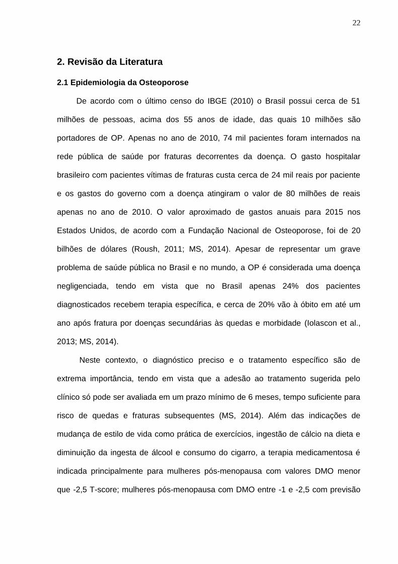
De acordo com a Organização Mundial de Saúde (OMS) a OP é definida como
uma doença sistêmica do esqueleto, caracterizada pela diminuição da densidade
mineral óssea (DMO) e deterioração da microarquitetura do tecido. Disso decorre o
aumento da fragilidade do osso e maior predisposição à fraturas (Kanis, 2008; Zaia,
2015; Rocha-Braz; Ferraz-de-Souza, 2016) (Figura 1). Apesar das técnicas de
prognóstico, diagnóstico e monitoramento estarem intimamente relacionadas à DMO
e aos níveis de estrógeno e calcemia, sabe-se que muitos são os fatores envolvidos
na patogenia da doença, classificados como fatores clínicos de origem óssea e não
óssea (Aspray, 2014).
Figura 1. Micrografia de osso. A: Osso normal. B: Osso osteoporótico.
Elementos como idade avançada, etnia caucasiana ou asiática, baixo peso corporal,
histórico familiar da doença, fumo, consumo excessivo de álcool, vida sedentária,
baixos níveis de vitamina D e ingesta insuficiente de cálcio compõem os principais
Fonte: Golob; Laya, 2015.
24
fatores de risco primário relacionados ao desenvolvimento da doença (Golob e Laya,
2015; Greenblatt et al., 2016). Nem sempre a OP tem início na alteração direta da
composição e disposição do tecido ósseo, como na OP primária pós-menopausa.
Em alguns casos ela pode ser secundária à outras patologias como síndromes
hipogonadais, alterações na tireóide, diabetes mellitus, artrite reumatoide, síndromes
de má absorção, doenças crônicas de fígado, rim e pulmão, desordens
hematológicas, além do consumo excessivo de medicamentos, em especial os
glicocorticoides (Pereira, 2009; Golob ; Laya, 2015).
Além destes fatores clínicos citados, componentes de outros sistemas
biológicos tem se mostrado relevantes em pesquisas recentes relacionadas à
desordens esqueléticas, a exemplo do perfil genético dos pacientes e do
comportamento do sistema imune e suas proteínas (Arron e Choi, 2000; Zupan et al.,
2013; Brincat et al., 2014). Com o mapeamento do genoma completo, vários genes
do sistema imune foram associados à diminuição da DMO e aumento da fragilidade
óssea, apresentando possíveis novos marcadores prognósticos e terapêuticos da
doença (Liu et al., 2014; Clark; Duncan, 2015; Qin et al., 2016).
A importância do aprofundamento nesses estudos de base fica ainda mais
evidente diante do quadro de subdiagnósticos e subtratamentos da OP, o que
agrava este problema na saúde pública (Roush, 2011; Golob; Laya, 2015). No grupo
de doenças negligenciadas, a OP primária pós-menopausa é particularmente
preocupante pelos seus altos índices de acometimento e diminuição significativa na
qualidade de vida de mulheres mais velhas, podendo inclusive, além dos sintomas
de dor intensa e aumento da morbidade, levar à morte (Iolascon et al., 2013;
Baccaro et al., 2015). Outro fator a ser considerado é a heterogeneidade de
25
metodologia utilizada para diagnóstico, o que contribui para imprecisão dos números
de prevalência da doença (Golob; Laya, 2015). Tomando como base alguns estudos
populacionais feitos no Brasil e Estados Unidos, a taxa média de mulheres
acometidas após os 50 anos de idade varia entre 15-35% (Pereira, 2009; Golob;
Laya, 2015). De acordo com a OMS até o ano de 2050 é esperado um aumento de
400% no número de fraturas de quadril, na população de até 60 anos, e de 700% na
população com idade superior a 65 anos, apenas para América Latina (Kanis, 2008).
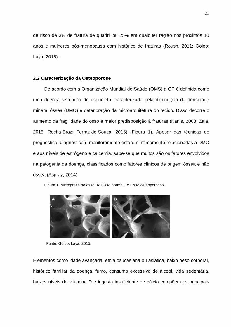
Por ser uma patologia de causa multifatorial e, em grande parte das vezes
assintomática, seu diagnóstico pode ser impreciso e de difícil predição. Inicialmente,
esse diagnóstico se baseava apenas nos índices de DMO obtidos através da
absormetria de dupla emissão com raios-X (DEXA), com resultado posteriormente
classificado de acordo com o número de desvios padrão superior ou inferior do
paciente em relação à DMO média de uma população jovem saudável de referência,
o T-score (Golob; Laya, 2015; Zaia, 2015). Os resultados de T-score superiores a -
1,0 são considerados como normais, entre -1,0 e -1,99 como osteopenia, entre -2,0
e 2,49 como osteopenia avançada, abaixo de -2,5 como OP e T-score abaixo de -2,5
associado a uma ou mais fraturas, são classificados como OP severa (Figura 2)
(Baccaro et al., 2015; Golob; Laya, 2015).
Figura 2: Valores de T-scores resultantes dos exames DEXA e seus respectivos diagnósticos
associados de acordo com a OMS.
Normal
Osteopenia
Osteoporose
- 1,0
- 2,5 T-score Diagnóstico
Fonte: Autor
26
No entanto, outros estudos tem revelado que a DMO analisada isoladamente
não possui alta eficácia diagnóstica, tendo em vista que não apenas a quantidade de
tecido ósseo é importante na homeostase do sistema, mas também a sua
microarquitetura, responsável por corrigir a fragilidade óssea decorrente da
reabsorção da matriz tecidual (Aspray, 2014; Zaia, 2015).
Outros componentes que podem auxiliar nesse contexto são os marcadores
bioquímicos do turnover osséo (Greenblatt et al., 2016). A variação dessas proteínas
decorrentes da atividade das células ósseas pode ser detectada mais rapidamente
que as mudanças na massa óssea (Chesnut et al., 1995; Greenblatt et al., 2016). Os
principais marcadores utilizados na clínica e pesquisa em OP são a fosfatase
alcalina (ALP), osteocalcina (OC), fosfatase ácida tartarato resistente (TRAP) e o
telopeptídeo carboxiterminal do colágeno tipo I (CDX) (Seibel, 2005; Zhou et al.,
2014; Zhou et al., 2015). Adicionalmente, níveis do hormônio paratireoide (PTH), 25
(OH) hidroxivitamina D e cálcio são monitorados nesse processo (Zhou et al., 2014;
Zhou et al., 2015). No entanto, esses elementos geralmente estão estritos ao
acompanhamento e não ao diagnóstico ou prognóstico da doença (Greenblatt et al.,
2016). Na tentativa de preencher essas lacunas, o conhecimento dos constituintes
celulares e inorgânicos e processos biológicos envolvidos na doença são relevantes,
não apenas para o diagnóstico e prognóstico, mas também para alvos terapêuticos
eficazes no combate à patologia.
2.2.1 O tecido ósseo
O osso é um tecido dinâmico de alto poder regenerativo constituído por
componentes celulares (2%) e matriz óssea (98%) com homeostase garantida por
27
diversos fatores como hormônios, vitaminas, níveis de cálcio e citocinas, que
coordenam a atividade dos três tipos celulares formadores do tecido: os osteoclastos,
osteoblastos e osteócitos (Sathy et al., 2015.; Lee; Shin 2007; Lewiecki, 2011; Sims;
Walsh, 2012; Faienza et al., 2013).
Os osteoclastos são grandes fagócitos multinucleados originados de células-
tronco hematopoiéticas que reabsorvem o osso através da ação de enzimas ácidas
secretadas na zona de vedação resultante da ligação dessas células na superfície
óssea por intermédio de integrinas com longos domínio extracelulares (Lewiecki,
2011). Sua ativação é iniciada pela ação do RANKL secretada por osteoblastos que
se liga ao receptor do fator nuclear kappa B (RANK) na superfície do osteoclasto,
sendo todo processo mediado pelo fator estimulador de colônia de macrófago (M-
CSF). Com a ativação, proliferação e aumento da sobrevivência dessas células, tem
início todo processo de reabsorção óssea que é modulado pela ação do receptor
solúvel osteoprotegerina (OPG), secretado também por osteoblastos, que inibe
competitivamente a ligação entre o RANKL e o seu receptor RANK. Os níveis de
expressão dessas citocinas e receptores pelas células ósseas é controlado e
mediado por fatores de crescimento, hormônios, drogas e principalmente pela ação
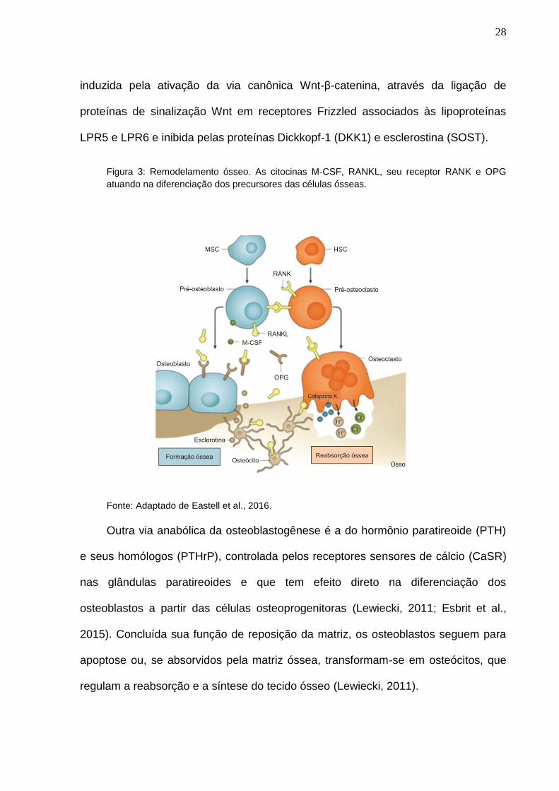
de outras citocinas (Figura 3) (Lewiecki, 2011; Drake et al., 2015).
A reabsorção é também mediada e compensada pelos osteoblastos, células
originadas de células-tronco mesenquimais, através da secreção de colágeno tipo I
e outras substâncias não colágenas, como a osteopontina, responsáveis por
constituir a matriz óssea do tecido retirada pelos osteoclastos. Esse processo ocorre
não apenas na patologia, mas também em processos fisiológicos normais (Seeman;
Delmas, 2006; Lewiecki, 2011). A estimulação para diferenciação de osteoblastos é
28
induzida pela ativação da via canônica Wnt-β-catenina, através da ligação de
proteínas de sinalização Wnt em receptores Frizzled associados às lipoproteínas
LPR5 e LPR6 e inibida pelas proteínas Dickkopf-1 (DKK1) e esclerostina (SOST).
Figura 3: Remodelamento ósseo. As citocinas M-CSF, RANKL, seu receptor RANK e OPG
atuando na diferenciação dos precursores das células ósseas.
Fonte: Adaptado de Eastell et al., 2016.
Outra via anabólica da osteoblastogênese é a do hormônio paratireoide (PTH)
e seus homólogos (PTHrP), controlada pelos receptores sensores de cálcio (CaSR)
nas glândulas paratireoides e que tem efeito direto na diferenciação dos
osteoblastos a partir das células osteoprogenitoras (Lewiecki, 2011; Esbrit et al.,
2015). Concluída sua função de reposição da matriz, os osteoblastos seguem para
apoptose ou, se absorvidos pela matriz óssea, transformam-se em osteócitos, que
regulam a reabsorção e a síntese do tecido ósseo (Lewiecki, 2011).
29
Os osteócitos se comunicam entre si e com as células da superfície através de
prolongamentos nervosos em canalículos do osso, respondendo à microfissuras e
pressões mecânicas (mechanosensation) e outras condições como alteração dos
níveis de hormônios e glicocorticoides. Essa regulação na remodelação óssea pode
favorecer não só a reabsorção mas também a reposição do tecido (Lewiecki, 2011;
Asada et al., 2013). Essas células produzem tanto substancias inibitórias da via Wnt
e estimuladoras da proliferação de osteoclastos, como a esclerostina, quanto
produtos anabólicos que atuam na sobrevivência dos osteoblastos, a exemplo das
prostaglandinas e óxido nítrico (Asada et al., 2013). Além dos componentes
celulares, os osteócitos também regulam a liberação de minerais da matriz óssea,
neste caso impedindo sua reabsorção (Pereira, 2009).
A matriz óssea possui uma composição mineral (70%), constituída de cristais
de cálcio e fosfato (hidroxipatita) e orgânica (30%), constituída de colágeno tipo I e
outras proteínas não colágenas. As glicoproteínas e os proteoglicanos formam a
substância amorfa sobre a qual os cristais de hidroxiapatita se ajustam às fibras
colágenas (Pereira, 2009).
O tecido ósseo mantem a estrutura do corpo humano, protege órgãos vitais,
confere movimentos às contrações musculares, além de ser uma reserva de íons
essenciais para a vida do indivíduo, como fósforo e cálcio. A desregulação entre
formação e reabsorção óssea provoca patologias decorrentes da diminuição de
DMO e perda da microarquitetura do tecido, a exemplo da OP. Apesar de todo esse
sistema ser multifatorialmente influenciado, a íntima relação do sistema imunológico
com a homeostase do tecido tornou-se destaque, principalmente após os estudos

30
envolvendo genes de citocinas e remodelamento ósseo, na emergente
Osteoimunologia (Arron; Choi, 2000; Pereira, 2009) .
2.2.2 Osteoimunologia
Apesar de várias citocinas já estarem comprovadamente relacionadas às
variações de DMO e a relação osso e células do sistema imune estar clara devido à
sua localização de origem, o termo Osteoimunologia foi utilizado pela primeira vez
por Arron e Choi no ano de 2000, em uma revisão onde os autores ressaltam que a
relação entre ambos vai além das células imunes se formarem na medula óssea. Os
linfócitos T e muitas de suas proteínas teriam relação direta com o remodelamento
ósseo, através do controle da ação dos osteoclastos, osteoblastos e osteócitos
(Takayanagi et al., 2000).
A via mais conhecida e abordada na osteoimunologia é a RANKL/RANK/OPG,
onde o RANKL é expresso nos linfócitos T ativados por inflamação ou queda de
estrógeno sérico, e se liga ao RANK nos osteoclastos, ativando o Fator 6 associado
ao receptor TNF (TRAF6), que por sua vez irá ativar o fator nuclear kappa B (NFkB),
Jun-c kinase N-terminal (JNK) e c-Src, responsáveis pela diferenciação, ativação e
sobrevivência dos osteoclastos (Figura 4).
31
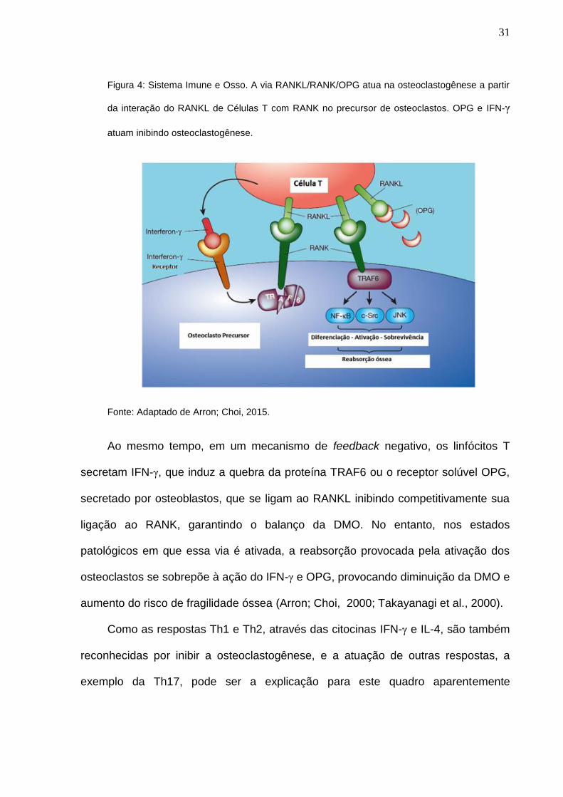
Figura 4: Sistema Imune e Osso. A via RANKL/RANK/OPG atua na osteoclastogênese a partir
da interação do RANKL de Células T com RANK no precursor de osteoclastos. OPG e IFN-γ
atuam inibindo osteoclastogênese.
Fonte: Adaptado de Arron; Choi, 2015.
Ao mesmo tempo, em um mecanismo de feedback negativo, os linfócitos T
secretam IFN-γ, que induz a quebra da proteína TRAF6 ou o receptor solúvel OPG,
secretado por osteoblastos, que se ligam ao RANKL inibindo competitivamente sua
ligação ao RANK, garantindo o balanço da DMO. No entanto, nos estados
patológicos em que essa via é ativada, a reabsorção provocada pela ativação dos
osteoclastos se sobrepõe à ação do IFN-γ e OPG, provocando diminuição da DMO e
aumento do risco de fragilidade óssea (Arron; Choi, 2000; Takayanagi et al., 2000).
Como as respostas Th1 e Th2, através das citocinas IFN-γ e IL-4, são também
reconhecidas por inibir a osteoclastogênese, e a atuação de outras respostas, a
exemplo da Th17, pode ser a explicação para este quadro aparentemente
32
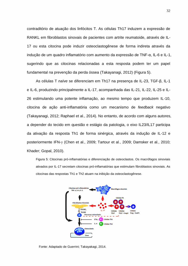
contraditório de atuação dos linfócitos T. As células Th17 induzem a expressão de
RANKL em fibroblastos sinovais de pacientes com artrite reumatoide, através de IL-
17 ou esta citocina pode induzir osteoclastogênese de forma indireta através da
indução de um quadro inflamatório com aumento da expressão de TNF-α, IL-6 e IL-1,
sugerindo que as citocinas relacionadas a esta resposta podem ter um papel
fundamental na prevenção da perda óssea (Takayanagi, 2012) (Figura 5).
As células T naïve se diferenciam em Th17 na presença de IL-23, TGF-β, IL-1
e IL-6, produzindo principalmente a IL-17, acompanhada das IL-21, IL-22, IL-25 e IL-
26 estimulando uma potente inflamação, ao mesmo tempo que produzem IL-10,
citocina de ação anti-inflamatória como um mecanismo de feedback negativo
(Takayanagi, 2012; Raphael et al., 2014). No entanto, de acordo com alguns autores,
a depender do tecido em questão e estágio da patologia, o eixo IL23/IL17 participa
da ativação da resposta Th1 de forma sinérgica, através da indução de IL-12 e
posteriormente IFN-γ (Chen et al., 2009; Tartour et al., 2009; Damsker et al., 2010;
Khader; Gopal, 2010).
Figura 5: Citocinas pró-inflamatórias e diferenciação de osteoclastos. Os macrófagos sinoviais
ativados por IL-17 secretam citocinas pró-inflamatórias que estimulam fibroblastos sinoviais. As
citocinas das respostas Th1 e Th2 atuam na inibição da osteoclastogênese.
Fonte: Adaptado de Guerrini; Takayakagi, 2014.
Fonte: Adaptado de Guerrini; Takayakagi, 2014.
33
Pela reconhecida atuação das citocinas na diferenciação, proliferação, ativação
e sobrevivência das células ósseas de forma direta ou indireta, essas proteínas
representam um potencial não apenas prognóstico, impedindo tratamentos
desnecessários e negligenciando intervenções necessárias, mas também
terapêutico, tendo em vista as diversas dificuldades encontradas nas diretrizes
terapêuticas atuais (Takayanagi, 2012; Guerrini; Takayanagi, 2014; Makras et al.,
2015).
2.3 Tratamento da Osteoporose
Os medicamentos indicados para OP são classificados em três grupos de
acordo com seu mecanismo de ação: os anti-catabólicos ou anti-reabsortivos,
representados pelo cálcio, vitamina D, bisfosfonatos, estrógeno, moduladores
seletivos do receptor de estrógeno, calcitonina e denosumab; os anabólicos,
representados pela teriparatida e hormônio paratireoide e, por último, os de ação
incerta, que provavelmente possuem ambas as atividades, como o ranelato de
estrôncio (Riancho; Hernández, 2012).
A administração de cálcio é indicada para todos os tipos de tratamentos, em
especial para riscos de fraturas vertebrais. No entanto, as mulheres com risco de
doença cardíaca e renal devem dar preferência à obtenção de cálcio na dieta, já que
alguns estudos sugerem que o mesmo eleva o risco de doenças nesses órgãos
(Baccaro et al., 2015; Golob; Laya, 2015). A primeira linha de medicamento de
escolha é a classe dos bisfosfonatos, representada pelo alendronato, risendronato,
ibandronato e ácido zolendrônico, que atuam impedindo a osteoclastogênese
através do acúmulo de análogos do ATP no citoplasma dos osteoclastos (não-
34
nitrogenados) ou interrompendo a via do mevalonato (nitrogenados) induzindo
apoptose e modulando a reabsorção óssea (Riancho e Hernández, 2012; MS, 2014).
O mecanismo de ação do estrógeno e dos moduladores seletivos do receptor de
estrógeno (SERMS), este último com menos efeitos colaterais, ainda não está bem
definido, mas acredita-se que não tenha ação direta na contenção da
osteoclatogênese, mas sim na modulação através da alteração dos níveis de
citocinas com consequente diminuição dos níveis de reabsorção óssea. Já a
calcitonina age diretamente nos osteoclastos, através de uma ligação via receptores
de membrana e consequente aumento do cAMP (Riancho; Hernandez, 2012). O
denosumabe, aprovado em 2010 para OP de alto risco, é um anticorpo humano anti-
RANKL que impede a ligação da proteína ao seu receptor e inibe a ativação e
proliferação de osteoclastos, diminuindo a reabsorção óssea (Cummings et al., 2009;
Lewiecki, 2011; Riancho; Hernández, 2012).
A classe dos anabólicos (Teriparatida ou PTH) atuam de forma diferente dos
antirreabsortivos, estimulando a remodelação óssea, aumentando não apenas a
quantidade de osso, mas promovendo a reestruturação da microarquitetura do
tecido através da melhora da conectividade trabecular (Esbrit et al., 2015; Yoshiki et
al., 2016). Seu mecanismo de ação é amplo indo desde estímulos diretos à
proliferação de osteoblastos quanto modulação da via Wnt e estimulo de expressão
de citocinas (Esbrit et al., 2015).
Em 2016, Eastell e cols. compilaram resultados de metanálises do efeito das
drogas utilizadas com a incidência de fraturas de quadril, vertebrais e não-vertebrais
(Figura 6). De acordo com a análise, dentre as terapias mais utilizadas, o uso
apenas de cálcio e vitamina D apresentaram riscos de fraturas em todos os sítios
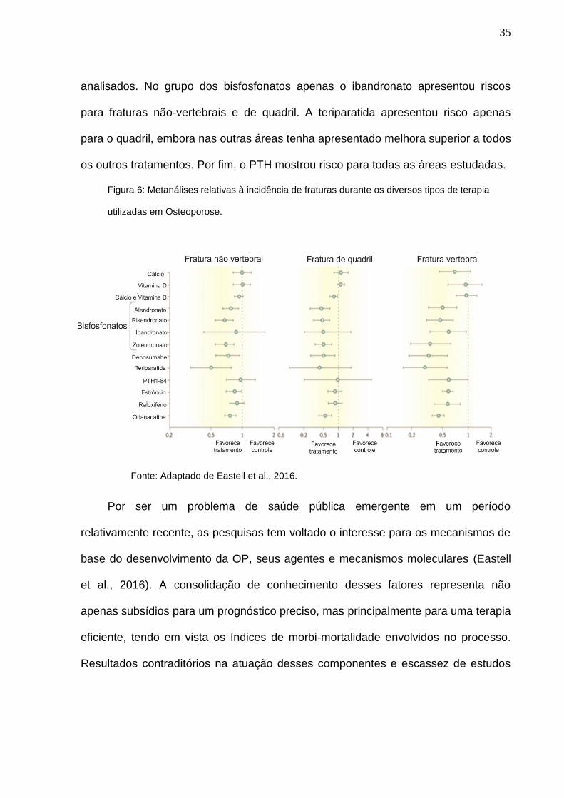
35
analisados. No grupo dos bisfosfonatos apenas o ibandronato apresentou riscos
para fraturas não-vertebrais e de quadril. A teriparatida apresentou risco apenas
para o quadril, embora nas outras áreas tenha apresentado melhora superior a todos
os outros tratamentos. Por fim, o PTH mostrou risco para todas as áreas estudadas.
Figura 6: Metanálises relativas à incidência de fraturas durante os diversos tipos de terapia
utilizadas em Osteoporose.
Por ser um problema de saúde pública emergente em um período
relativamente recente, as pesquisas tem voltado o interesse para os mecanismos de
base do desenvolvimento da OP, seus agentes e mecanismos moleculares (Eastell
et al., 2016). A consolidação de conhecimento desses fatores representa não
apenas subsídios para um prognóstico preciso, mas principalmente para uma terapia
eficiente, tendo em vista os índices de morbi-mortalidade envolvidos no processo.
Resultados contraditórios na atuação desses componentes e escassez de estudos
Fonte: Adaptado de Eastell et al., 2016.
36
funcionais envolvendo estes elementos fornecem ainda mais subsídios para estes
estudos (Blumenfeld et al., 2014).
2.4 Genética da Osteoporose
Um dos principais fatores envolvidos no estabelecimento da OP é a
hereditariedade, especialmente para variações de DMO, risco de fraturas e a área
de ocorrência desses eventos (Rivadeneira et al., 2009; Eastell et al. 2016; Karasik
et al., 2016; Rocha-Braz; Ferraz-de-Souza, 2016). Nos últimos anos a tecnologia
array e os sequenciamentos de nova geração tem permitido a ampliação do número
de genes estudados e sua relação com aspectos clínicos da OP, sugerindo assim
possíveis vias atuantes no processo de remodelamento do tecido ósseo (Riancho;
Hernández, 2012; Karasik et al., 2016). O reconhecimento do componente genético
na doença cresceu de tal forma que 2 consórcios internacionais foram criados ainda
na primeira década do séc XXI com objetivo de identificar variantes genéticas de
risco para a doença: o Consórcio de Marcadores Genéticos para OP (GENOMOS,
do inglês Genetic Markers for Osteoporosis), em 2003 e o Consórcio de Fatores
Genéticos para OP (GEFOS do inglês Genetic Factors for Osteoporosis Consortium),
em 2008. O GEFOS conduziu 3 estudos de metanálises a partir de GWAS nos quais
36.016 SNPs em mais de 150 genes foram avaliados. Nesses estudos um total de
mais de 240 SNPs localizados em 23 diferentes genes apresentaram associação
com variações de DMO, dentre eles os genes do RANKL (TNFSF11), RANK
(TNFRSF11A) e OPG (TNFRSF11B). Em relação à fraturas, foi reportado um total
de 60 SNPs em 5 genes, dentre eles o RANKL (TNFSF11) e RANK (TNFRSF11A).
37
Esta iniciativa tem sido essencial para os avanços nos conhecimentos dos
fatores genéticos relacionados à doença, pois apesar de variantes raras ou genes
relacionados à doenças autossômicas como SOST e Wnt terem sido associados à
OP, a patologia sendo uma doença complexa, parece ser mais influenciada por
variantes comuns que contribuem conjuntamente para a susceptibilidade e
alterações fenótipicas (Riancho e Hernández, 2012; Karasik et al., 2016). A DMO,
por exemplo, é controlada em 60 a 80% dos casos por diferentes genes atuando em
conjunto, de acordo com estudos realizados em gêmeos idênticos (Flicker et al.
1995; Zhou et al., 2014).
Em 2014, Liu e cols. conduziram um update dos GWAS realizados em OP,
avaliando 19 estudos dos quais 5 foram metanálises e dentre essas, 2 realizadas
pelo GEFOS. Os GWAS identificaram vários polimorfismos genéticos em mais de 60
genes associados à variações de DMO, em especial em genes de receptores de
vitamina D (VDR), receptores de estrógeno (ESR-α e ESR-β) e de componentes das
vias de osteoclasto e osteoblastogênese, como as vias RANKL/RANK/OPG, Wnt e
MVK (Riancho e Hernández, 2012; Urano e Inoue, 2014; Clark e Duncan, 2015;
Karasik et al., 2016). Além disso, cerca de 20 genes foram identificados como
relacionados ao risco de fraturas e mais de 100 à susceptibilidade à OP (Liu et al.,
2014; Clark; Duncan, 2015; Karasik et al., 2016).
Dentre esses genes relacionados à doença, pode-se destacar os genes do
sistema imune. O gene do receptor da IL-21 (IL21R) e receptor do fator de
transformação do crescimento beta III (TGFBR3) foram associados à mudanças de
DMO em mulheres caucasianas chinesas, enquanto o genes do TNFSF11,
TNFRSF11A e TNFRSF11B foram relacionados com o processo de
38
osteoclastogênese em mulheres caucasianas chinesas e europeias (Liu et al., 2014;
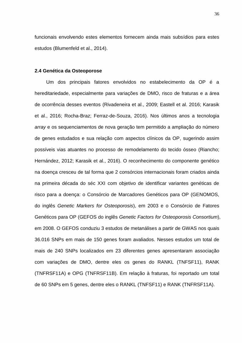
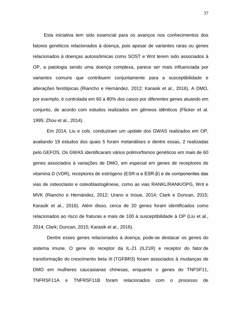
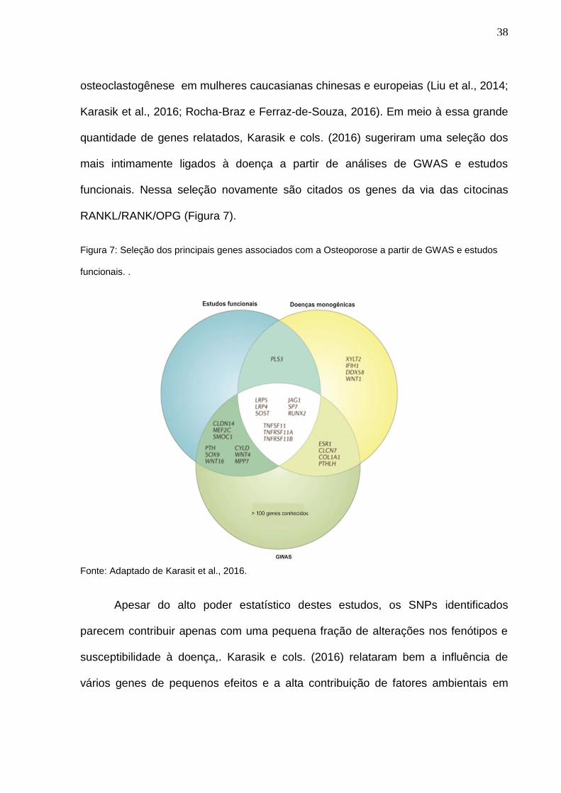
Karasik et al., 2016; Rocha-Braz e Ferraz-de-Souza, 2016). Em meio à essa grande
quantidade de genes relatados, Karasik e cols. (2016) sugeriram uma seleção dos
mais intimamente ligados à doença a partir de análises de GWAS e estudos
funcionais. Nessa seleção novamente são citados os genes da via das citocinas
RANKL/RANK/OPG (Figura 7).
Figura 7: Seleção dos principais genes associados com a Osteoporose a partir de GWAS e estudos
funcionais. .
Fonte: Adaptado de Karasit et al., 2016.
Apesar do alto poder estatístico destes estudos, os SNPs identificados
parecem contribuir apenas com uma pequena fração de alterações nos fenótipos e
susceptibilidade à doença,. Karasik e cols. (2016) relataram bem a influência de
vários genes de pequenos efeitos e a alta contribuição de fatores ambientais em
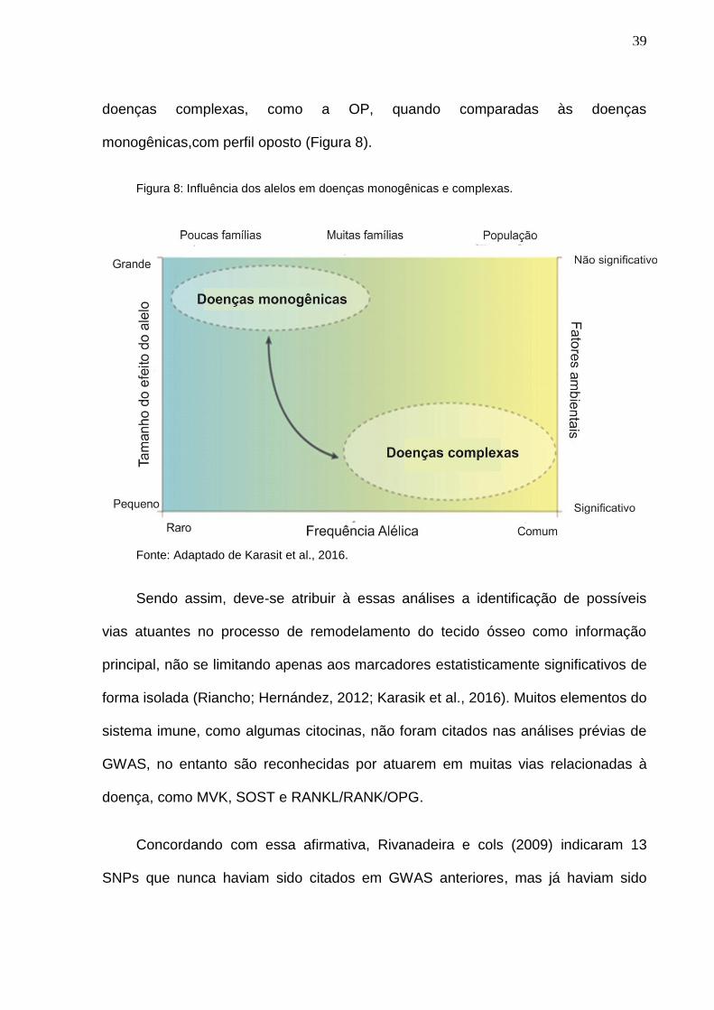
39
doenças complexas, como a OP, quando comparadas às doenças
monogênicas,com perfil oposto (Figura 8).
Figura 8: Influência dos alelos em doenças monogênicas e complexas.
Fonte: Adaptado de Karasit et al., 2016.
Sendo assim, deve-se atribuir à essas análises a identificação de possíveis
vias atuantes no processo de remodelamento do tecido ósseo como informação
principal, não se limitando apenas aos marcadores estatisticamente significativos de
forma isolada (Riancho; Hernández, 2012; Karasik et al., 2016). Muitos elementos do
sistema imune, como algumas citocinas, não foram citados nas análises prévias de
GWAS, no entanto são reconhecidas por atuarem em muitas vias relacionadas à
doença, como MVK, SOST e RANKL/RANK/OPG.
Concordando com essa afirmativa, Rivanadeira e cols (2009) indicaram 13
SNPs que nunca haviam sido citados em GWAS anteriores, mas já haviam sido
40
reportados em estudos de associação comuns. Esses dados juntamente com as
descobertas em Osteoimunologia encorajam as pesquisas com SNPs em genes de
citocinas associadas a essas vias, mesmo que estas não tenham sido citados em
GWAS prévios. Estudos recentes tem descrito citocinas como o IFN-γ inibindo a via
RANKL/RANK/OPG bem como a IL-17 induzindo essa mesma via, ambas afetando
indiretamente os processos de homeostase óssea (Guerrini; Takayanagi, 2014;
Talaat et al., 2015). SNPs funcionais em genes dessas citocinas, bem como nas
suas principais indutoras, a IL-23 e a IL-12, respectivamente, tem sido reportados
em diversas doenças, inclusive em desordens ósseas (Wong et al., 2012; Emami et
al., 2015; Marques et al., 2015; Zhang et al., 2015; Leng et al., 2016).
Embora seja reconhecido a importância dessas proteínas do sistema imune no
estabelecimento da OP, poucos estudos tem sido feitos relacionando funcionalmente
esses SNPs e a susceptibilidade à OP pós-menopausa e sua resposta terapêutica.
2.4.1. SNPs em genes de citocinas
As citocinas são proteínas com papeis essenciais não apenas na imunidade
inata e adaptativa, mas também influenciando e sendo influenciada por diversos
sistemas e órgãos como o osso, além de estarem associadas ao desenvolvimento
de diversas patologias (Bhushan; Perumal, 2012; Takayanagi, 2012). SNPs podem
alterar tanto a estrutura (éxon e íntrons em regiões de splincing) quanto os níveis
dessas proteínas (regiões regulatórias: 5’ e 3’ UTR e promotoras) e muitos estudos,
inclusive os de GWAS anteriormente citados, tem identificado marcadores
importantes na susceptibilidade e manifestações de diversas doenças multifatoriais,
como a OP (Richards et al., 2008; Rivadeneira et al., 2009; Mah et al., 2011; Qin et
41
al., 2016). Por outro lado, por não apresentar associação com patologias em outra
parcela de estudos, muitos questionamentos são feitos em relação ao papel desses
SNPs e as interpretações decorrentes dos estudos com associações
estatisticamente significativas (Bhushan; Perumal 2012).
Com intuito de derivar parâmetros para relação de causalidade entre SNPs e
doenças, foi criado em 2012 o DACS-DB, do inglês Disease Associated Cytokine
SNP Database (http://www.iupui.edu/~cytosnp). Nesse banco público de dados
constam as informações de 453 genes de citocinas, 63.000 SNPs e 853 doenças
associadas à esses SNPs. Outros bancos de dados mais gerais que podem ser
consultado para estudos são o 1000 genomes, Ensembl Genome Browser e a seção
dbSNSP do National Center for Biotechnology Information (NCBI). Os sites de livre
acesso fornecem informações de localização dos polimorfismos, frequência do alelo
menor de acordo com as diferentes populações e estudos científicos publicados. Os
quatro SNPs avaliados nesse estudo, IL23R +2284 (C>A, rs10889677), IL17A +672
(G>A, rs7747909), IL12B +1188 (T>G, rs3212227) e IFNG -1616 (G>A, rs2069705),
estão cadastrados em todos os bancos de dados citados.
O IL23R +2284 (C>A, rs10889677) está localizado na região regulatória 3’UTR
do gene, no cromossomo 1, já foi citado em 98 estudos e mostrou associação com o
aumento do risco de doença de Crohn, espondilite anquilosante e Doença de Graves.
O IL17A +672 (G>A, rs7747909), também localizado na região regulatória 3’UTR do
gene no cromossomo, foi citado em 8 estudos, dentre os quais conferiu aumento de
risco para arterite, panuveíte e degeneração macular. O IL12B +1188 (T>G,
rs3212227), também da 3’UTR do gene no cromossomo 5, foi citado em 147
estudos e mostrou associação com a psoríase e artrite psoriática. E por fim o IFNG -
42
1616 (G>A, rs2069705), que embora seja citado em estudos prévios como
localizado na região promotora do gene, está atualmente registrado com localização
intrônica no cromossomo 12 e foi mencionado em 41 estudos publicados. O SNP foi
reportado como associado à desfechos de mortalidade em pacientes norte-
americanos (1000 Genomes - http://www.internationalgenome.org).
Lima e cols. (2017) relataram pela primeira vez a relação entre esses SNPs e
marcadores bioquímicos de remodelagem óssea e variações de DMO em pacientes
sob tratamento de bisfosfonatos. No estudo, os SNPs IL17A rs7747909 juntamente
com IL12 rs3212227 estiveram associados à variações nos níveis de vitamina D, e
IL23R rs10889677 com alterações na calcemia. O SNP IFNG rs2069705 foi o que
apresentou maior relação com a resposta terapêutica, estando associado à
alterações nos valores de PTH e DMO durante os 4 anos de tratamento. Esses
dados sugerem um importante papel desses SNPs nas decisões terapêuticas em
OP.
Algumas ferramentas computacionais também tem sido uma abordagem
utilizada para medida dos efeitos dos SNPs nas suas proteínas correspondentes,
além de simulações de respostas terapêuticas. Essas análises in silico representam
uma saída rápida e relativamente de fácil execução, comparadas aos experimentos
que precisariam ser executados para a predição de efeitos fenotípicos da quantidade
crescente de genes previamente associados à doenças (Mah et al., 2011; Singh;
Mistry, 2016). Esses algoritmos mensuram as diferenças de energia livre,
solubilidade e dinâmica das proteínas originadas dos diferentes SNPs e suas
variantes selvagens e mutantes (Singh; Mistry, 2016). As sequencias a serem
estudas podem ser obtidas a partir dos bancos de dados disponíveis e as
43
simulações feitas em softwares escolhidos de acordo com a análise desejada. Para
análises inicias, programas com I-Mutant e SNP&Go podem predizer o efeito dos
SNPs na estabilidade da proteína, enquanto ferramentas como o Swiss-PdbViewer
mensuram o efeito dessas variantes a partir da análise da energia de forças de
campo (Force Field Energies) e energia de minimização (Minimization Energies).
Valores mais altos de energia correspondem a proteínas mais instáveis (Singh;
Mistry, 2016; Swiss-PdbViewer tutorial ).
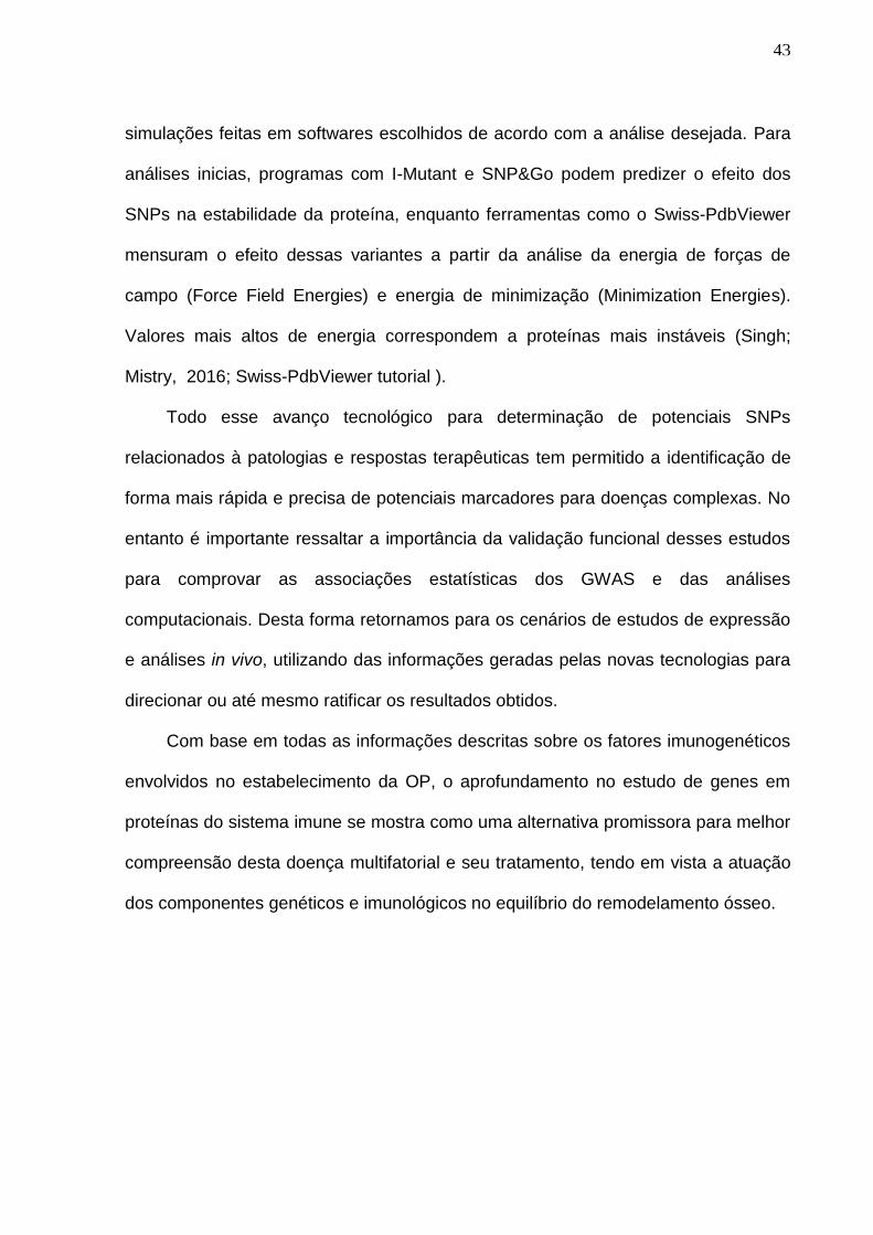
Todo esse avanço tecnológico para determinação de potenciais SNPs
relacionados à patologias e respostas terapêuticas tem permitido a identificação de
forma mais rápida e precisa de potenciais marcadores para doenças complexas. No
entanto é importante ressaltar a importância da validação funcional desses estudos
para comprovar as associações estatísticas dos GWAS e das análises
computacionais. Desta forma retornamos para os cenários de estudos de expressão
e análises in vivo, utilizando das informações geradas pelas novas tecnologias para
direcionar ou até mesmo ratificar os resultados obtidos.
Com base em todas as informações descritas sobre os fatores imunogenéticos
envolvidos no estabelecimento da OP, o aprofundamento no estudo de genes em
proteínas do sistema imune se mostra como uma alternativa promissora para melhor
compreensão desta doença multifatorial e seu tratamento, tendo em vista a atuação
dos componentes genéticos e imunológicos no equilíbrio do remodelamento ósseo.
44
3. Objetivos
3.1 Objetivo geral
Investigar a influência das citocinas pró-inflamatórias IL-23, IL-17, IL-12 e IFN-γ com
a susceptibilidade e resposta terapêutica à Osteoporose primária pós-menopausa
3.2 Objetivos específicos
1. Determinar a frequência dos SNPs IL23R +2284 (C>A) (rs10889677), IL17A +672
(G>A) (rs7747909), IL12B +1188 (T>G) (rs3212227) e IFNG -1616 (G>A)
(rs2069705) em pacientes com Osteoporose primária pós-menopausa e indivíduos
saudáveis do estado de Pernambuco;
2. Avaliar o grau de associação desses SNPs com a susceptibilidade à OP primária
pós-menopausa e com a resposta terapêutica aos bifosfonatos em pacientes de
Osteoporose primária pós-menopausa;
3. Validar genes de referência para estudos funcionais em Osteoporose;
4. Avaliar comparativamente a expressão, em amostras de sangue periférico, da(s)
citocina(s) associada à susceptibilidade, se existente, em pacientes com
Osteoporose primária pós-menopausa e indivíduos saudáveis;
5. Avaliar a atuação da(s) citocina(s) associada(s) ao desenvolvimento da OP
primária pós-menopausa, se existente, na atividade de células humanas de
formação óssea.

45
4. Capítulo I
Artigo aceito para publicação na revista Inflammopharmacology
Fator de Impacto: 2,3
Qualis CB1: B1
Guia para autores: Anexo A
46
Polymorphisms in key bone modulator cytokines genes influence
bisphosphonates therapy in postmenopausal women
Lima, CAD*1,2; Javorski, NR1,2; Souza, APO2; Barbosa, AD2,3; Valença, APMC4;
Crovella, S1,2; Souza, PRE2,5; De Azevêdo Silva, J1,2 and Sandrin-Garcia, P1,2
1 – Department of Genetics - Federal University of Pernambuco (UFPE), Recife, Pernambuco, Brazil. 2 – Laboratory of Immunopathology Keizo Asami – Federal University of Pernambuco (UFPE), Recife, Pernambuco, Brazil. 3 - Division of Rheumatology, Clinical Hospital, Federal University of Pernambuco (UFPE), Recife, Pernambuco, Brazil. 4 – Department of Oceanography - Federal University of Pernambuco (UFPE), Recife, Pernambuco, Brazil. 5 – Department of Genetics – Rural Federal University of Pernambuco (UFRPE), Recife, Pernambuco, Brazil.
*Corresponding author: Camilla AD Lima
Email address: [email protected]
Accepted for publication in Inflammopharmacology Journal. DOI: 10.1007/s10787-017-0322-7
47
Abstract
Osteoporosis is a multifactorial and debilitating disease resulting from decreased bone mineral
density (BMD) and loss of tissue microarchitecture. Ineffective therapies may lead to bone
fractures and subsequent death. Single nucleotide polymorphisms (SNPs) in key immune
regulator genes have been associated with therapeutic response to bisphosphonates, which are
the first therapeutic line of choice for osteoporosis. However, cytokine pathways and their
relation with therapeutic adhesion remain to be fully elucidated. Aimed at better
understanding these processes, we investigated the response to bisphosphonate therapy in
postmenopausal women and four SNPs in key proinflammatory cytokines genes: IL23R
+2284 (C>A) (rs10889677), IL17A +672 (G>A) (rs7747909), IL12B +1188 (T>G)
(rs3212227) and IFNG -1616 (G>A) (rs2069705). A total of 69 patients treated with
bisphosphonate were followed for a period of 1 up to 4 years, genotyped and compared
according their changes in bone mineral density (BMD) and level of biochemical markers
during their treatment. The IFNG -1616 G/G associated with increased BMD values in
femoral neck (GG/AA, p=0.016) and decreased BMD values in total hip (GG/GA, p= 0.019;
GG/AA, p= 0.011). In relation to biochemical markers, IFNG -1616 SNP associated with
increased alkaline phosphatase (GG/AA; p<
0.0001) and parathyroid hormone levels
(AA/GA; p= 0.017). Vitamin D values changes were related to IL17A +672 (GG/GA,
p=0.034) and to IL12B +1188 (TT/TG, p= 0.046) SNPs. Besides, significant differences in
changes of calcium levels correlated with IL23R +2284 (CC/CA, p= 0.016) genotypes.
Altogether, we suggest that these polymorphisms may play an important role for therapeutic
decisions in osteoporosis treatment.
Keywords: Osteoimmunology, Osteoporosis Therapy, Bone Mineral Density, Bone Biochemical
Markers, Genetics of osteoporosis.
48
1. Introduction
Osteoporosis (OP) is a systemic disease characterized by the decrease of bone mineral
density (BMD) and by loss of tissue microarchitecture. OP main clinical manifestations are
fragility fracture and injuries related to bone break (Sun et al. 2014; Tella and Gallagher 2014;
Drake et al. 2015; Tastan et al. 2016). According to World Health Organization (WHO), OP
diagnosis is determined by BMD measures with T-scores less than -2.5 (Kanis 2008). Despite
being a major problem for global public health, osteoporosis is considered under diagnosed
and, consequently, an undertreated disorder by health care systems (Kerschan-Schindl 2016;
Miller 2016). About half of fractures diagnosed in OP patients are due to unspecific or
inefficient drug therapies, increasing morbidity and mortality rates, since about 20% of
patients die during the first year of the treatment due to bone fractures (Roush 2011; Iolascon
et al. 2013; Drake et al. 2015; Miller 2016).
The bisphosphonates (BPs) are a class of drugs able to decrease bone loss, known as
the anticatabolic therapy of choice for postmenopausal women, mainly due to its low cost
(Zhou et al. 2014; Baccaro et al. 2015). The idea of "one fits all" is increasing morbidity,
severity and overall costs with this disease and can even lead patient’s death due to non-
specific and general treatment (Hiligsmann et al. 2015; Abrahamsen and Prieto-Alhambra
2016). The fracture location and genetic information from patients are pivotal in treating these
women since the phenotypic features in OP are modulated by genetic factors in about 50-80%
from all cases (Riancho and Hernández 2012b).
The influence of genetic polymorphisms in genes involved in the main pathways of
BPs action mechanisms have been well characterized, pointing out the importance of specific
genes such as Wnt, MVK and RANK in the modulation of BPs therapeutic response (Marini
49
and Brandi 2014; Zhou et al. 2014; Wang et al. 2015a; Zheng et al. 2015; Zhou et al. 2015).
Even though genetic polymorphisms within the above-mentioned genes are known to
influence BPs therapeutic response, there are important pathways remaining to be elucidated,
particularly the immune related ones. Noteworthy, cytokines and their network display several
essential functions in the bone remodeling and are closely related to osteoporosis treatment
(Yuan et al. 2012; Marini and Brandi 2014; Talaat et al. 2015). Recently, cytokines such as
IFN-γ have been described as inhibitor of RANKL/RANK/OPG pathway through TRAF 6
protein degradation and IL-17 as inducer of RANK-L and Th17 cells production, both
affecting bone homeostasis processes (Guerrini and Takayanagi 2014; Talaat et al. 2015).
Therefore, we investigated whether there is an association between single nucleotide
polymorphisms (SNPs) within IL23R, IL17A, IL12B and IFN-γ genes from Th1 and Th17
pathways and the response to BPs therapy in a population-based sample of postmenopausal
women from Northeast Brazil treated over a period of 1- 4 years.
2. Materials and methods
2.1 Subjects
In the present study we collected data from 240 postmenopausal osteoporotic women
from the Division of Rheumatology of Clinical Hospital in Federal University of
Pernambuco, Brazil enrolled within 2011 and 2015. The patients were diagnosed according
WHO criteria (Kanis 2008). All therapies included in this study followed the clinical
guidelines in health supplement from National Agency of Health of Brazil (Cunha et al.
2011). Dosage guidelines and oral doses administration recommended were alendronate (70
mg/week), risendronate (35 mg/week), ibandronate (150 mg/week) or intravenous infusion of
zolendronic acid (5 mg/year). Additionally, all women were supplemented with 600 mg/day
50
of elemental calcium and 400 IU/day of vitamin D3. Patients received one of these above-
mentioned treatments for at least 1 year, and only BPs’ class were included in all subjects in
this study. The exclusion criteria in patients’ group were osteopenia, inflammatory disease,
autoimmune diseases, cancer, use of anti-inflammatories and other drugs for bone disorders
such as anabolic agents, hormone replacement therapy or selective estrogen receptor
modulators at any moment of the treatment period.
All the participants provided a written informed consent. This study was approved by
the Research Ethics Committee of the Center for Health Sciences, Federal University of
Pernambuco (CEP/CCS/UFPE nº 513/11) and is in accordance with the Declaration of
Helsinki.
2.2 SNPs selection and genotyping
We selected four SNPs from IL23R, IL17A, IL12B and IFN-γ genes: IL23R +2284
(C>A, rs10889677), IL17A +672 (G>A, rs7747909), IL12B +1188 (T>G, rs3212227) and
IFN-γ -1616 (G>A, rs2069705). SNPs information was obtained from SNP Browser software
4.0 (Applied Biosystems, Foster City, CA, USA) and 1000 Genomes
(http://browser.1000genomes.org/index.html). The selection criteria were minor allele
frequency (MAF) higher than 10% in African (YRI subpopulation) and European (CEU
subpopulation) populations and SNPs positions in regulatory regions such as promoter
regions, 5’ or 3’ UTR.
Genomic DNA extraction was performed from peripheral leukocytes using the rapid
salting out method (Bignon and Viña 1995). Samples were genotyped with specific
fluorogenic probes (Taqman Probes, Applied Biosystems, Foster City, CA, USA), using Real-
51
Time PCR performed with ABI 7500 detection system (Applied Biosystems, Foster City, CA,
USA).
2.3 Assessment of BMD changes in response to BPs Therapy
The BMD areas assessed were: lumbar spine (LS: L1-L4), femoral neck (FN) and total
hip (TH) at the beginning of treatment and after 1, 2, 3 and 4 years. The BPs’ effects in BMD
values were measured by percentage changes of BMD, calculated according to Zhou et al.
(2015) following the formula: % changes of BMD = [(BMDafter treatment – BMDbaseline) /
BMDbaseline ] × 100%.
BMD values were measured by dual-energy x-ray absorptiometry (Hologic or Lunar).
The standardization method for values correction from different machines was described by
Genant et al. (1993) enabling the comparison between the measures from both x-rays (Genant
et al. 1993; Zhou et al. 2015). For BMD values in lumbar spine, the formula applied was:
Hologic BMD (g/cm2) = (Lunar BMD - 0.054)/1.074 (r = 0.987, p < 0.05, standard error of
the estimate = ± 0.030 g/cm2) and for BMD values in both femoral neck and total hip:
Hologic BMD (g/cm2) = (Lunar BMD - 0.142)/1.013 (r = 0.920, p< 0.05, standard error = ±
0.051 g/cm2).
2.4 Assessment of biochemical markers of bone turnover changes
The levels of alkaline phosphatase (ALP) in peripheral blood and calciuria in urine
sample (24-hour collection) were used as indicators of bone activity. The vitamin D, calcium
and parathyroid hormone (PTH) levels in peripheral blood were also analyzed. All measures
were performed on AU680 (Beckman Coulter) and BT 3000 plus (Winer Lab.) equipments
using specific kits. The BPs’ effects in these biomarkers were assessed by percentage changes
52
of biomarkers, calculated according to Zhou et al. (2015) in the following formula (e.g.
Vitamin D): % changes of Vitamin D = [(Vitamin Dafter treatment – Vitamin Dbaseline) / Vitamin
Dbaseline ] × 100%.
2.5 Statistical analyses
Allelic and genotypic frequencies and Hardy–Weinberg equilibrium were performed
using the SNPStats tool (http://bioinfo.iconcologia.net/SNPstats). Comparisons among the
genotypes related to BMD and biochemical markers changes were performed using Analyses
of Variance (ANOVA) or Kruskal-Wallis tests as appropriate. When a significant difference
was observed, Tukey test was applied for pairwise comparisons of genotypes. All statistical
analyses were conducted using the SPSS 18.0 statistical package (SPSS Inc., Chicago, IL,
USA). Differences were accepted as significant at p-values < 0.05.
3. Results
3.1 Patients characteristics
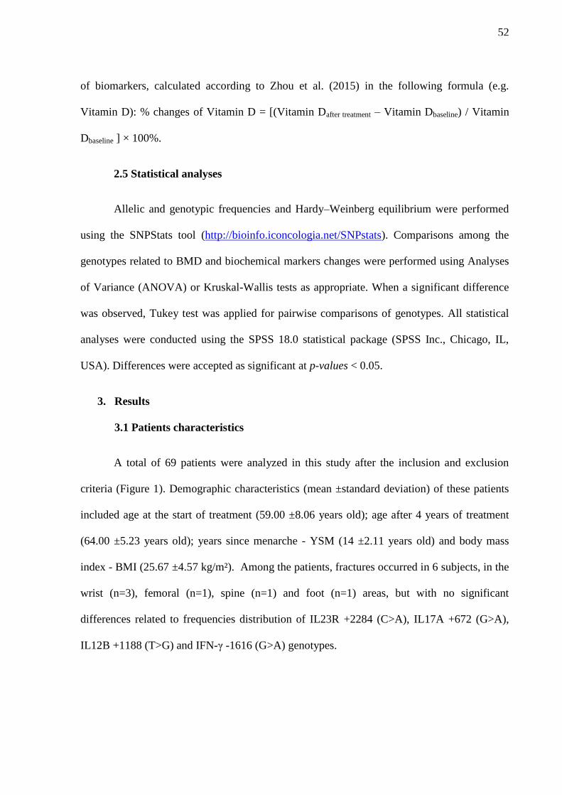
A total of 69 patients were analyzed in this study after the inclusion and exclusion
criteria (Figure 1). Demographic characteristics (mean ±standard deviation) of these patients
included age at the start of treatment (59.00 ±8.06 years old); age after 4 years of treatment
(64.00 ±5.23 years old); years since menarche - YSM (14 ±2.11 years old) and body mass
index - BMI (25.67 ±4.57 kg/m²). Among the patients, fractures occurred in 6 subjects, in the
wrist (n=3), femoral (n=1), spine (n=1) and foot (n=1) areas, but with no significant
differences related to frequencies distribution of IL23R +2284 (C>A), IL17A +672 (G>A),
IL12B +1188 (T>G) and IFN-γ -1616 (G>A) genotypes.
53
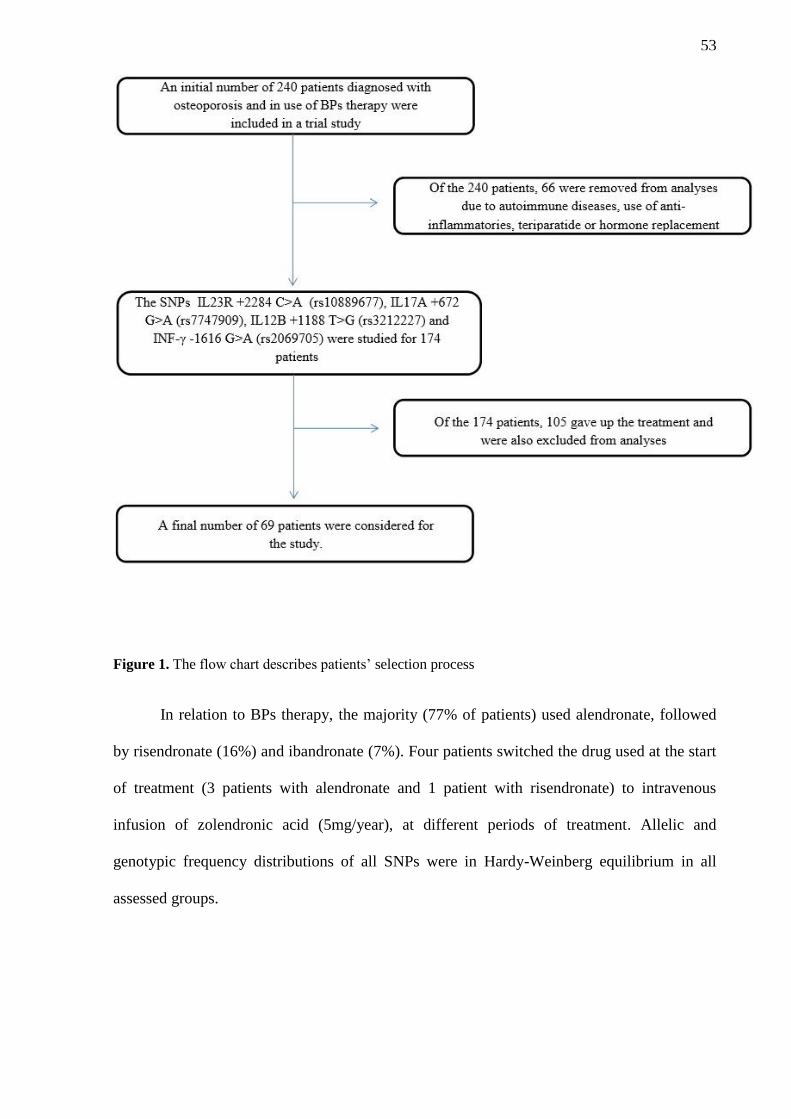
Figure 1. The flow chart describes patients’ selection process
In relation to BPs therapy, the majority (77% of patients) used alendronate, followed
by risendronate (16%) and ibandronate (7%). Four patients switched the drug used at the start
of treatment (3 patients with alendronate and 1 patient with risendronate) to intravenous
infusion of zolendronic acid (5mg/year), at different periods of treatment. Allelic and
genotypic frequency distributions of all SNPs were in Hardy-Weinberg equilibrium in all
assessed groups.
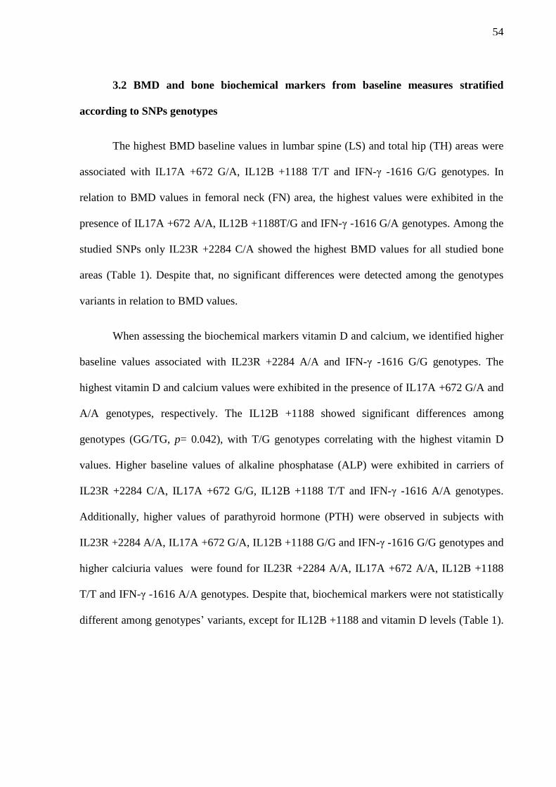
54
3.2 BMD and bone biochemical markers from baseline measures stratified
according to SNPs genotypes
The highest BMD baseline values in lumbar spine (LS) and total hip (TH) areas were
associated with IL17A +672 G/A, IL12B +1188 T/T and IFN-γ -1616 G/G genotypes. In
relation to BMD values in femoral neck (FN) area, the highest values were exhibited in the
presence of IL17A +672 A/A, IL12B +1188T/G and IFN-γ -1616 G/A genotypes. Among the
studied SNPs only IL23R +2284 C/A showed the highest BMD values for all studied bone
areas (Table 1). Despite that, no significant differences were detected among the genotypes
variants in relation to BMD values.
When assessing the biochemical markers vitamin D and calcium, we identified higher
baseline values associated with IL23R +2284 A/A and IFN-γ -1616 G/G genotypes. The
highest vitamin D and calcium values were exhibited in the presence of IL17A +672 G/A and
A/A genotypes, respectively. The IL12B +1188 showed significant differences among
genotypes (GG/TG, p= 0.042), with T/G genotypes correlating with the highest vitamin D
values. Higher baseline values of alkaline phosphatase (ALP) were exhibited in carriers of
IL23R +2284 C/A, IL17A +672 G/G, IL12B +1188 T/T and IFN-γ -1616 A/A genotypes.
Additionally, higher values of parathyroid hormone (PTH) were observed in subjects with
IL23R +2284 A/A, IL17A +672 G/A, IL12B +1188 G/G and IFN-γ -1616 G/G genotypes and
higher calciuria values were found for IL23R +2284 A/A, IL17A +672 A/A, IL12B +1188
T/T and IFN-γ -1616 A/A genotypes. Despite that, biochemical markers were not statistically
different among genotypes’ variants, except for IL12B +1188 and vitamin D levels (Table 1).
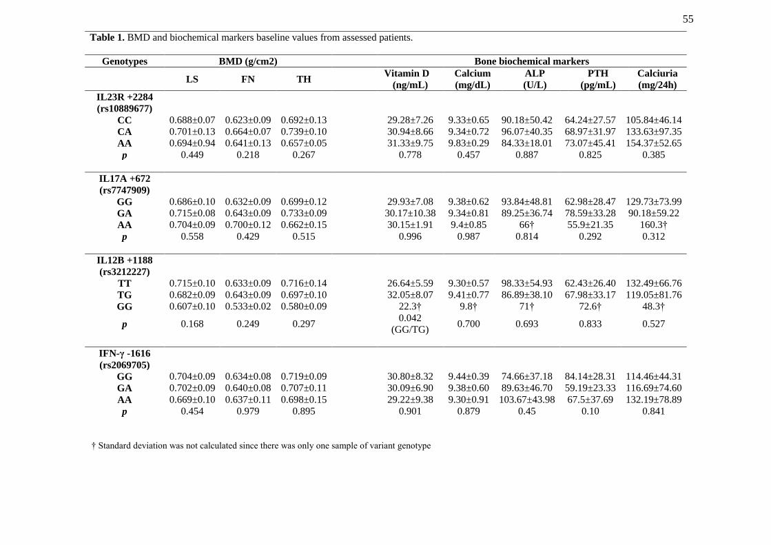
55
Table 1. BMD and biochemical markers baseline values from assessed patients.
Genotypes BMD (g/cm2) Bone biochemical markers
LS FN TH Vitamin D
(ng/mL)
Calcium
(mg/dL)
ALP
(U/L)
PTH
(pg/mL)
Calciuria
(mg/24h)
IL23R +2284
(rs10889677)
CC 0.688±0.07 0.623±0.09 0.692±0.13 29.28±7.26 9.33±0.65 90.18±50.42 64.24±27.57 105.84±46.14
CA 0.701±0.13 0.664±0.07 0.739±0.10 30.94±8.66 9.34±0.72 96.07±40.35 68.97±31.97 133.63±97.35
AA 0.694±0.94 0.641±0.13 0.657±0.05 31.33±9.75 9.83±0.29 84.33±18.01 73.07±45.41 154.37±52.65
p 0.449 0.218 0.267 0.778 0.457 0.887 0.825 0.385
IL17A +672
(rs7747909)
GG 0.686±0.10 0.632±0.09 0.699±0.12 29.93±7.08 9.38±0.62 93.84±48.81 62.98±28.47 129.73±73.99
GA 0.715±0.08 0.643±0.09 0.733±0.09 30.17±10.38 9.34±0.81 89.25±36.74 78.59±33.28 90.18±59.22
AA 0.704±0.09 0.700±0.12 0.662±0.15 30.15±1.91 9.4±0.85 66† 55.9±21.35 160.3†
p 0.558 0.429 0.515 0.996 0.987 0.814 0.292 0.312
IL12B +1188
(rs3212227)
TT 0.715±0.10 0.633±0.09 0.716±0.14 26.64±5.59 9.30±0.57 98.33±54.93 62.43±26.40 132.49±66.76
TG 0.682±0.09 0.643±0.09 0.697±0.10 32.05±8.07 9.41±0.77 86.89±38.10 67.98±33.17 119.05±81.76
GG 0.607±0.10 0.533±0.02 0.580±0.09 22.3† 9.8† 71† 72.6† 48.3†
p 0.168 0.249 0.297 0.042
(GG/TG) 0.700 0.693 0.833 0.527
IFN-γ -1616
(rs2069705)
GG 0.704±0.09 0.634±0.08 0.719±0.09 30.80±8.32 9.44±0.39 74.66±37.18 84.14±28.31 114.46±44.31
GA 0.702±0.09 0.640±0.08 0.707±0.11 30.09±6.90 9.38±0.60 89.63±46.70 59.19±23.33 116.69±74.60
AA 0.669±0.10 0.637±0.11 0.698±0.15 29.22±9.38 9.30±0.91 103.67±43.98 67.5±37.69 132.19±78.89
p 0.454 0.979 0.895 0.901 0.879 0.45 0.10 0.841
† Standard deviation was not calculated since there was only one sample of variant genotype
† Standard deviation was not calculated since there was only one sample of variant genotype
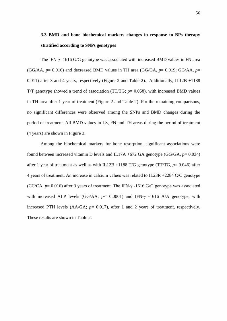
56
3.3 BMD and bone biochemical markers changes in response to BPs therapy
stratified according to SNPs genotypes
The IFN-γ -1616 G/G genotype was associated with increased BMD values in FN area
(GG/AA, p= 0.016) and decreased BMD values in TH area (GG/GA, p= 0.019; GG/AA, p=
0.011) after 3 and 4 years, respectively (Figure 2 and Table 2). Additionally, IL12B +1188
T/T genotype showed a trend of association (TT/TG; p= 0.058), with increased BMD values
in TH area after 1 year of treatment (Figure 2 and Table 2). For the remaining comparisons,
no significant differences were observed among the SNPs and BMD changes during the
period of treatment. All BMD values in LS, FN and TH areas during the period of treatment
(4 years) are shown in Figure 3.
Among the biochemical markers for bone resorption, significant associations were
found between increased vitamin D levels and IL17A +672 GA genotype (GG/GA, p= 0.034)
after 1 year of treatment as well as with IL12B +1188 T/G genotype (TT/TG, p= 0.046) after
4 years of treatment. An increase in calcium values was related to IL23R +2284 C/C genotype
(CC/CA, p= 0.016) after 3 years of treatment. The IFN-γ -1616 G/G genotype was associated
with increased ALP levels (GG/AA; p< 0.0001) and IFN-γ -1616 A/A genotype, with
increased PTH levels (AA/GA; p= 0.017), after 1 and 2 years of treatment, respectively.
These results are shown in Table 2.
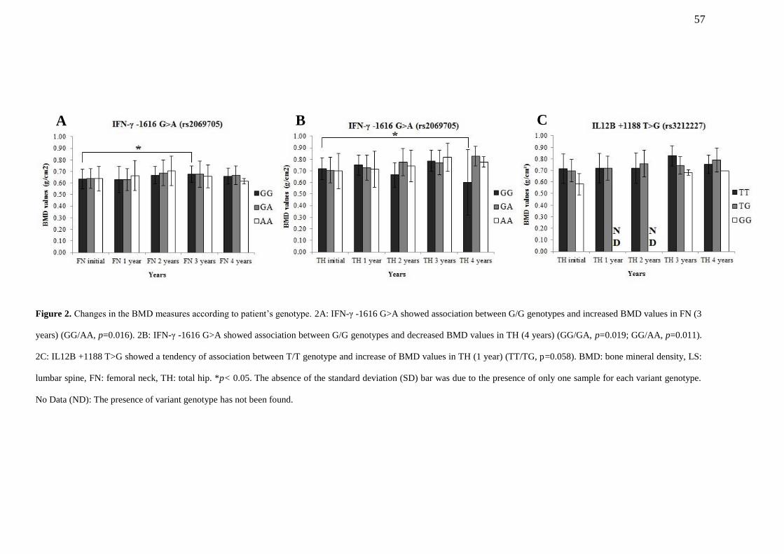
57
Figure 2. Changes in the BMD measures according to patient’s genotype. 2A: IFN-γ -1616 G>A showed association between G/G genotypes and increased BMD values in FN (3
years) (GG/AA, p=0.016). 2B: IFN-γ -1616 G>A showed association between G/G genotypes and decreased BMD values in TH (4 years) (GG/GA, p=0.019; GG/AA, p=0.011).
2C: IL12B +1188 T>G showed a tendency of association between T/T genotype and increase of BMD values in TH (1 year) (TT/TG, p=0.058). BMD: bone mineral density, LS:
lumbar spine, FN: femoral neck, TH: total hip. *p< 0.05. The absence of the standard deviation (SD) bar was due to the presence of only one sample for each variant genotype.
No Data (ND): The presence of variant genotype has not been found.
A B C
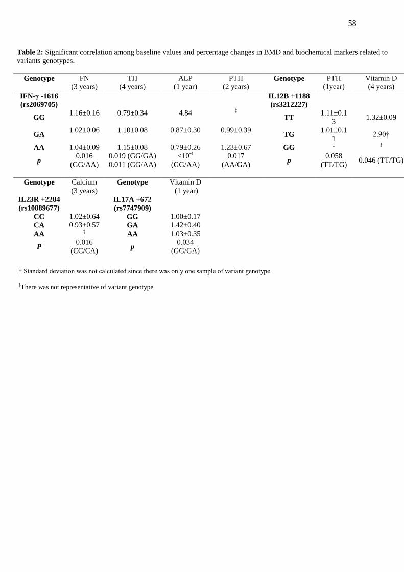
58
Table 2: Significant correlation among baseline values and percentage changes in BMD and biochemical markers related to
variants genotypes.
Genotype FN
(3 years)
TH
(4 years)
ALP
(1 year)
PTH
(2 years) Genotype PTH
(1year)
Vitamin D
(4 years)
IFN-γ -1616
(rs2069705)
IL12B +1188
(rs3212227)
GG 1.16±0.16 0.79±0.34 4.84
‡
TT 1.11±0.1
3 1.32±0.09
GA 1.02±0.06 1.10±0.08 0.87±0.30 0.99±0.39
TG 1.01±0.1
1 2.90†
AA 1.04±0.09 1.15±0.08 0.79±0.26 1.23±0.67 GG ‡ ‡
p 0.016
(GG/AA)
0.019 (GG/GA)
0.011 (GG/AA)
<10-4
(GG/AA)
0.017
(AA/GA) p
0.058
(TT/TG) 0.046 (TT/TG)
Genotype Calcium
(3 years) Genotype Vitamin D
(1 year)
IL23R +2284
(rs10889677)
IL17A +672
(rs7747909)
CC 1.02±0.64 GG 1.00±0.17
CA 0.93±0.57 GA 1.42±0.40
AA ‡
AA 1.03±0.35
P 0.016
(CC/CA) p
0.034
(GG/GA)
† Standard deviation was not calculated since there was only one sample of variant genotype
‡There was not representative of variant genotype
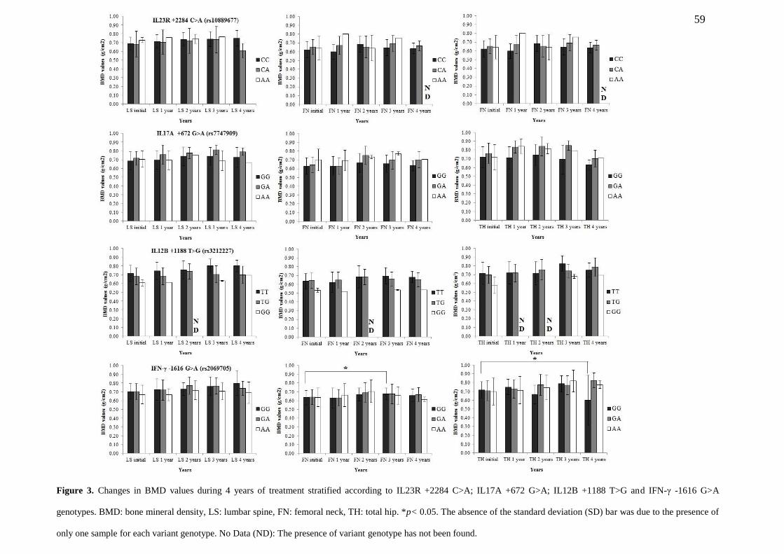
59
Figure 3. Changes in BMD values during 4 years of treatment stratified according to IL23R +2284 C>A; IL17A +672 G>A; IL12B +1188 T>G and IFN-γ -1616 G>A
genotypes. BMD: bone mineral density, LS: lumbar spine, FN: femoral neck, TH: total hip. *p< 0.05. The absence of the standard deviation (SD) bar was due to the presence of
only one sample for each variant genotype. No Data (ND): The presence of variant genotype has not been found.
60
4. Discussion
In our study we showed that cytokine genes SNPs within were associated with BMD
changes in response to BPs therapy. To our knowledge, this is the first study showing these
polymorphisms as related to BPs response and maybe shed some influence upon further OP
therapy strategies.
Our results are consistent with previously reported studies which related SNPs in
cytokine genes to BMD changes and metabolic bone disorders such as psoriatic arthritis and
gout (Czerny et al. 2010; Jadon et al. 2013; Liu et al. 2015). Additionally, polymorphisms in
genes related to bone disorders immune response and to variations in BMD levels showed
different results according to the studied population (Swanberg et al. 2012; Boroń et al. 2014;
Louahchi et al. 2016). In our population all SNPs analyzed showed differences related to any
biochemical markers changes whereas only IFN-γ -1616 exhibited association regarding
BMD changes.
In relation to the biochemical markers, our results showed that IL17A +672 G/A and
IL12B +1188 T/G genotypes were associated with higher vitamin D levels. Vitamin D
presents pleiotropic actions in bone environment: in osteoprogenitors cells it is responsible for
increasing RANKL levels and in mature osteoblasts acts decreasing RANKL expression
(Christakos et al. 2016). Therefore, it is possible that IL-17A and IL-12B may be related to
induction of bone formation or resorption upon osteoblast maturation stage. Additionally,
IL23R +2284 C/C genotype showed statistically significant correlation with higher calcium
levels, a key element for mineralization in bone matrix and tissue homeostasis (Carmeliet et
al. 2015). The relation between IL-23R and calcium mineralization has been cited by Tu et al.
(2015) in osteogenic differentiation assays, which suggested that IL-23/IL-23R/β-catenin
% C
ha
ng
es B
MD
va
lues
61
pathway induces bone formation. These authors observed that IL-23R silencing suppressed
calcium mineralization and modified the expression of osteogenic markers such as Runt-
related transcription factor 2 (RUNX2), ALP, Osteocalcin (OCN) and Dickkopf-related
protein 1 (DKK1) in mesenchymal stem cells in osteoblasts differentiation process. Besides,
IFN-γ -1616 A/A genotype was also related to higher PTH levels, indicating bone resorption,
and IFN-γ G/G genotype to higher ALP levels, denoting bone formation. PTH acts regulating
bone resorption through matrix calcium release whereas alkaline phosphatase is a bioproduct
of osteoblast activities related to bone calcification (Sharma et al. 2014; Carmeliet et al. 2015;
Christakos et al. 2016).
Herein we also identified changes in BMD values. After 3 years of treatment IFN-γ
G/G genotype continued associated with indicators of bone formation in FN area. Conversely,
individuals with this genotype showed decreased BMD values in TH area. Therefore, a
possible pleiotropic action of this cytokine is hypothesized, dependent on time/period of
treatment and/or body-area assessed. Furthermore, TH area showed higher BMD values than
FN area (t-test= -6.60; p< 0.001) suggesting that IFN-γ may act in BMD value-dependent as
an anabolic or catabolic factor, depending on the number of cells (low or high quantities,
respectively). The impact of this cytokine in the bone homeostasis has been widely
recognized in the context of osteoimmunology (Arron and Choi 2000). Some authors suggest
that lower levels of IFN-γ cytokine may increase the chances of survival, activation and
differentiation in osteoclasts (Arron and Choi 2000; Nasr et al. 2014). According to Arron and
Choi (2000) the binding of IFN-γ and its receptor induces TNF receptor-associated factor 6
(TRAF6) degradation, impairing RANKL action. The role of RANKL/RANK/OPG pathway
has been established in the bone homeostasis: RANKL is secreted by osteoblast and bind to
its receptor RANK in osteoclast, acting in osteoclastogenesis, whereas OPG act as
62
competitive antagonist for RANKL (Riancho and Hernández 2012; Makras et al. 2015).
Furthermore, IFN-γ increases the bone morphogenetic protein 2 (BMP-2), which participates
of Wnt pathway inducing bone mineralization and osteoblast differentiation (Raisz 2005).
SNPs within genes from Wnt pathway have been associated with antireabsortive therapies’
response as alendronate, risendronate and tamoxifen (Kruk et al. 2009; Hartmaier et al. 2012;
Zhou et al. 2014; López-Delgado et al. 2016). Nevertheless, it is worth mentioning that some
studies indicate that IFN-γ present contradictory functions in the bone environment when
comparing their in vivo and in vitro actions (Takayanagi et al. 2005; Gao et al. 2007; Zhang et
al. 2015a). Despite the in vivo impact described above, in vitro IFN-γ present a pleiotropic
action by inducing both the increase and decrease of the osteoclastogenesis by different
pathways (Gao et al. 2007; Zhang et al. 2015a). An important physiologic role for IFN-γ in
terms of bone homeostasis was explored by Duque et al. (2011) with ovariectomized mice,
which suggested this cytokine as a new anabolic agent against osteoporosis. On the other
hand, Gao et al. (2007) showed that IFN-γ promotes the osteoclastogenesis by inducing
specific cytokines and T cell activation. Our results suggest that IFN-γ may act as both
anabolic and catabolic agent in bone environment depending on period of treatment or body-
area assessed. Therefore the IFN-γ function may be influenced by these parameters as well as
by BMD values. It is difficult to predict which major pathway is involved in the process
taking into consideration that this cytokine acts in several routes. The IFN-γ is the main
cytokine induced by IL-12 in the Th1 response (Raphael et al. 2014; Annunziato et al. 2015).
Some authors pointed out that IL-12/ IFN-γ pathway from Th1 response is related to Th17
response since it is likely that IL-17 can induce IL-12 (Damsker et al. 2010; Khader and
Gopal 2010). There is a general consensus about IL-17 being stimulated by IL-23 in the
presence of TGF-β and IL-6, initiating the Th17 response (Iwakura and Ishigame 2006;
63
Sutton et al. 2009). The SNPs selection in genes of IL-23, IL-17, IL-12 and IFN-γ cytokines
was performed in our study and analyzed in both pathways, Th1 and Th17, in therapeutic
adhesion of OP patients. However, due to different responses in BMD and biochemical
markers changes after BPs treatment, we suggest that cytokines from both pathways may have
different roles in terms of bone homeostasis.
Additionally, we propose possible pathways involved in the bone resorption, as
showed in Figure 4. In the JAK-STAT – dependent pathway, STAT dimers translocate to the
nucleus being able to regulate Gamma Activated Sequences (GAS) and IFN Stimulated
Regulatory Elements (ISRE), inducing the expression of Interferon regulatory factor 1 (IRF1)
and other regulatory proteins from cell cycle and apoptosis (Ramana et al. 2002; Wong et al.
2002). Other JAK-STAT – independent pathway, studied in mice, showed that osteoclasts
activate the expression of IRF9 gene through mitogen activated protein kinase kinase kinase 1
(Map3K1 in humans and MEKK1 in murine), mitogen activated protein kinase kinase 1
(Map2K1 in humans and MEK1), mitogen activated protein kinases 1 and 2 (ERK1/2) and
C/Ebpbeta cascade, also involved in cell cycle activities (Roy et al. 2002; Haynes 2006; Pham
et al.2014). Consequently, it is possible that the IFN-γ participates in bone remodeling by
directly inducing osteoclasts and osteoblasts apoptosis during treatment.
The aminobisphosphonates (aminoBPs) used in the treatment of OP patients also
induce apoptosis in osteoclasts by inhibiting the mevalonate kinase (MVK) pathway (Riancho
and Hernández 2012b). Furthermore, it is important to highlight that aminoBPs may prevent
apoptosis in osteoblasts by a cell survival mechanism through extracellular signal-regulated
kinases that affects the production of bone mass (Riancho and Hernández 2012b).
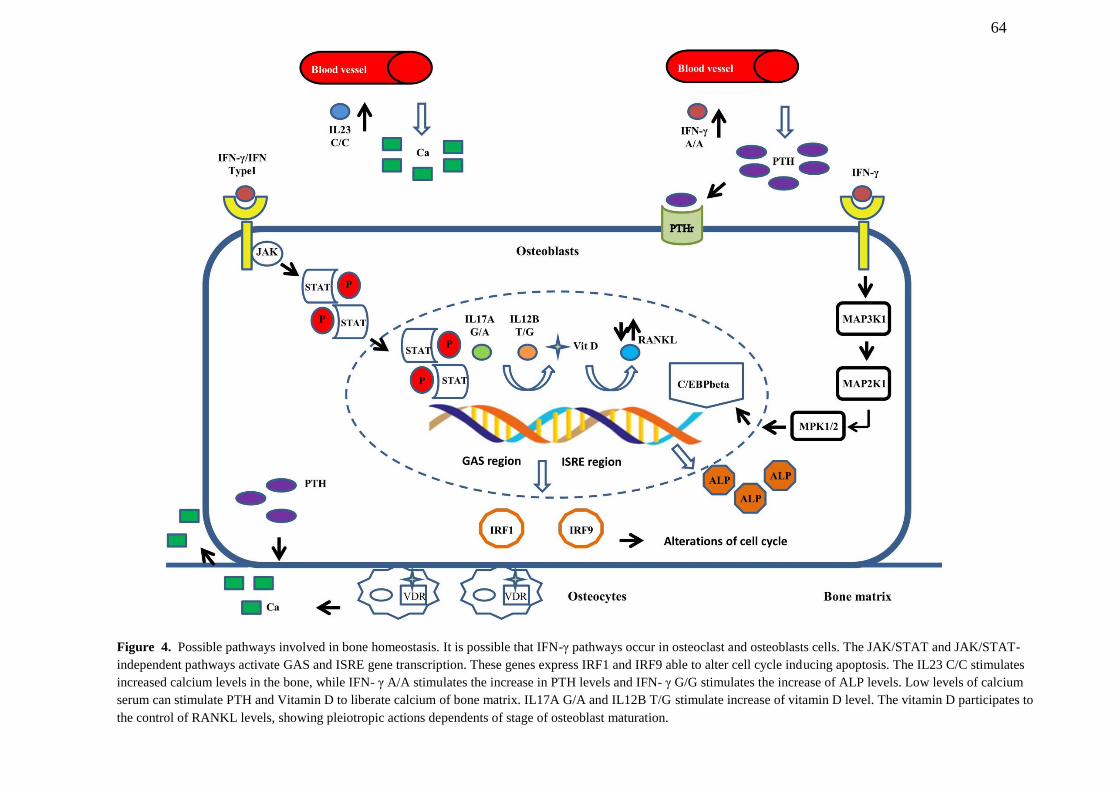
64
Figure 4. Possible pathways involved in bone homeostasis. It is possible that IFN-γ pathways occur in osteoclast and osteoblasts cells. The JAK/STAT and JAK/STAT-
independent pathways activate GAS and ISRE gene transcription. These genes express IRF1 and IRF9 able to alter cell cycle inducing apoptosis. The IL23 C/C stimulates
increased calcium levels in the bone, while IFN- γ A/A stimulates the increase in PTH levels and IFN- γ G/G stimulates the increase of ALP levels. Low levels of calcium
serum can stimulate PTH and Vitamin D to liberate calcium of bone matrix. IL17A G/A and IL12B T/G stimulate increase of vitamin D level. The vitamin D participates to
the control of RANKL levels, showing pleiotropic actions dependents of stage of osteoblast maturation.
65
Some cytokines such as RANKL and Nuclear Factor kappa B (NFk-B) cannot only
influence osteoclastogenesis and osteoblastogenesis, but also act directly in MVK pathway.
RANKL and NFk-B play a role in differentiation, activation and survival osteoclasts and are
regulated by cytokines such as IFN-γ and IL-17 (Arron and Choi 2000; Riancho and
Hernández 2012b).
We analyze simultaneously the association between IL23R, IL17A, IL12B and IFN-γ
SNPs and BPs therapy. The SNPs in cytokine genes may change their levels and protein
structure and this may interfere in the bone disease environment and response to BPs
treatment. Understanding the genetic contribution in multifactorial diseases, such as the
osteoporosis, is essential for providing specific therapeutic and diagnostic procedures,
allowing customized treatments based on different patients’ genetic profiles. Besides, our
findings highlight the importance of other factors different than RANKL/RANK/OPG
pathway associated with OP. It is acknowledged that other molecules as leptin or leptin
receptors may activate the secretion of proinflammatory cytokines and regulate via Janus
kinases downstream events (Legiran and Brandi 2012; Ye and Lu 2013; Lee et al. 2014).
Additionally, SNPs in other genes related to immune systems such as IL-1, IL-6, IL-4, TNF-
α, STATs, ERAP, HLA and TRAF proteins are already recognized as influencing bone
environment (Takayanagi et al. 2005; Yuan et al. 2012; Jadon et al. 2013), possibly presenting
potential action in OP therapeutic adhesion.
Conclusions
A relationship among SNPs in IL23R, IL17A, IL12B, IFN-γ genes and changes in
BMD values or biomarkers bone turnover in BPs treatment are described for the first time in
this study. The IFN-γ -1616 (G>A) genotype was associated with BMD changes while IL23R
66
+2284 (C>A), IL17A +672 (G>A) and IL12B +1188 (T>G) genotypes were associated with
bone turnover, based on biochemical markers changes. This suggests that these SNPs may act
as potential indicators in therapeutic decisions used in the treatment of osteoporosis, related to
choice of BPs as first-line treatment, its therapy period and the use of supplements, such as
vitamin D and calcium. SNPs in cytokines genes may have an important role in therapy’s
success and in paving the way for personalized medicine in osteoporosis.
Acknowledgments
We thank the following Brazilian funding agencies: CAPES (Coordenação de
Aperfeiçoamento de Pessoal de Nível Superior), CNPq (Conselho Nacional de
Desenvolvimento Científico e Tecnológico), and FACEPE (Fundação de Amparo à Ciência e
Tecnologia de Pernambuco) for financial support.
Conflict of interest
None of the authors declared any conflict of interest related to this manuscript.
67
5. Capítulo II
Artigo submetido na revista Molecular and Celular Endocrinology
Fator de Impacto: 3,8
Guia para autores: Anexo A
68
Stability of Candidate Reference Genes for Osteoporosis: Selection
and Validation for Quantitative Real-time PCR assays
Lima, CAD1,2*; Lima, SC2; Barbosa, AD2,3; Crovella, S1,2; Sandrin-Garcia, P1,2; Pita,
WB4; and De Azevêdo Silva, J1,2
1 – Department of Genetics - Federal University of Pernambuco (UFPE), Recife, Pernambuco, Brazil.
2 – Laboratory of Immunopathology Keizo Asami – Federal University of Pernambuco (UFPE), Recife, Pernambuco, Brazil.
3 - Division of Rheumatology, Clinical Hospital, Federal University of Pernambuco (UFPE), Recife, Pernambuco, Brazil.
4 – Department of Antibiotics - Federal University of Pernambuco (UFPE), Recife, Pernambuco, Brazil.
*Corresponding author: Camilla AD Lima
Email address: [email protected]
Submitted in Molecular and Cellular Endocrinology Journal
69
Abstract
Osteoporosis (OP) is a disease influenced by genetic factors in more than half of the cases. In
spite of the efforts to clarify the relationship among these factors and OP development, many
discoveries need to be further functionally validated. Real-time reverse transcription
quantitative PCR (RT-qPCR) is the main used method for quantification of gene expression
studies. However, some limitations as the choice of stably expressed reference genes (RG) are
required to ensure the efficiency and quality of gene expression assays. By our knowledge,
validation study for RG in OP is still missing. Therefore, this study compared the expression
levels of 10 RG (G6PD, B2M, GUSB, HSP90, EF1A, RPLP0, GAPDH, ACTB, 18S and
HPRT1) to assess their suitability in OP analysis by using of GeNorm, Normfinder,
BestKeeper and RefFinder programs. A minimal number of two RG was recommended by
GeNorm to obtain a reliable normalization. RPLP0 and B2M were identified as the most
stable genes in OP studies while ACTB, 18S and HPRT1 were inadequate for normalization
in our data. Moreover, we showed the dramatic effects of suboptimal RG choice on the
quantification of a target gene highlighting the importance in identificating the most
appropriate reference gene to specific diseases. The used algorithms showed different results
among them in relation to top ranked genes, thus suggesting an individualized and carefully
choice in the used packages and reference genes selection.
Keywords: Reference Genes, RT-qPCR, Genetics of Osteoporosis, Gene Expression
Analysis.
70
1. Introduction
Osteoporosis (OP) is a multifactorial and complex-traits disease, characterized by low
bone mineral density (BMD) and loss of tissue microarchitecture (Clark and Duncan 2015;
Rocha-Braz and Ferraz-de-Souza 2016). The disease is influenced by genetic factors around
50-85% of cases, especially in early or idiopathic osteoporosis (Riancho and Hernández
2012b; Karasik et al. 2016; Rocha-Braz and Ferraz-de-Souza 2016). The occurrence of
fractures, the most important clinical manifestation of OP, shows heritability estimated in the
range of 25-68%, presenting the highest values for younger patients and fractures that occur
before 70 years old (Albagha and Ralston 2006; Rocha-Braz and Ferraz-de-Souza 2016).
Recent Genome-Wide Association Studies (GWAS) already have identified over 60 genes
associated with BMD variations and 14 genes related to fractures risk, however these
discoveries remain to be further functionally validated (Clark and Duncan 2015; Karasik et al.
2016).
Gene expression analyses performed by quantitative real-time polymerase chain
reaction (qPCR) is a sensitive, accurate and commonly used approach in molecular biological
studies (Wang et al. 2015b; Wang et al. 2016). In 2009, in view of resolving technical
challenges and to standardize qPCR experiments, the Minimum Information for Publication
of Quantitative Real-Time PCR experiments (MIQE) guidelines was published (Bustin et al.
2009). One of the points highlighted by those guidelines were the need for validating
reference genes for specific tissues, thereby ensuring the efficiency and quality of gene
expression assays (Bustin et al. 2009). Regardless of Glyceraldehyde 3-phosphate
dehydrogenase (GAPDH) and Beta-actin (ACTB) being traditionally used as reference genes,
it is necessary to choose constitutive and stably expressed genes for different tissues and
different experimental conditions (Wang et al. 2015b; Normann et al. 2016).
71
In spite of the efforts of Genetic Markers for Osteoporosis (GENOMOS) and Genetic
Factors of Osteoporosis (GEFOS) consortiums to clarify the genetic factors involving in OP
development, to our knowledge, none of the studies and efforts brought out specific reference
genes in OP gene expression assays were performed up until now. Therefore, we conducted
an evaluation of 10 candidate reference genes commonly used in previous studies of
rheumatic diseases to establish a set of specific and validated reference genes for expression
analysis in OP-related genes contributing to the advance in the field of bone research.
2. Material and Methods
2.1. Subjects
Peripheral blood samples of ten osteoporosis patients diagnosed according WHO
criteria and ten healthy controls in postmenopausal period and without osteometabolic and/or
inflammatory and autoimmune diseases and cancer were selected for this study. The present
study was approved by the Research Ethics Committee of the Center for Health Sciences,
Federal University of Pernambuco (CEP/CCS/UFPE nº 513/11), performed according the
Declaration of Helsinki and all the participants who accepted to participate of this research
provided a written informed consent.
2.2. RNA Extraction and cDNA Synthesis
Total RNA was extracted by TRIzol Reagent (Invitrogen, USA) according to the
manufacturer’s instructions. The integrity of RNA was verified by 1.5% agarose gel
electrophoresis and the quantification and RNA quality was checked by Nanodrop ND 1000
spectrophotometer (Nanodrop Technologies Inc, Delaware, USA). The samples that showed
integrity of 23S and 16S fragments and the absorbance ratio OD260/280 values from 1.8 to
2.0 were considered able to proceed to the analysis. The cDNA synthesis was performed from
72
each RNA sample (500 ng) using GoScript™ Reverse Transcription System (Promega, USA)
following the manufacturer’s instructions.
2.3. Selection of Candidate Reference Genes and PCR Primer Design
The selection of candidate reference genes was performed by using panels of the
mainly used reference genes from researchers in humans (Applied Biosystems 2008; Roche
Applied Science 2009). Besides, reference genes commonly used in rheumatic diseases were
added to the analysis. A total of 10 genes were selected: glucose-6-phosphate dehydrogenase
(G6PD), β2-microglobulin (B2M), β-glucuronidase (GUSB), heat shock protein 90 (HSP90),
elongation factor 1 alpha (EF1A), ribosomal protein P0 (RPLP0), GAPDH, ACTB, 18S
ribosomal RNA (18S) and hypoxanthine phosphoribosyltransferase 1 (HPRT1). All gene
sequences were obtained from GenBank (www.ncbi.nlm.nih.gov/genbank/) and all primers
were designed by NCBI/Primer-BLAST (www.ncbi.nlm.nih.gov/tools/primer-blast/). For
ACTB and 18S were used Taqman ® probes. Sequence details are showed in Table 1. The
optimal annealing temperature and amplicon size were tested by gradient PCR and 1.5%
agarose gel electrophoresis.
2.4. Amplification Efficiency Testing
The efficiency value for each primer (E) was determined by slopes of standard curves
from five 10-fold serial dilution points for each cDNA sample. The acceptable values were
defined as between 95% and 105%. These data and the correlation coefficients (R2) were
calculated by ABI 7500 system software. For ACTB and 18S (Taqman® probes), it was
considered a reaction efficiency of 100% ensured by manufactures’ information.
2.5. Quantitative Real-Time PCR Assay
The qPCR was performed in the ABI 7500 (Applied Biosystems, Foster City, CA,
USA) using 1 μL diluted cDNA (1:5) from cDNA synthesis previous experiments , 10 µM of
73
each primer, 5 μL SYBR® Green Master Mix (1X) (Thermo Fischer Scientific; former Savant,
MA, USA) and ultrapure water in sufficient quantity to a final volume of 10 μL. The
Taqman® assays were performed following the manufactures’ instructions. PCR
amplifications were performed in technical triplicate and negative controls without templates
were added for each reaction. Thermo cycling conditions used were: initial denaturing at 50ºC
for 2 min and 95°C for 10 minutes followed by 45 cycles of 15 s at 95°C and 1 min at 60°C.
The melting curve was analyzed to confirm the specificities of the amplification reactions.
2.6. Data analysis of gene expression stability
Three statistical algorithms from Excel based free software packages were used to
evaluate the expression stability of 10 candidate reference genes to OP: GeNorm (Version
v3.5) (Vandesompele et al. 2002), NormFinder (Version 20) (Andersen et al. 2004) and
BestKeeper (Version 1) (Pfaffl et al. 2004). Additionally, the web-based online tool
RefFinder (Xie et al. 2012) and GeNorm were used to assist in the ranking and to calculate
the optimal combination and minimal number of the candidate reference genes, respectively.
The relative quantities used to GeNorm and Normfinder input were calculated and corrected
to amplifications efficiencies according the equation suggested by Spiegelaere et al. (2015):
RQ = E-(minCq-sampleCq)
.
2.6.1. GeNorm
The GeNorm uses the geometric means to determine gene expression normalization
factor and stability value (M) for each gene (Vandesompele et al. 2002). M values less than
1.5 are acceptable and the lowest values are considered most stable. Then, the software
performs a pairwise comparison (Vn/n+1) adding genes, one by one, in order to set the most
stable reference genes until a cut-off less than 0.15 for determining the minimal number of the
74
candidate reference genes (Vandesompele et al. 2002; Liu et al. 2012; Wang et al. 2015b;
Normann et al. 2016).
2.6.2. NormFinder
The NormFinder uses the standard curve or the delta-Ct method from the
transformation of Ct values in a linear scale (Andersen et al. 2004). The program estimates
the expression variation of the candidate genes, providing a stability value (M) for each gene,
and the variation between sample subgroups of the sample e.g. patient and healthy groups
(Andersen et al. 2004). It is required at least 8 sample per analysis and the algorithm
recommends the best combination of two reference genes, providing the M values of this
combination. Similar to GeNorm, the lowest M values are considered most stable (Andersen
et al. 2004; De Spiegelaere et al. 2015; Normann et al. 2016).
2.6.3. BestKeeper
The BestKeeper uses the geometric mean of the candidate reference genes and the
software provides a correlation coefficient (r) of each gene besides to calculate standard
deviation (SD) and coefficient of variation (CV) from the samples Ct-values (Pfaffl et al.
2004). It is recommended SD to be [± CP] < 1, SD [± x-fold] <2 and correlation coefficient to
be as higher as possible, close to the value 1 (Pfaffl et al. 2004; Liu et al. 2012). Low SD and
CV associated to high correlation coefficient (r) values indicate a stable reference gene,
however the program does not perform a ranking order of the analyzed genes (Pfaffl et al.
2004; Normann et al. 2016). Spiegelaere et al. (2015) highlighted that correlation coefficient
(r) is a better parameter to assess the most stable genes than the standard deviation because
the first one is able to analyze the correlation of each gene with the BestKeeper Index from
the geometric mean from the studied reference genes. Thus we chose this measurement to
evaluate and to perform the rank of reference genes in BestKeeper analysis.
75
2.6.4. RefFinder
The RefFinder is a web-based platform (http://fulxie.0fees.us/?type=reference) which
integrates the three software packages abovementioned and additionally performs the
comparative ∆∆Ct method (Xie et al. 2012; De Spiegelaere et al. 2015; Ma et al. 2015). The
RefFinder only uses Cq to perform the reference genes rank through of geometric mean
values (GM), without any possibility to include PCR efficiency as the Genorm and
NormFinder (De Spiegelaere et al. 2015). Due to the comparison among algorithms to be
impossible, the RefFinder platform was used as complementary tool to assess reference gene
stability and its result interpreted from particularities of each program.
2.7. Reference and target genes analysis
After the determination of optimal combination and minimal number of the candidate
reference genes by software analysis, the most stable combination and the recommended
candidate reference genes were used to perform the normalization factor from geometric
media and ∆∆Ct analysis. To validate these potential reference genes, the IFNG was used as
target gene (Table 1, SM1). This gene codes the interferon gamma protein recognized to
playing an essential role in bone environmental homeostasis.
3. Results
3.1. Specificity and amplification efficiencies
The specificity of the primers was verified by using agarose gel electrophoresis, in
which a single band was detected, followed by a single peak in the melting curve from qPCR
analysis. The melting curves of all tested primers are showed in Supplementary material 1.
The R2 and E values of the 10 candidate reference genes ranged from 0.999 – 1.000 and
95.12% - 104.79%, respectively (Table 1). These values were in accordance to the MIQE
guidelines instructions.
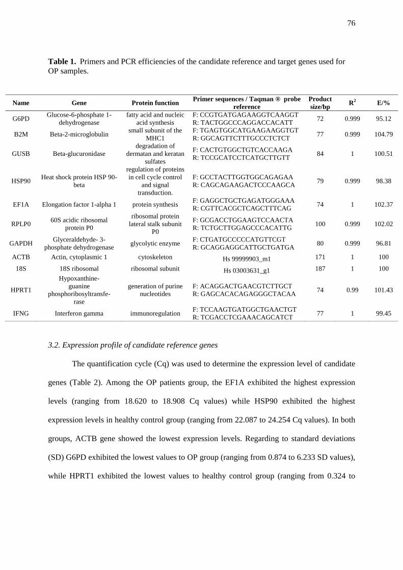
76
Table 1. Primers and PCR efficiencies of the candidate reference and target genes used for
OP samples.
3.2. Expression profile of candidate reference genes
The quantification cycle (Cq) was used to determine the expression level of candidate
genes (Table 2). Among the OP patients group, the EF1A exhibited the highest expression
levels (ranging from 18.620 to 18.908 Cq values) while HSP90 exhibited the highest
expression levels in healthy control group (ranging from 22.087 to 24.254 Cq values). In both
groups, ACTB gene showed the lowest expression levels. Regarding to standard deviations
(SD) G6PD exhibited the lowest values to OP group (ranging from 0.874 to 6.233 SD values),
while HPRT1 exhibited the lowest values to healthy control group (ranging from 0.324 to
Name Gene Protein function Primer sequences / Taqman ® probe
reference
Product
size/bp R
2 E/%
G6PD Glucose-6-phosphate 1-
dehydrogenase
fatty acid and nucleic
acid synthesis
F: CCGTGATGAGAAGGTCAAGGT
R: TACTGGCCCAGGACCACATT 72 0.999 95.12
B2M Beta-2-microglobulin small subunit of the
MHC1
F: TGAGTGGCATGAAGAAGGTGT
R: GGCAGTTCTTTGCCCTCTCT 77 0.999 104.79
GUSB Beta-glucuronidase
degradation of
dermatan and keratan
sulfates
F: CACTGTGGCTGTCACCAAGA
R: TCCGCATCCTCATGCTTGTT 84 1 100.51
HSP90 Heat shock protein HSP 90-
beta
regulation of proteins
in cell cycle control
and signal
transduction.
F: GCCTACTTGGTGGCAGAGAA
R: CAGCAGAAGACTCCCAAGCA 79 0.999 98.38
EF1A Elongation factor 1-alpha 1 protein synthesis F: GAGGCTGCTGAGATGGGAAA
R: CGTTCACGCTCAGCTTTCAG 74 1 102.37
RPLP0 60S acidic ribosomal
protein P0
ribosomal protein
lateral stalk subunit
P0
F: GCGACCTGGAAGTCCAACTA
R: TCTGCTTGGAGCCCACATTG 100 0.999 102.02
GAPDH Glyceraldehyde- 3-
phosphate dehydrogenase glycolytic enzyme
F: CTGATGCCCCCATGTTCGT
R: GCAGGAGGCATTGCTGATGA 80 0.999 96.81
ACTB Actin, cytoplasmic 1 cytoskeleton Hs 99999903_m1 171 1 100
18S 18S ribosomal ribosomal subunit Hs 03003631_g1 187 1 100
HPRT1
Hypoxanthine-
guanine
phosphoribosyltransfe-
rase
generation of purine
nucleotides
F: ACAGGACTGAACGTCTTGCT
R: GAGCACACAGAGGGCTACAA 74 0.99 101.43
IFNG Interferon gamma immunoregulation F: TCCAAGTGATGGCTGAACTGT
R: TCGACCTCGAAACAGCATCT 77 1 99.45
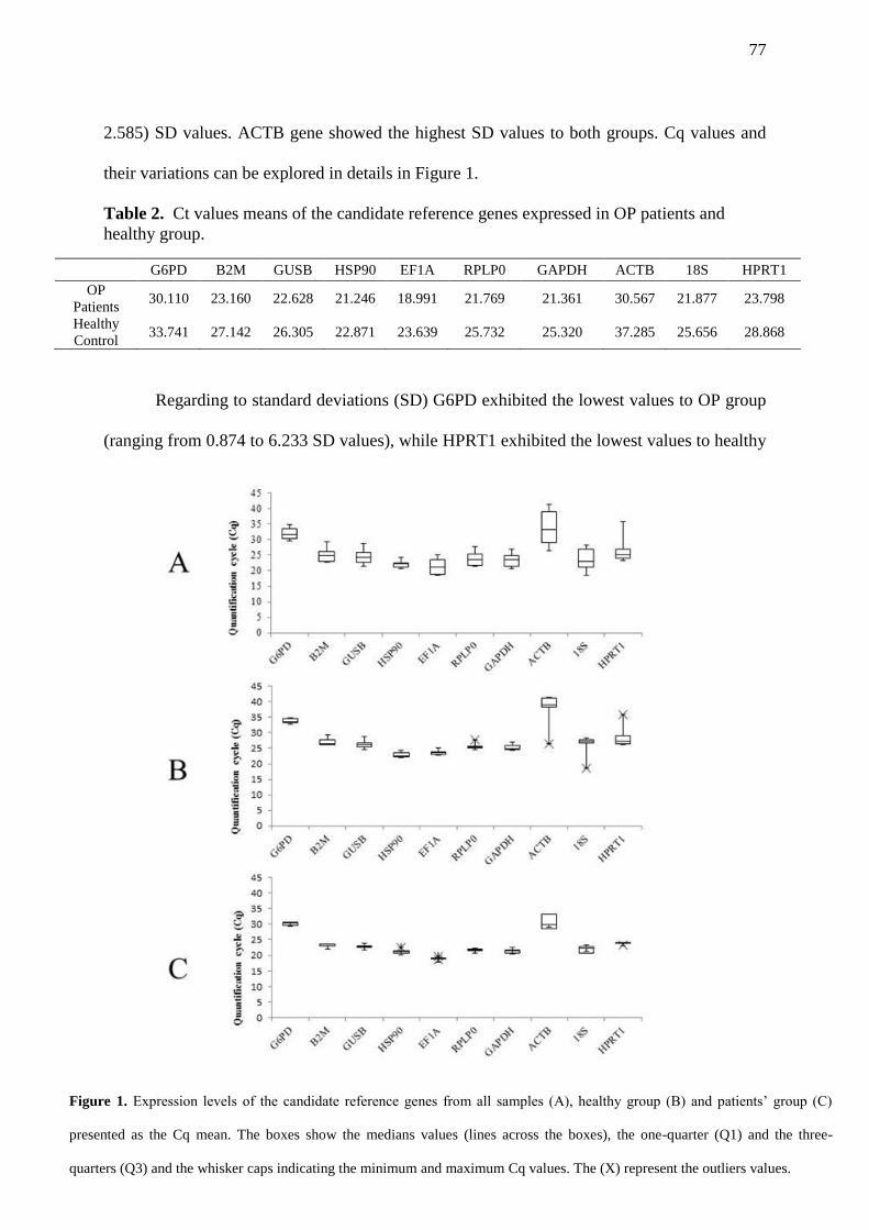
77
2.585) SD values. ACTB gene showed the highest SD values to both groups. Cq values and
their variations can be explored in details in Figure 1.
Table 2. Ct values means of the candidate reference genes expressed in OP patients and
healthy group.
G6PD B2M GUSB HSP90 EF1A RPLP0 GAPDH ACTB 18S HPRT1
OP
Patients 30.110 23.160 22.628 21.246 18.991 21.769 21.361 30.567 21.877 23.798
Healthy
Control 33.741 27.142 26.305 22.871 23.639 25.732 25.320 37.285 25.656 28.868
Regarding to standard deviations (SD) G6PD exhibited the lowest values to OP group
(ranging from 0.874 to 6.233 SD values), while HPRT1 exhibited the lowest values to healthy
Figure 1. Expression levels of the candidate reference genes from all samples (A), healthy group (B) and patients’ group (C)
presented as the Cq mean. The boxes show the medians values (lines across the boxes), the one-quarter (Q1) and the three-
quarters (Q3) and the whisker caps indicating the minimum and maximum Cq values. The (X) represent the outliers values.

78
control group (ranging from 0.324 to 2.585) SD values. ACTB gene showed the highest SD
values to both groups. Cq values and their variations can be explored in details in Figure 1.
3.3. GeNorm analysis
In accordance to GeNorm ranking generated from M values, RPLP0 and B2M were
the most stable reference genes (M = 0.823) for the present study (Figure 2).
The pairwise variations (Vn/n+1) showed values below the cut-off (0.15) to V2/V3,
V3/V4, V4/V5, V5/V6, V6/V7 and V9/V10 values (Figure 3).
Figure 2. Candidate reference gene stability analyzed using GeNorm. Low M values predicts high stability while high M values
indicate low stability
Figure 3. Pairwise variation (Vn/Vn+1) to determine the optimal number of reference genes required for accurate normalization
by GeNorm. In this OP study, the pairwise variation value less than the cut-off (0.15) is reached with two reference genes.
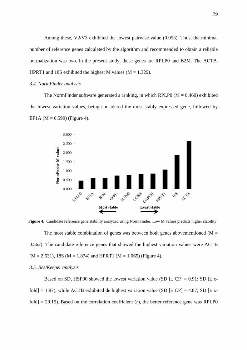
79
Among these, V2/V3 exhibited the lowest pairwise value (0.053). Thus, the minimal
number of reference genes calculated by the algorithm and recommended to obtain a reliable
normalization was two. In the present study, these genes are RPLP0 and B2M. The ACTB,
HPRT1 and 18S exhibited the highest M values (M = 1.329).
3.4. NormFinder analysis
The NormFinder software generated a ranking, in which RPLP0 (M = 0.460) exhibited
the lowest variation values, being considered the most stably expressed gene, followed by
EF1A (M = 0.599) (Figure 4).
The most stable combination of genes was between both genes abovementioned (M =
0.562). The candidate reference genes that showed the highest variation values were ACTB
(M = 2.631), 18S (M = 1.874) and HPRT1 (M = 1.065) (Figure 4).
3.5. BestKeeper analysis
Based on SD, HSP90 showed the lowest variation value (SD [± CP] = 0.91; SD [± x-
fold] = 1.87), while ACTB exhibited de highest variation value (SD [± CP] = 4.87; SD [± x-
fold] = 29.15). Based on the correlation coefficient (r), the better reference gene was RPLP0
Figure 4. Candidate reference gene stability analyzed using NormFinder. Low M values predicts higher stability.
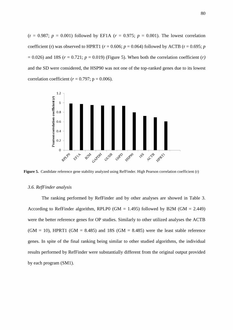
80
(r = 0.987; p = 0.001) followed by EF1A (r = 0.975; p = 0.001). The lowest correlation
coefficient (r) was observed to HPRT1 (r = 0.606; p = 0.064) followed by ACTB (r = 0.695; p
= 0.026) and 18S (r = 0.721; p = 0.019) (Figure 5). When both the correlation coefficient (r)
and the SD were considered, the HSP90 was not one of the top-ranked genes due to its lowest
correlation coefficient (r = 0.797; p = 0.006).
3.6. RefFinder analysis
The ranking performed by RefFinder and by other analyses are showed in Table 3.
According to RefFinder algorithm, RPLP0 (GM = 1.495) followed by B2M (GM = 2.449)
were the better reference genes for OP studies. Similarly to other utilized analyses the ACTB
(GM = 10), HPRT1 (GM = 8.485) and 18S (GM = 8.485) were the least stable reference
genes. In spite of the final ranking being similar to other studied algorithms, the individual
results performed by RefFinder were substantially different from the original output provided
by each program (SM1).
Figure 5. Candidate reference gene stability analyzed using RefFinder. High Pearson correlation coefficient (r)
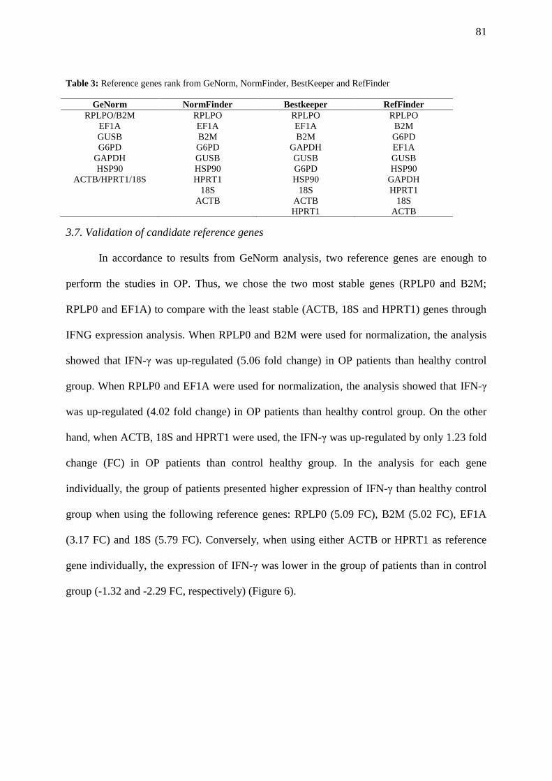
81
Table 3: Reference genes rank from GeNorm, NormFinder, BestKeeper and RefFinder
3.7. Validation of candidate reference genes
In accordance to results from GeNorm analysis, two reference genes are enough to
perform the studies in OP. Thus, we chose the two most stable genes (RPLP0 and B2M;
RPLP0 and EF1A) to compare with the least stable (ACTB, 18S and HPRT1) genes through
IFNG expression analysis. When RPLP0 and B2M were used for normalization, the analysis
showed that IFN-γ was up-regulated (5.06 fold change) in OP patients than healthy control
group. When RPLP0 and EF1A were used for normalization, the analysis showed that IFN-γ
was up-regulated (4.02 fold change) in OP patients than healthy control group. On the other
hand, when ACTB, 18S and HPRT1 were used, the IFN-γ was up-regulated by only 1.23 fold
change (FC) in OP patients than control healthy group. In the analysis for each gene
individually, the group of patients presented higher expression of IFN-γ than healthy control
group when using the following reference genes: RPLP0 (5.09 FC), B2M (5.02 FC), EF1A
(3.17 FC) and 18S (5.79 FC). Conversely, when using either ACTB or HPRT1 as reference
gene individually, the expression of IFN-γ was lower in the group of patients than in control
group (-1.32 and -2.29 FC, respectively) (Figure 6).
GeNorm NormFinder Bestkeeper RefFinder
RPLPO/B2M RPLPO RPLPO RPLPO
EF1A EF1A EF1A B2M
GUSB B2M B2M G6PD
G6PD G6PD GAPDH EF1A
GAPDH GUSB GUSB GUSB
HSP90 HSP90 G6PD HSP90
ACTB/HPRT1/18S HPRT1 HSP90 GAPDH
18S 18S HPRT1
ACTB ACTB 18S
HPRT1 ACTB
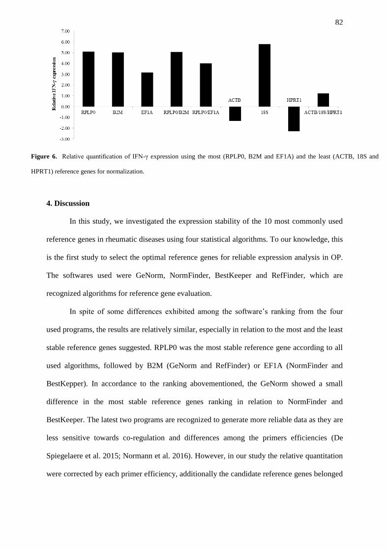
82
4. Discussion
In this study, we investigated the expression stability of the 10 most commonly used
reference genes in rheumatic diseases using four statistical algorithms. To our knowledge, this
is the first study to select the optimal reference genes for reliable expression analysis in OP.
The softwares used were GeNorm, NormFinder, BestKeeper and RefFinder, which are
recognized algorithms for reference gene evaluation.
In spite of some differences exhibited among the software’s ranking from the four
used programs, the results are relatively similar, especially in relation to the most and the least
stable reference genes suggested. RPLP0 was the most stable reference gene according to all
used algorithms, followed by B2M (GeNorm and RefFinder) or EF1A (NormFinder and
BestKepper). In accordance to the ranking abovementioned, the GeNorm showed a small
difference in the most stable reference genes ranking in relation to NormFinder and
BestKeeper. The latest two programs are recognized to generate more reliable data as they are
less sensitive towards co-regulation and differences among the primers efficiencies (De
Spiegelaere et al. 2015; Normann et al. 2016). However, in our study the relative quantitation
were corrected by each primer efficiency, additionally the candidate reference genes belonged
Figure 6. Relative quantification of IFN-γ expression using the most (RPLP0, B2M and EF1A) and the least (ACTB, 18S and
HPRT1) reference genes for normalization.
83
to different functional groups, which helped to correct this deficiency from GeNorm.
Similarly, the RefFinder also showed in its final rank RPLP0 and B2M as the best
combination of reference genes. However the calculated ranking by RefFinder to each
program were very discrepant in relation to original results calculated by programs
individually, which probably occurred due to the fact that the program uses raw Cq values and
did not accept corrections to reactions efficiencies (SM2) (De Spiegelaere et al. 2015).
Because of that we do not recommend this software as a sole tool in order to validate
reference genes as the output may be biased. On the other hand, the ΔΔCt and normalization
analysis showed more similar values between RPLP0 and B2M than RPLP0 and EF1A. Thus,
based in GeNorm output and these last two analyses, we suggest the combination RPLP0 and
B2M as the most stable for OP gene expression studies.
RPLP0 encodes one out of approximately 80 ribossomal proteins in human, which are
involved in protein synthesis and induction of proliferation and apoptosis processes (Artero-
Castro et al. 2011). Previously studies already have demonstrated RPLP0 as one of the most
stable reference genes among a set of candidate genes in inflammatory bowel disease (Bamias
et al. 2013), ovarian (Kolkova et al. 2013) and gastrointestinal stromal tumors (Fassunke et al.
2010). In addition, Ragni et al. (2013) classified the RPLP0 in the most stable reference gene
in assays of mesenchymal stem cell differentiation, the precursor cells of osteoblasts. The
second most stable gene, B2M, encodes β2-microglobulin, which has not been cited by
GWAS as a potential gene involved in OP, however, previous studies related its association
with bone metabolism in tumor processes (Josson et al. 2011; Hsu and Kiel 2012; Levy et al.
2015). In spite of that, similarly to our study, Stephens et al. (2011) and Li et al. (2015)
showed B2M as the one of the most stable reference genes in expression analysis in mouse
bone cells and mesenchymal stem cells cultures, respectively. It appears that the relation
84
between bone system and B2M variation is more related to cancer cells than health bone ones
(Josson et al. 2011; Rienzo et al. 2013).
In contrast, for the low stability analysis, all the programs showed the ACTB, HPRT1
and 18S as the three least stable reference genes. ACTB, as well as GAPDH, are used as
internal controls in more than 70% of expression analyses performed by RT-qPCR (Wang et
al. 2015b; Klenke et al. 2016). However, due to the very low stability in all used programs,
ACTB was not recommended as reference gene for our analysis. ACTB gene encodes for β-
actin, one of six different actin isoforms in vertebrates and ubiquitously expressed in cell
cytoplasm (Rubenstein 1990; Bunnell et al. 2011). Actins compose the cytoskeleton, which
plays critical roles in cell motility, structure, and integrity, besides to act in the regulation of
gene expression (Bunnell et al. 2011). Tai et al. (2015) showed for the first time that the
factor osteoinductive simvastatin acts in the bone regeneration by increasing actin filament
organization and cell rigidity. Similarly, Elsafadi et al. (2016) related in regenerative medicine
studies the involvement of distribution of the actin filament and changes in cytoskeletal
organization in the osteoblastic and adipocytic differentiation of stem cells (hMSC) in vitro.
This new approach of actin beta actin mechanism is a possible explanation to variation of
ACTB gene expression in OP samples, as the disease is strictly related to the balance between
bone formation and resorption.
HPRT1 gene encodes for hypoxanthine-guanine phosphoribosyl transferase (HPRT),
recognized by transferase activity, able to catalyze purine bases guanine and hypoxanthine
into their respective monophosphate nucleoside (Kelley and Andersson 2014). In spite of
being acceptably stable in osteoblasts and osteoclasts (Stephens et al. 2011) and osteosarcoma
(Rienzo et al. 2013) cell culture studies, Yan et al. (2015) suggested that HPRT1 might
regulate bone metabolism. This study suggested that this protein might be involved in the
85
development of osteoporosis though the process of the transferase activity, which may
contribute to the generation of free radical species and oxidative stress affecting the bone
metabolism. Additionally, Isomura et al. (Isomura et al. 2004) also showed in postmenopausal
rats studies that oxidative stress could be involved in the OP pathogenesis.
18S gene encodes for ribosome 18S rRNA subunit and to our knowledge there is no
study report of alteration in expression levels of this gene or its protein in bone cells or
diseases related to changes of bone mineral density. Nevertheless, studies have described
several difficulties associated, for instance, with its expression levels being higher than the
target gene (Paolacci et al. 2009; Kozera and Rapacz 2013) and also the regulation of their
transcription by biological and chemical agents (Nicot et al. 2005). These alterations,
similarly to the other candidate genes in our analysis, are not in accordance to criteria of
constant level of the expression and the no influence by environmental factors required for an
acceptable reference gene (Kozera and Rapacz 2013). Besides, Ragni et al. (2013) and
Stephens et al. (2011) considered the 18S inadequate for normalization in osteogenic and
chondrogenic differentiation and mouse osteoblasts and osteoclasts analysis, respectively.
The impact of using these reference genes might be visualized in the expression levels
of the target gene, which ranged greatly when different reference genes combinations were
used. The ACTB and HPRT1 showed that IFN-γ was down regulated in OP patients group,
while according to all the other analyzed genes the IFN-γ was up regulated in the same group.
These results highlight the importance of the validation of reference genes to specific studies
and diseases and showed the aggravating effect of ACTB to be used in most expression
analysis, which suggests that its validation has been often disregarded in expression studies
(Bustin et al. 2009; Wang et al. 2015b). The qPCR is a technique recognized by high
sensitivity and sequence-specificity, however, the conclusions about the mRNA expression
86
analysis may be performed only after the appropriate reference gene selection (Wong and
Medrano 2005; Bustin et al. 2009; Klenke et al. 2016).
Therefore, according to our results, we recommend the use of RPLP0 and B2M as the
most stable reference genes to OP studies, as we showed their lower variation impact and
influence on the evaluation of a target gene expression. On the other hand, we do not
recommend the use of the least stable reference genes (ACTB, HPRT1 and 18S) in OP
expression assays. Finally, we emphasize a carefully choice of software packages used in
reference gene selection.
Acknowledgments
We thank the following Brazilian funding agencies: CAPES (Coordenação de
Aperfeiçoamento de Pessoal de Nível Superior), CNPq (Conselho Nacional de
Desenvolvimento Científico e Tecnológico), and FACEPE (Fundação de Amparo à Ciência e
Tecnologia de Pernambuco) for financial support.
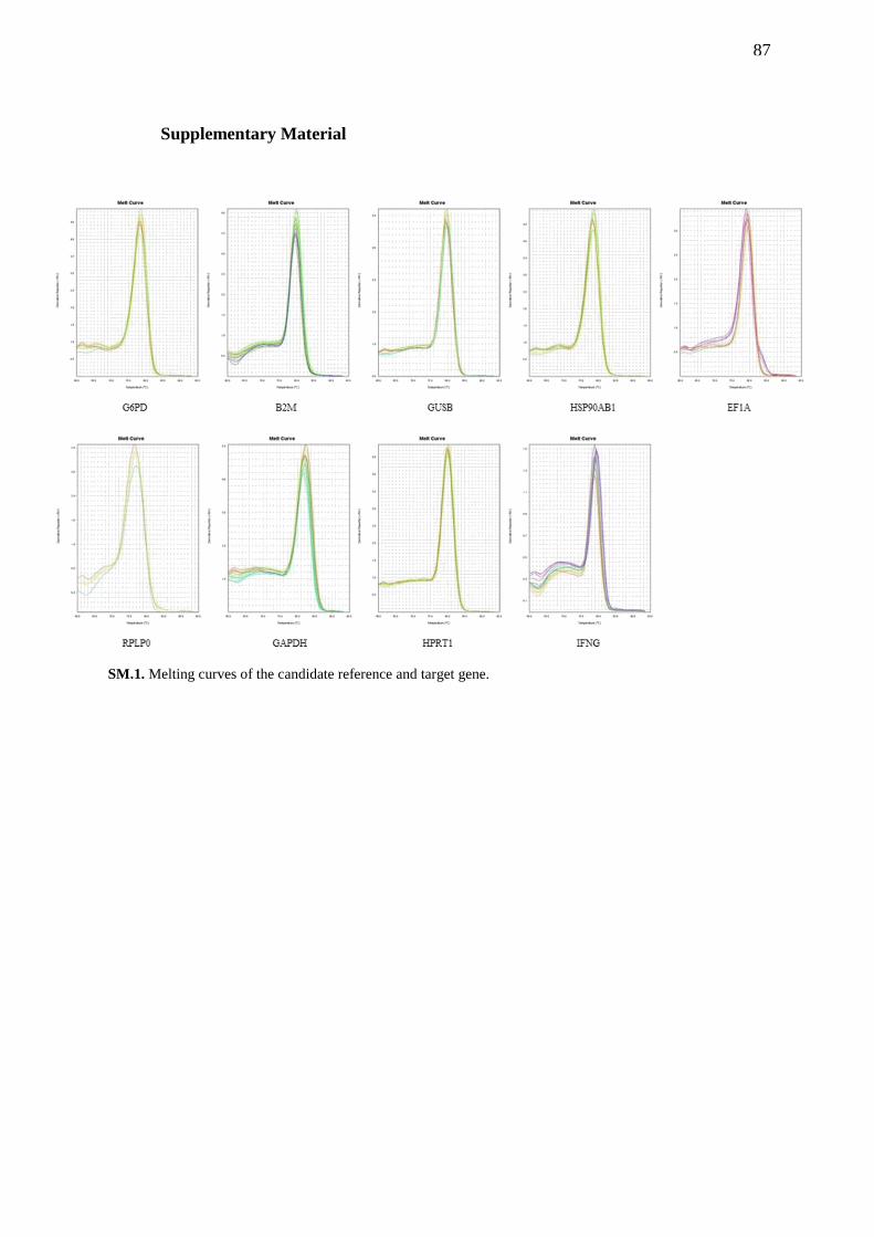
87
Supplementary Material
SM.1. Melting curves of the candidate reference and target gene.
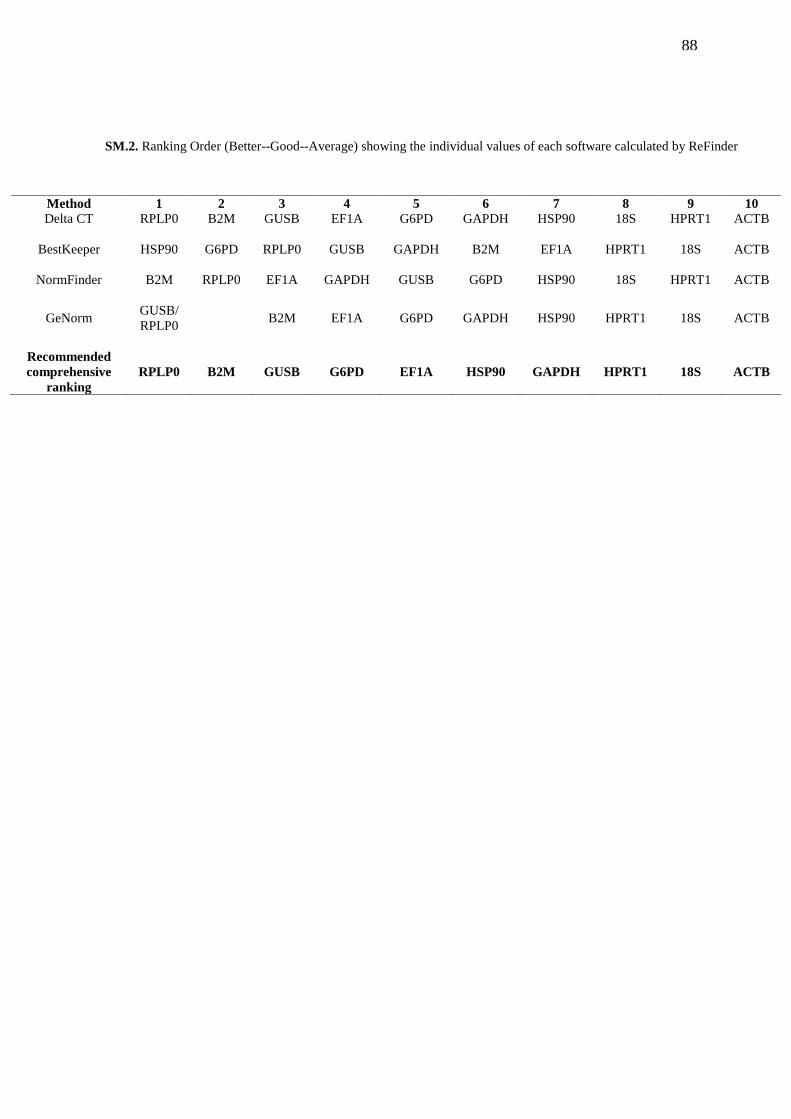
88
Method 1 2 3 4 5 6 7 8 9 10
Delta CT RPLP0 B2M GUSB EF1A G6PD GAPDH HSP90 18S HPRT1 ACTB
BestKeeper HSP90 G6PD RPLP0 GUSB GAPDH B2M EF1A HPRT1 18S ACTB
NormFinder B2M RPLP0 EF1A GAPDH GUSB G6PD HSP90 18S HPRT1 ACTB
GeNorm GUSB/
RPLP0 B2M EF1A G6PD GAPDH HSP90 HPRT1 18S ACTB
Recommended
comprehensive
ranking
RPLP0 B2M GUSB G6PD EF1A HSP90 GAPDH HPRT1 18S ACTB
SM.2. Ranking Order (Better--Good--Average) showing the individual values of each software calculated by ReFinder
89
6. Capítulo III
Artigo a ser submetido na revista European Journal of Human Genetics
Fator de Impacto: 4,5
Guia para autores: Anexo A
90
Interferon-γ plays a role in postmenopausal osteoporosis and in
human bone cells calcification: A genetic and functional study
LIMA, CAD1,2; SOUZA, APO2; SEABRA, MALB2; SILVA, HHSA2; GUARANÁ, W2;
JAVORSKI, NR1,2; BARBOSA, AD2,3; LORENA, MVB4; SANTOS, PEA2; CROVELLA,
S1,2; De AZEVÊDO SILVA, J1,2; SANDRIN-GARCIA; P1,2.
1 – Department of Genetics - Federal University of Pernambuco (UFPE), Recife, Pernambuco, Brazil.
2 – Laboratory of Immunopathology Keizo Asami – Federal University of Pernambuco (LIKA - UFPE), Recife, Pernambuco, Brazil.
3 - Division of Rheumatology, Clinical Hospital, Federal University of Pernambuco (HC - UFPE), Recife, Pernambuco, Brazil.
4 - Aggeu Magalhães Research Center (CPqAM - Fiocruz) Department of Immunology, Pernambuco, Brazil.
*Corresponding author: Camilla AD Lima
Email address: [email protected]
To be submitted for publication in European Journal of Human Genetics
91
Abstract
Osteoporosis (OP) is a complex disease in which several genes from immune system are
recognized to contribute in the susceptibility to disease’s establishment. In spite of the efforts
to be functionally validated, many of these genes, as interferon-γ (IFNG), remain with
unrevealed mechanisms. The aim of the present study was to examine whether IFN-γ -1616
(G>A, rs2069705) polymorphism was associated to postmenopausal OP. A total of 366
patients were genotyped, followed by gene expression and IFN-γ levels measured.
Furthermore, the IFN-γ action in calcifications and viability of osteoblast-line cells were
analyzed in human bone osteosarcoma (SaOs-2) cell culture. We found that IFNG rs2069705
G allele is able to decrease the IFN-γ protein stability and thus increase risk for OP (OR =
1.45, 95% CI = (1.03 – 2.05), p = 0.03). Furthermore, IFN-γ stimulus in SaOs-2 cell culture
seems to favor the calcification and viability in dose dependent manner. These findings
suggested that IFNG rs2069705 G SNP may increase OP risk and IFN-γ might favor the bone
formation. However, we suggest that further functional studies are required to confirm the
action of this cytokine in osteoblasts-like cells.
Keywords: SNP, rs2069705, Bone environment, SaOs-2 cells.
92
1. Introduction
Osteoporosis (OP) is a multifactorial metabolic bone disease influenced by genetic
factors in around 50 to 85% of all cases (Clark and Duncan 2015; Rocha-Braz and Ferraz-de-
Souza 2016). Similarly to other complex diseases many genes, in a smaller effect, contribute
to the overall phenotype such as bone mineral density (BMD) variation, fracture risk, bone
turnover rate and bone geometry (Clark and Duncan 2015). In 2008, Genome-Wide
Association Studies (GWAS) confirmed a close relationship between bone and immune
system proposed in the basis of Osteoimmunology by Arron and Choi (2000), showing
cytokines and its pathways involved in BMD, fractures and OP processes ( Richards et al.
2008; Styrkarsdottir et al. 2008).
Although technological advances have revealed novel candidate genes and numerous
polymorphisms associated to osteoporosis, biological function of many cytokines with a
recognized role in the bone remodeling remain with unrevealed mechanisms (Karasik et al.
2016; Rocha-Braz and Ferraz-de-Souza 2016). Interferon-γ (IFN-γ), one of the main example,
presents contradictory functions in bone environment (Takayanagi et al. 2005; Gao et al.
2007; Zhang et al. 2015) and its action as anabolic or catabolic agent yet raises many
questions.
IFN-γ is recognized to influence in the survival, activation and differentiation of the
osteoclasts by Tumor Necrosis Factor receptor-associated factor 6 (TRAF6) degradation and
blockade of RANK/RANKL pathway (Arron and Choi 2000). On the other hand, previous
studies showed that IFN-γ is able to promote the osteoclastogenesis by inducing specific
cytokines and T cell activation (Gao et al. 2007; Yun gang et al. 2016). In spite of GWAS
discoveries about immune systems and bone remodeling, a few functional studies with IFN-γ
polymorphisms in osteoporosis were performed up to this date.
93
Thus, in the present study aimed to assess the role of IFN-γ by studying of IFN-γ -
1616 (G>A, rs2069705) polymorphism, previously classified as a single nucleotide
polymorphism (SNP) in the promoter region and its impact in the cytokine serum levels and
consequent susceptibility to OP. Furthermore, the effects of this cytokine in the bone
mineralization were assessed by calcifying human bone osteosarcoma (SaOs-2) cell line.
2. Materials and methods
2.1 Subjects
In the present study 366 patients from the Division of Rheumatology of Clinical
Hospital in Federal University of Pernambuco, Brazil were analyzed. All the women were
submitted to BMD measurements by dual energy X-ray absorptiometry (Hologic or Lunar)
for OP diagnosis at lumbar spine (vertebrae L1-L4), femoral neck (FN) and total femur (TF).
The women classified as osteoporotics according to World Health Organization criteria were
included in patients group while the classified as healthy were included in healthy control
group.
The patients group was evaluated for secondary diseases according health assessment
questionnaire. Patients with osteopenia, inflammatory disease, autoimmune diseases, cancer,
under the use of anti-inflammatories and other rheumatic diseases were excluded from this
study. This study was approved by the Research Ethics Committee of the Center for Health
Sciences, Federal University of Pernambuco (CEP/CCS/UFPE nº 513/11) and all the
participants signed a written informed consent.
2.2 SNP prediction
SNP IFN-γ -1616 (G>A, rs2069705) have been cited in previous studies as localized
in a promoter region (Kumar and Ghosh 2008; Kanchan et al. 2015; Leng et al. 2016). To
94
confirm this information we conducted a research in 1000 Genomes
(http://browser.1000genomes.org/index.html) to analyze the SNP position. The Genome
Reference Consortium (GRC) was used to analyze the SNP position in the CRCh38.p7 and
CRCh37.p13 (for the 2016 and 2013 sequencing, respectively). Protein structure was
analyzed by the forward orientation of the SNP (FwdSNP) (C>T, R160Q, rs201359065). The
Three-Dimensional structure of the IFN-γ was downloaded from the Protein Database
(RSCB-PDB, 1EKU). Finally Swiss-PdbViewer v4.1.0 (Guex, N. and Peitsch, M.C. 1997)
software was used to visualize the protein structure, calculate their force field energy and its
minimization energy caused by the polymorphism. The software is also capable to change a
specific residue in FwdSNP, in this case, the residue of the FwdSNP, R160Q. More positive
values indicate low stability.
2.3 DNA isolation and determination of IFN-γ genotypes
Genomic DNA extraction was performed from human peripheral blood monocyte cells
(PBMCs) according to the rapid salting out method (Bignon and Viña 1995). Genotyping of
extracted DNA were performed by Real-Time PCR ABI 7500 detection system (Applied
Biosystems, Foster City, CA, USA) using specific fluorogenic probes (Taqman Probes,
Applied Biosystems, Foster City, CA, USA).
2.4 RNA isolation and gene expression analysis
Total RNA from OP patients and healthy women was extracted from human PBMCs
with TRIzol reagent (Invitrogen, USA) according to the manufacturer’s instructions. RNA
integrity, concentration and purity were determined by agarose gel (1.5%) and Nanodrop ND
1000 spectrophotometer (Nanodrop Technologies Inc, Delaware, USA), respectively. Only
95
samples that exhibited 23S and 16S fragments and the absorbance ratio OD260/280 values
from 1.9 to 2.0 were included in the analysis
cDNA synthesis was performed by GoScript™ Reverse Transcription System kit
(Promega, USA) according to the manufacturer’s instructions. β2-microglobulin (B2M),
ribosomal protein P0 (RPLP0) and glyceraldehyde 3-phosphate dehydrogenase (GAPDH)
were used as reference genes to data normalization. Gene expression was determined, in
triplicate, by Real-Time PCR ABI 7500 detection system (Applied Biosystems, Foster City,
CA, USA) using specific primers designed by NCBI/Primer-BLAST
(www.ncbi.nlm.nih.gov/tools/primer-blast/) from gene sequences obtained in GenBank
(www.ncbi.nlm.nih.gov/genbank/) (S1).
2.5 Measurement of IFN-γ serum levels from OP patients and Th1/Th2 cytokines from
cell culture
IFN-γ serum levels from OP patients and healthy women as well Th1 and Th2
cytokines (IL-2, IL-4, IL-6, IL-10, TNF-α) supernatants from SaOs-2 cell line were measured
by Cytometric Bead Array (CBA) using CBA Human Th1/Th2 Kit, according to the
manufacturer’s protocol. All data were obtained in a Becton Dickinson FACScalibur flow
cytometer and analyses were performed by BD CBA software (BD Biosciences, USA). The
results are showed by mean intensity of fluorescence (MIF).
2.6 In vitro assays
SaOs-2 cell line derived from an osteosarcoma of 11-year old Caucasian female
patient from the public tissue bank at the Federal University of Rio de Janeiro (UFRJ) and
maintained in a humidified incubator at 37°C with 5% CO2. This cell line were used as in
vitro model for bone mineralization assay as it is recognized to calcify matrix in vivo besides
expressing cytokines greatly similar to human osteoblastic cells (Querques F and Colella G
96
2015). For experiments, cells were cultured in 6-well plates and maintained for 15 days in
hMSC Osteogenic Differentiation BulletKit™ Medium to induce calcification. The media
changes were performed every 3 days.
Subsequently to this calcifying process, SaOs-2 cells confluent cultures were seeded at
2 ×10⁵ cells/well and then were treated in presence or absence of recombinant human IFN-γ
(Peprotech, Germany). The maximum (1000 U/ml) and minimum (20 U/ml) concentrations
were used based on Pedersen et al. (2015) (Pedersen et al. 2004) with modifications. After
IFN-γ stimulation, calcification and viability analysis were performed with 24 hours, while
the cytokines levels were measured after 6, 8 and 24 hours.
2.7Alizarin red staining and quantification
Calcified SaOs-2 cells were plated in 24-well plates and incubated at 37 ºC and 5 %
CO2 for 24 h in presence or absence of IFN-γ (20U/mL and 1000U/mL) for 24 h. Cells were
stained with Alizarin Red (pH 4.2, Sigma cat# A5533) for 20 minutes then washed 5 times for
5 minutes with deionized water. 5% of Cetylpyridinium Chloride (Sigma cat# C0732)
solution was used to dissolve the alizarin red bound to calcium deposits and their
concentration was measured by using a spectrophotometer at a λ=570 nm wavelength
(Varioskan Flash - Thermo Scientific, Rockford, IL,USA).
2.8 Cell viability assay
Calcified cells were stimulated for 24 h in presence of IFN-γ in 24-well plates
(20U/mL and 1000U/mL). Mitochondrial activity assay was measured by methyl-tetrazolium
bromide (MTT) according to Yao et al. (2017) (Yao et al. 2017). Solution of MTT reagent
(Sigma Chemical Co, St Louis, Missouri) (5 mg/mL) was added and incubated at 37 ºC and 5
% CO2 (under inspection of color change to brown). After 3 hours, the medium was removed
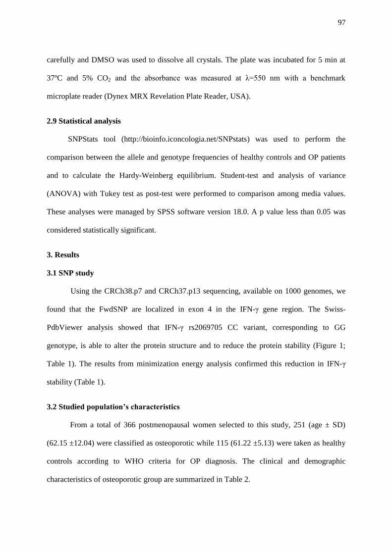
97
carefully and DMSO was used to dissolve all crystals. The plate was incubated for 5 min at
37ºC and 5% CO2 and the absorbance was measured at λ=550 nm with a benchmark
microplate reader (Dynex MRX Revelation Plate Reader, USA).
2.9 Statistical analysis
SNPStats tool (http://bioinfo.iconcologia.net/SNPstats) was used to perform the
comparison between the allele and genotype frequencies of healthy controls and OP patients
and to calculate the Hardy-Weinberg equilibrium. Student-test and analysis of variance
(ANOVA) with Tukey test as post-test were performed to comparison among media values.
These analyses were managed by SPSS software version 18.0. A p value less than 0.05 was
considered statistically significant.
3. Results
3.1 SNP study
Using the CRCh38.p7 and CRCh37.p13 sequencing, available on 1000 genomes, we
found that the FwdSNP are localized in exon 4 in the IFN-γ gene region. The Swiss-
PdbViewer analysis showed that IFN-γ rs2069705 CC variant, corresponding to GG
genotype, is able to alter the protein structure and to reduce the protein stability (Figure 1;
Table 1). The results from minimization energy analysis confirmed this reduction in IFN-γ
stability (Table 1).
3.2 Studied population’s characteristics
From a total of 366 postmenopausal women selected to this study, 251 (age ± SD)
(62.15 ±12.04) were classified as osteoporotic while 115 (61.22 ±5.13) were taken as healthy
controls according to WHO criteria for OP diagnosis. The clinical and demographic
characteristics of osteoporotic group are summarized in Table 2.
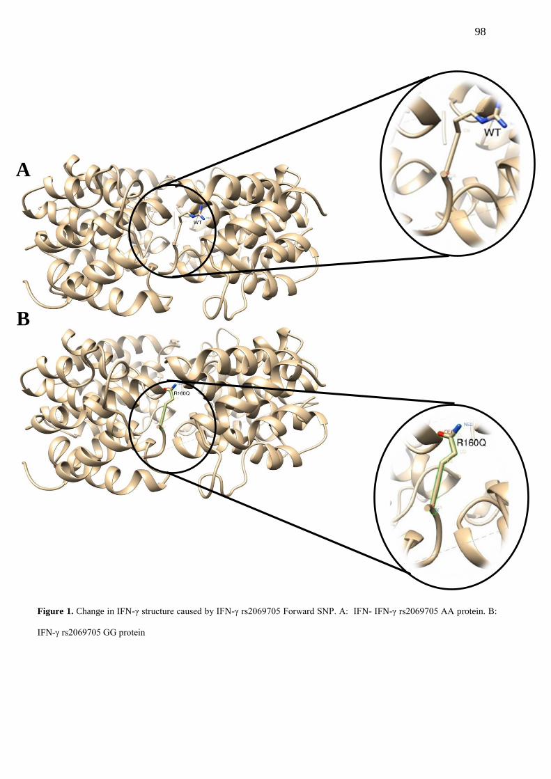
98
Figure 1. Change in IFN-γ structure caused by IFN-γ rs2069705 Forward SNP. A: IFN- IFN-γ rs2069705 AA protein. B:
IFN-γ rs2069705 GG protein
A
B
A
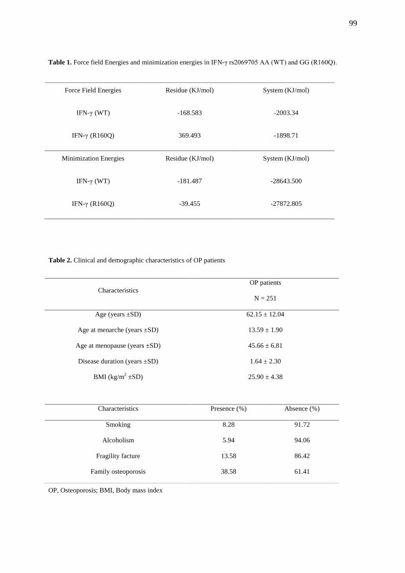
99
OP, Osteoporosis; BMI, Body mass index
Force Field Energies Residue (KJ/mol) System (KJ/mol)
IFN-γ (WT) -168.583 -2003.34
IFN-γ (R160Q) 369.493 -1898.71
Minimization Energies Residue (KJ/mol) System (KJ/mol)
IFN-γ (WT) -181.487 -28643.500
IFN-γ (R160Q) -39.455 -27872.805
Characteristics
OP patients
N = 251
Age (years ±SD) 62.15 ± 12.04
Age at menarche (years ±SD) 13.59 ± 1.90
Age at menopause (years ±SD) 45.66 ± 6.81
Disease duration (years ±SD) 1.64 ± 2.30
BMI (kg/m2 ±SD) 25.90 ± 4.38
Characteristics Presence (%) Absence (%)
Smoking 8.28 91.72
Alcoholism 5.94 94.06
Fragility facture 13.58 86.42
Family osteoporosis 38.58 61.41
Table 1. Force field Energies and minimization energies in IFN-γ rs2069705 AA (WT) and GG (R160Q).
Table 2. Clinical and demographic characteristics of OP patients
100
3.3 Distributions of IFN-γ genotypes and risk of osteoporosis
The genotype and allele frequencies of the IFN-γ -1616 (G>A, rs2069705)
polymorphisms for all the studied groups are shown in Table 3. All genotype frequencies of
OP patients and healthy control groups were found to be in the Hardy–Weinberg equilibrium.
There were significant differences in the allele frequencies of IFN-γ rs2069705 between
osteoporosis cases and healthy controls. Compared with the healthy control group, the OP
patients exhibited a higher frequency of allele G, showing this allele significantly associated
with increased risk for osteoporosis (OR = 1.45, 95% CI = (1.03 – 2.05), p = 0.03).
In spite of the OP patients group have showed higher IFN-γ rs2069705 GG genotype
frequency than healthy control (22.4% vs 14.6%) only the dominant model exhibited a trend
of statistically significant association (OR = 0.62, 95% CI = (0.38–1.01), p = 0.056) as shown
in Table 3.
3.4 Expression levels of IFN-γ in OP patients and healthy controls
IFN-γ expression analyses using the normalization factor as quantification method
showed cytokine levels increased significantly by 3.38 fold in OP patients when compared
with healthy control group (p = 0.002). No statistically significant differences were found
among studied genotypes and IFN-γ mRNA levels (Figure 2).
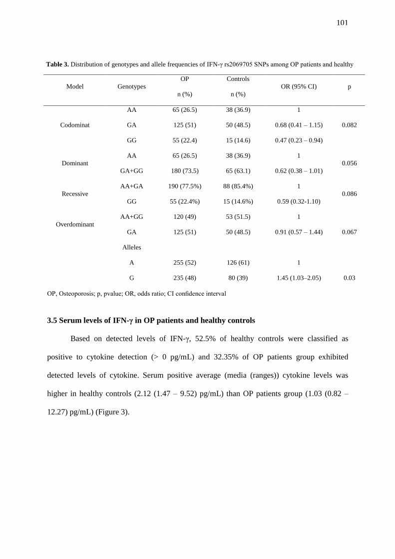
101
OP, Osteoporosis; p, pvalue; OR, odds ratio; CI confidence interval
3.5 Serum levels of IFN-γ in OP patients and healthy controls
Based on detected levels of IFN-γ, 52.5% of healthy controls were classified as
positive to cytokine detection (> 0 pg/mL) and 32.35% of OP patients group exhibited
detected levels of cytokine. Serum positive average (media (ranges)) cytokine levels was
higher in healthy controls (2.12 (1.47 – 9.52) pg/mL) than OP patients group (1.03 (0.82 –
12.27) pg/mL) (Figure 3).
Model Genotypes
OP
n (%)
Controls
n (%)
OR (95% CI) p
Codominat
AA 65 (26.5) 38 (36.9) 1
0.082 GA 125 (51) 50 (48.5) 0.68 (0.41 – 1.15)
GG 55 (22.4) 15 (14.6) 0.47 (0.23 – 0.94)
Dominant
AA 65 (26.5) 38 (36.9) 1
0.056
GA+GG 180 (73.5) 65 (63.1) 0.62 (0.38 – 1.01)
Recessive
AA+GA 190 (77.5%) 88 (85.4%) 1
0.086
GG 55 (22.4%) 15 (14.6%) 0.59 (0.321.10)
Overdominant
AA+GG 120 (49) 53 (51.5) 1
GA 125 (51) 50 (48.5) 0.91 (0.57 – 1.44) 0.067
Alleles
A 255 (52) 126 (61) 1
G 235 (48) 80 (39) 1.45 (1.03–2.05) 0.03
Table 3. Distribution of genotypes and allele frequencies of IFN-γ rs2069705 SNPs among OP patients and healthy
subjects (p = OP vs. controls)
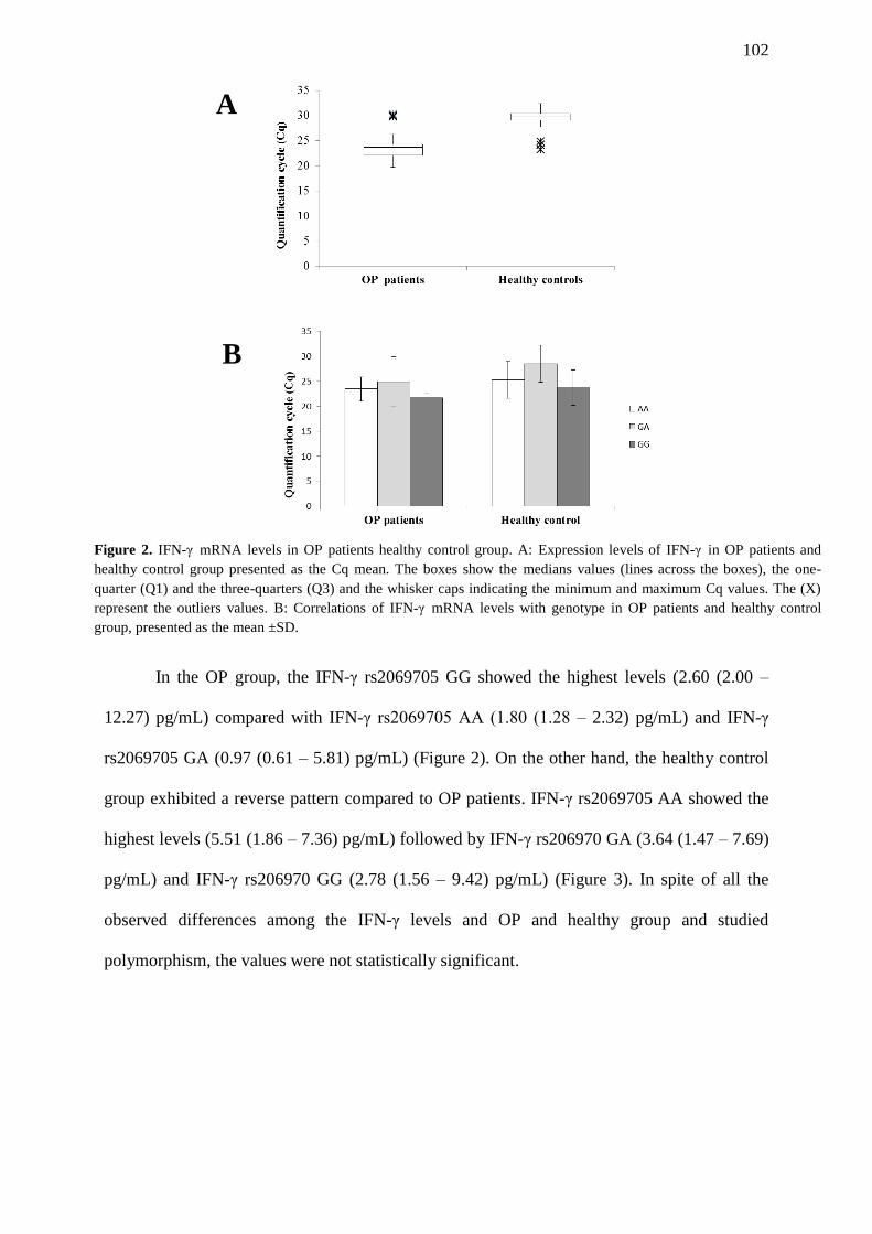
102
In the OP group, the IFN-γ rs2069705 GG showed the highest levels (2.60 (2.00 –
12.27) pg/mL) compared with IFN-γ rs2069705 AA (1.80 (1.28 – 2.32) pg/mL) and IFN-γ
rs2069705 GA (0.97 (0.61 – 5.81) pg/mL) (Figure 2). On the other hand, the healthy control
group exhibited a reverse pattern compared to OP patients. IFN-γ rs2069705 AA showed the
highest levels (5.51 (1.86 – 7.36) pg/mL) followed by IFN-γ rs206970 GA (3.64 (1.47 – 7.69)
pg/mL) and IFN-γ rs206970 GG (2.78 (1.56 – 9.42) pg/mL) (Figure 3). In spite of all the
observed differences among the IFN-γ levels and OP and healthy group and studied
polymorphism, the values were not statistically significant.
A
B
Figure 2. IFN-γ mRNA levels in OP patients healthy control group. A: Expression levels of IFN-γ in OP patients and
healthy control group presented as the Cq mean. The boxes show the medians values (lines across the boxes), the one-
quarter (Q1) and the three-quarters (Q3) and the whisker caps indicating the minimum and maximum Cq values. The (X)
represent the outliers values. B: Correlations of IFN-γ mRNA levels with genotype in OP patients and healthy control
group, presented as the mean ±SD.
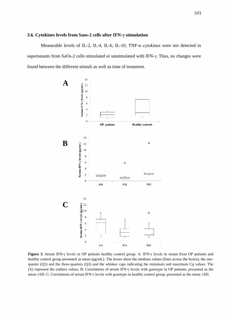
103
3.6. Cytokines levels from Saos-2 cells after IFN-γ stimulation
Measurable levels of IL-2, IL-4, IL-6, IL-10, TNF-α cytokines were not detected in
supernatants from SaOs-2 cells stimulated or unstimulated with IFN-γ. Thus, no changes were
found between the different stimuli as well as time of treatment.
A
B
C
Figure 3. Serum IFN-γ levels in OP patients healthy control group. A: IFN-γ levels in serum from OP patients and
healthy control group presented as mean (pg/mL). The boxes show the medians values (lines across the boxes), the one-
quarter (Q1) and the three-quarters (Q3) and the whisker caps indicating the minimum and maximum Cq values. The
(X) represent the outliers values. B: Correlations of serum IFN-γ levels with genotype in OP patients, presented as the
mean ±SD. C: Correlations of serum IFN-γ levels with genotype in healthy control group, presented as the mean ±SD.
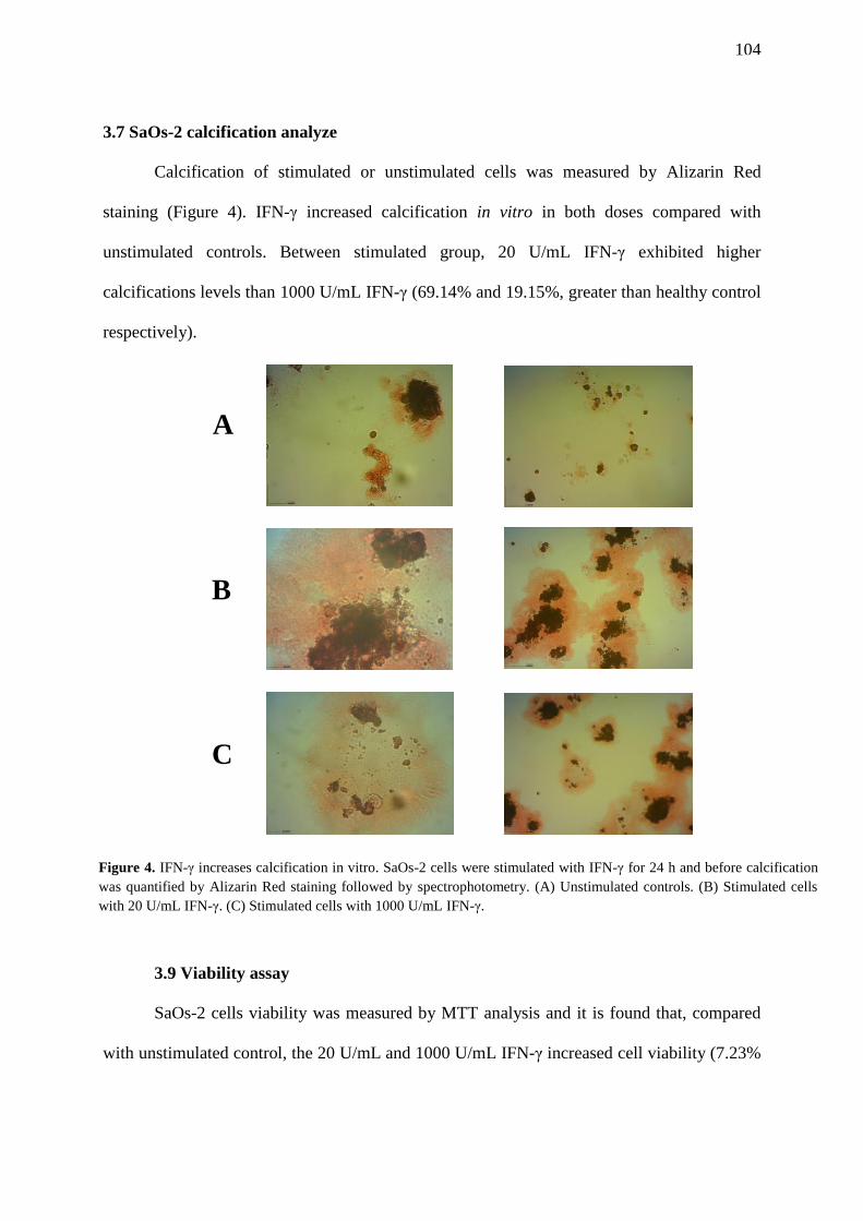
104
3.7 SaOs-2 calcification analyze
Calcification of stimulated or unstimulated cells was measured by Alizarin Red
staining (Figure 4). IFN-γ increased calcification in vitro in both doses compared with
unstimulated controls. Between stimulated group, 20 U/mL IFN-γ exhibited higher
calcifications levels than 1000 U/mL IFN-γ (69.14% and 19.15%, greater than healthy control
respectively).
3.9 Viability assay
SaOs-2 cells viability was measured by MTT analysis and it is found that, compared
with unstimulated control, the 20 U/mL and 1000 U/mL IFN-γ increased cell viability (7.23%
A
B
C
Figure 4. IFN-γ increases calcification in vitro. SaOs-2 cells were stimulated with IFN-γ for 24 h and before calcification
was quantified by Alizarin Red staining followed by spectrophotometry. (A) Unstimulated controls. (B) Stimulated cells
with 20 U/mL IFN-γ. (C) Stimulated cells with 1000 U/mL IFN-γ.
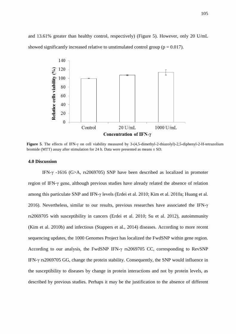
105
and 13.61% greater than healthy control, respectively) (Figure 5). However, only 20 U/mL
showed significantly increased relative to unstimulated control group (p = 0.017).
4.0 Discussion
IFN-γ -1616 (G>A, rs2069705) SNP have been described as localized in promoter
region of IFN-γ gene, although previous studies have already related the absence of relation
among this particulate SNP and IFN-γ levels (Erdei et al. 2010; Kim et al. 2010a; Huang et al.
2016). Nevertheless, similar to our results, previous researches have associated the IFN-γ
rs2069705 with susceptibility in cancers (Erdei et al. 2010; Su et al. 2012), autoimmunity
(Kim et al. 2010b) and infectious (Stappers et al., 2014) diseases. According to more recent
sequencing updates, the 1000 Genomes Project has localized the FwdSNP within gene region.
According to our analysis, the FwdSNP IFN-γ rs2069705 CC, corresponding to RevSNP
IFN-γ rs2069705 GG, change the protein stability. Consequently, the SNP would influence in
the susceptibility to diseases by change in protein interactions and not by protein levels, as
described by previous studies. Perhaps it may be the justification to the absence of different
Figure 5. The effects of IFN-γ on cell viability measured by 3-(4,5-dimethyl-2-thiazolyl)-2,5-diphenyl-2-H-tetrazolium
bromide (MTT) assay after stimulation for 24 h. Data were presented as means ± SD.
106
protein levels among genotypes although there is a relationship among these variants and
susceptibility for several diseases.
In functional analysis, we found that OP patients groups exhibited 3-fold higher of
mRNA IFN-γ levels than healthy control groups. This alteration appears to be an increasing
protein levels compensatory mechanism in OP patients group, which showed the highest
frequency of the more instable allelic variant IFN-γ rs2069705 G. Taken all these data
together, it was observed that IFN-γ stimuli favored the calcification and viability of SaOs-2
cells and appeared to be involved to bone mass increasing. Similar results related to IFN-γ
action in bone formation were found by Duque et al. (2011) (Duque et al. 2011) in in vivo
studies with ovariectomized mice, which suggested that alterations in IFN-γ signaling
pathway may be used advantageously to improve bone strength. Zhang et al (2015) (Zhang et
al. 2015a) also associated the cytokine with inhibition of bone resorption in postmenopausal
women. However, the IFN-γ dual role in bone environmental is already recognized (Ferrari-
Lacraz and Ferrari 2007; Gao et al. 2007; Duque et al. 2011). Previous studies highlighted the
direct anti-resorptive action of IFN-γ through inhibition of the RANK/RANKL pathway and
blocks osteoclast formation. At the same time its indirect pro-resorptive action stimuli T cells
and cytokines such as TNF-α and RANKL (Takayanagi et al. 2002; Cenci et al. 2003;
Takayanagi et al. 2005; Ferrari-Lacraz and Ferrari 2007; Gao et al. 2007; Guerrini and
Takayanagi 2014). Nevertheless, it is important to mention that the most of these results are
related to studies with osteoclasts and murine cells and a few studies have been performed to
clarify this role in human bone formation cells and their pathway. Among these studies with
osteoblast-like cells, Wimbauer et al. (2012) (Wimbauer et al. 2012) described the IFN-γ
associated with 2-methoxyestradiol treatment showed anti-proliferative effects in
osteosarcoma cells by IFN-stimulated response element (ISRE) sequence-dependent
107
transcription and gamma-activated sequence (GAS)-dependent transcription. However, our
results showed that IFN-γ increased calcification and viability in SaOs-2 cells. It is important
to emphasize that the lowest concentration of IFN-γ was more favorable to osteoblasts
activity than highest concentration. Additionally, we did not verify alterations in other
cytokines levels, the main indirect action pro-resorptive associated with IFN-γ. We highlight
that further studies should be conducted for confirmation of relationship between osteoblasts
activity and cytokines levels. On the other hand, it is possible that other pathways different of
observed in osteoclasts are involved in human osteoblasts survival process.
The direct effect of IFN-γ in osteoblastogenesis have been already reported by Urizar
et al. (2015) (Ibarra Urizar et al. 2015) which related that 5 ng/mL (50 U/mL) IFN-γ was able
to stimulate bone morphogenetic proteins 2 (BMP-2) expression in pancreatic beta cells,
although had verified that this cytokine did not affect the expression of protein in rats islets.
BMP-2 is recognized by stimulating the differentiation of progenitor cells in pre-osteoblasts
besides to have a critical role for osteoblast function together with Wnt pathway and its
inhibitors (Stein and Lian 1993; Raisz 2005; Bunnell et al. 2011). Additionally, previous
studies have related IFN-γ together with interleukin 1 (IL-1) stimulates nitric oxid (NO) and
indirectly induce increasing bone mass (Ralston et al. 1994; Van’t Hof and Ralston 2001;
Duque et al. 2011). The endothelial isoform of nitric oxide synthase (eNOS) and its NO
derived acts as a mediator of the effects of estrogen in bone and associated with
prostaglandins promote bone formation and suppress bone resorption (Van’t Hof and Ralston
2001). In accordance to Duque et al. (2011) (Duque et al. 2011), we suggested that this IFN-γ
anabolic activity is dose-dependent. High IFN-γ doses seems to contribute to bone loss (Mann
et al. 1994; Tohkin et al. 1994; Duque et al. 2011). IFN-γ variable effects in bone homeostasis
also appears to be dependents on the experimental model and other conditions used in the
108
analysis (Ferrari-Lacraz and Ferrari 2007; Duque et al. 2011). We verified that human bone
cells and in vivo studies have showed different results when compared with cells murine
analysis (Ferrari-Lacraz and Ferrari 2007; Gao et al. 2007; Duque et al. 2011).
To the best of our knowledge, this is the first time the IFN-γ -1616 (G>A, rs2069705)
was analyzed in OP patients. In our study, we found a significantly increased risk for OP
associated with the IFN-γ rs2069705 G allele. The FwdSNP is localized in exon region and
according to our analysis is able to decrease the protein stability, but not its levels.
Consequently, no significant differences have been observed in genotype variants. The high
levels observed in mRNA IFN-γ levels seems to be associated with a compensatory response
since IFN-γ favored the calcification and viability in the SaOs-2 study related to used doses.
We also observed that previous studies showed differences according to their used cells and
models. Therefore we strongly indicated that this cytokine concentration and the choice of a
model in vivo and in vitro must be into consideration for future analysis. Finally, we suggest
that further functional studies are required to examine the IFN-γ activity in osteoblast-like
cells bearing in mind that this is one of the few studies that performs this approach.
Acknowledgments
We thank the following Brazilian funding agencies: CAPES (Coordenação de
Aperfeiçoamento de Pessoal de Nível Superior), CNPq (Conselho Nacional de
Desenvolvimento Científico e Tecnológico), and FACEPE (Fundação de Amparo à Ciência e
Tecnologia de Pernambuco) for financial support. We are also grateful to Dr. João Ricardo de
Oliveira for SaOs-2 cells donation.
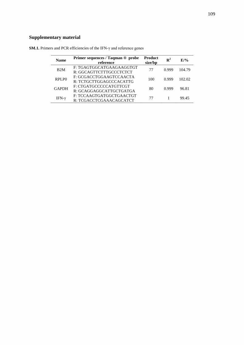
109
Supplementary material
SM.1. Primers and PCR efficiencies of the IFN-γ and reference genes
Name Primer sequences / Taqman ® probe
reference
Product
size/bp R
2 E/%
B2M F: TGAGTGGCATGAAGAAGGTGT
R: GGCAGTTCTTTGCCCTCTCT 77 0.999 104.79
RPLP0 F: GCGACCTGGAAGTCCAACTA
R: TCTGCTTGGAGCCCACATTG 100 0.999 102.02
GAPDH F: CTGATGCCCCCATGTTCGT
R: GCAGGAGGCATTGCTGATGA 80 0.999 96.81
IFN-γ F: TCCAAGTGATGGCTGAACTGT
R: TCGACCTCGAAACAGCATCT 77 1 99.45
110
7. Discussão geral
Para o nosso conhecimento, este é o primeiro estudo que verificou a relação
entre os SNPs IL23R +2284 (C>A) (rs10889677), IL17A +672 (G>A) (rs7747909),
IL12B +1188 (T>G) (rs3212227) e IFN-γ -1616 (G>A) (rs2069705) e a OP primária
pós-menopausa. Na análise de associação entre os SNPs e a resposta terapêutica
aos bisfosfonatos encontramos os genótipos IL17A rs7747909 G/A e IL12B
rs3212227 T/G associados a altos níveis de vitamina D, IL23R rs10889677 C/C
associado a altos níveis de cálcio, IFN-γ rs2069705 A/A associado a altos níveis de
PTH e o IFN-γ rs2069705 G/G a altos níveis de ALP. Adicionalmente, verificamos
relação entre o genótipo IFN-γ rs2069705 G/G e o aumento da DMO na cabeça do
fêmur e diminuição no quadril total, após 3 e 4 anos de tratamento, respectivamente.
Por fim selecionamos para nosso terceiro estudo o SNP associado à
susceptibilidade a OP, o IFN-γ -1616 (G>A) (rs2069705). Na confirmação da
localização do SNP verificamos que o FwdSNP correspondente ao nosso
polimorfismo de estudo se encontra no éxon 4 e não na região promotora do gene,
como relatado por estudos anteriores. Nessa análise encontramos que a variante G,
associada à susceptibilidade à OP no nosso estudo, confere maior instabilidade à
proteína. Esses resultados foram reforçados pela análise in vitro com células SaOs-
2 , que sugeriram o IFN-γ como proteína pró-absortiva, embora o grupo de pacientes
tenha apresentando maiores níveis de mRNA da citocina comparados ao grupo
controle saudável.
Os SNPs aqui estudados foram analisados pela primeira vez em OP, mas já
haviam sido relacionados à outras doenças reumatológicas como artrite reumatoide
(Faragó et al., 2008; Shen et al., 2015); espondilite anquilosante (Wong et al., 2012),
111
arterite (Márquez et al., 2014) e lúpus eritematoso sistêmico (LES) (Kim et al., 2010).
Semelhante às nossas análises, muitos desses estudos não encontraram alterações
nos níveis do IFN-γ em relação aos diferentes genótipos genótipo (Erdei et al. 2010;
Kim et al. 2010; Huang et al. 2016). As citocinas estudadas também já mostraram
influências nas vias da homeostase óssea. A via IL-23/IL-23R/β-catenina já foi citada
por induzir formação óssea (Tu et al., 2015), a IL-17A por estimular RANKL, a IL-12B
por inibir a formação óssea in vivo (Xu et al., 2016) e o IFN-γ com relatos
contraditórios sobre sua participação na formação e na reabsorção óssea (Gao et al.,
2007; Duque et al., 2011). O nosso trabalho também mostrou essa ação pleiotrópica
da citocina na resposta terapêutica aos bisfosfonatos, com o mesmo genótipo IFN-γ
rs2069705 G/G associado à diferentes respostas na DMO. Além disso, encontramos
concentrações menores da citocina mais favoráveis à calcificação e viabilidade em
células da linhagem osteoblástica que concentrações maiores induzidas no mesmo
ensaio. Esses dados associados aos estudos anteriores, nos possibilitaram sugerir
uma ação da citocina dependente dos valores de DMO, local de atuação e
quantidade da proteína disponibilizada. Adicionalmente, levantamos a discussão
sobre os processos de apoptose como fator importante nessa atuação antagônica
do IFN-γ. Um outro componente importante que verificamos em estudos anteriores é
o modelo selecionado para essas avaliações. O resultado das análises parece estar
também relacionado a esses modelos in vivo e in vitro utilizados (Ferrari-Lacraz e
Ferrari, 2007; Duque et al., 2011). E por fim, uma das contribuições que deve ser
destacada no nosso estudo é a validação dos genes de referência que conduziu
nossa posterior análise funcional e deve ser levada em consideração para os
próximos ensaios de expressão em OP. Nesse estudo pioneiro, além de definirmos
112
os genes mais estáveis para análises de expressão, RPLP0 e B2M, enumeramos o
18S, ACTB e HPRT1 como não recomendados para estudos na doença.
Reportamos a instabilidade do ACTB para nossa análise em OP, embora ele seja
usado em mais de 70% das quantificações de mRNA.
A despeito do fato de que muito ainda tenha que ser feito para um completo
esclarecimento dos fatores genéticos e osteoimunológicos envolvidos no
estabelecimento da OP, acreditamos que os estudos avaliando SNPs e conduzindo
suas validações funcionais são de grande importância na identificação de fatores de
risco e marcadores precoces para diagnóstico e adesão terapêutica. Desta forma,
além de tratamentos preventivos e personalizados baseados na susceptibilidade
genética, poderíamos também evitar o óbito dos 20% pacientes de OP que não
aderem ao tratamento ainda no primeiro ano de terapia (Roush, 2011; Iolascon et al.,
2013; Drake et al., 2015; Miller, 2016). Além disso, nós acreditamos que essas
contribuições em conjunto encorajam outras pesquisas proeminentes como a
influência desses polimorfismos localizados em regiões 3’UTR na ligação de
miRNAs e consequente susceptibilidade à OP (Coronnello et al., 2012; Qin et al.,
2016).
113
8. Conclusões
Após os estudos realizados neste trabalho, alguns pontos em relação à
Osteoporose e seus fatores genéticos e funcionais foram esclarecidos, de acordo
com os objetivos inicialmente propostos.
Verificamos que SNPs nos genes das citocinas estudadas podem contribuir
para decisões terapêuticas em OP. SNPs nos genes IL23R +2284 (C>A)
(rs10889677), IL17A +672 (G>A) (rs7747909), IL12B +1188 (T>G) (rs3212227) e
IFN-γ -1616 (G>A) (rs2069705) exibiram associação estatisticamente significativa
com mudanças nos marcadores bioquímicos do metabolismo ósseo em pacientes
submetidos à terapia com bisfofonatos. Além disso, o IFN-γ rs2069705 G/G mostrou
associação com mudanças de DMO nesses pacientes. Adicionalmente sugerimos
que um mecanismo de apoptose em osteoblastos relacionado ao IFN-γ pode
explicar alguns casos em que ele está, contraditoriamente, associado à diminuição
de DMO.
Para os estudos funcionais, validamos os genes de referência RPLP0 e B2M
como os mais estáveis para nosso estudo de expressão em OP. Nesse estudo
também verificamos que o ACTB, 18S e HPRT1 são inadequados para
normalização dos dados de expressão na doença.
Por fim concluímos que O SNP IFN-γ -1616 (G>A) (rs2069705) parece alterar
a estabilidade da proteína e não seus níveis. Esta informação está de acordo com o
último sequenciamento de 2016 (CRCh38.p7) que comprova sua posição fora da
região promotora e seu FwdSNP correspondente localizado no éxon 4. Além disso,o
alelo IFN-γ rs2069705 G, que confere maior instabilidade à proteína esteve
associado com susceptibilidade à OP na nossa população de estudo. Esses dados
114
nos fazem sugerir que o IFN-γ tenha um papel protetor na OP. Concordando com
esses resultados, a proteína aumentou calcificação e viabilidade em células SaOs-2.
No entanto, acreditamos que essa indução é dose-dependente, tendo em vista que a
maior dose (1000 U/mL) utilizada para os estímulos celulares apresentaram níveis
de calcificação e viabilidade menores que o valor mínimo utilizado da citocina (20
U/mL).
Além de responder à alguns dos nossos questionamentos iniciais, esses
resultados encorajam pesquisas funcionais em Osteoimunologia, em busca de
marcadores diagnósticos e terapêuticos, além de modelos de células ósseas e
concentrações ótimas de estímulos com citocinas, a fim de nos aproximarmos cada
vez mais do entendimentos do papel dessas proteínas na OP pós-menopausa.
115
Referências Bibliográficas
Abrahamsen B and Prieto-Alhambra D (2016) Patients with Atypical Femur Fractures have the Same Mortality as the Background Population - Drug-Channelling Bias, Bisphosphonate Effects and Public Health Implications. J Bone Miner Res 31: 31(3):488-490. doi: 10.1002/jbmr.2801
Albagha OME and Ralston SH (2006) Genetics and Osteoporosis. Rheum Dis Clin North Am 32:659–680. doi: 10.1016/j.rdc.2006.08.001
Andersen CL, Jensen JL and Ørntoft TF (2004) Normalization of real-time quantitative reverse transcription-PCR data: A model-based variance estimation approach to identify genes suited for normalization, applied to bladder and colon cancer data sets. Cancer Res 64:5245–5250. doi: 10.1158/0008-5472.CAN-04-0496
Annunziato F, Romagnani C and Romagnani S (2015) The 3 major types of innate and adaptive cell-mediated effector immunity. J Allergy Clin Immunol 135:626–635. doi: 10.1016/j.jaci.2014.11.001
Applied Biosystems (2008) Guide to Performing Relative Quantitation of Gene Expression Using Real-Time Quantitative PCR. Gene Expr 2009:1–60. doi: 10.1007/978-3-642-31107-9
Arron JR and Choi Y (2000) Bone versus immune system. Nature 408:535–536. doi: 10.1038/35046196
Artero-Castro A, Castellvi J, García A, Hernández J, Cajal SRY and Lleonart ME (2011) Expression of the ribosomal proteins Rplp0, Rplp1, and Rplp2 in gynecologic tumors. Hum Pathol 42:194–203. doi: 10.1016/j.humpath.2010.04.020
Asada N, Katayama Y, Sato M, Minagawa K, Wakahashi K, Kawano H, Kawano Y, Sada A, Ikeda K, Matsui T et al. (2013) Matrix-embedded osteocytes regulate mobilization of hematopoietic stem/progenitor cells. Cell Stem Cell 12:737–747. doi: 10.1016/j.stem.2013.05.001
Aspray TJ (2014) Fragility fracture: recent developments in risk assessment. Ther Adv Musculoskelet Dis 7:17–25. doi: 10.1177/1759720X14564562
Baccaro LF, Conde DM, Costa-Paiva L and Pinto-Neto AM (2015) The epidemiology and management of postmenopausal osteoporosis: A viewpoint from Brazil. Clin Interv Aging 10:583–591. doi: 10.2147/CIA.S54614
Bamias G, Goukos D, Laoudi E, Balla IG, Siakavellas SI, Daikos GL and Ladas SD (2013) Comparative study of candidate housekeeping genes for quantification of target gene messenger RNA expression by real-time PCR in patients with inflammatory bowel disease. Inflamm Bowel Dis 19:2840–2847. doi: 10.1097/01.MIB.0000435440.22484.e8
Bhushan S and Perumal NB (2012) Disease associated cytokine SNPs database: An annotation and dissemination model. Cytokine 57:107–112. doi: 10.1016/j.cyto.2011.10.009
116
Bignon JD, Viña MF (1995) 12th IHWC HLA Class II Reference Protocol. In: Fauchet R and Charron D (ed) Technical Handbook of the 12th International Histocompatibility Workshop. Paris, HLA et Médecine.
Blumenfeld O, Williams FMK, Valdes A, Hart DJ, Malkin I, Spector TD and Livshits G (2014) Association of interleukin-6 gene polymorphisms with hand osteoarthritis and hand osteoporosis. Cytokine 69:94–101. doi: 10.1016/j.cyto.2014.05.012
Boroń D, Author E, Agnieszka S, Daniel K, Anna B and Adam K (2014) Polymorphism of interleukin17 and its relation to mineral density of bones in perimenopausal women. Eur J Med Res. doi: 10.1186/s4000101400691
Bunnell TM, Burbach BJ, Shimizu Y and Ervasti JM (2011) β-Actin specifically controls cell growth, migration, and the G-actin pool. Mol Biol Cell 22:4047–58. doi: 10.1091/mbc.E11-06-0582
Bustin SA, Benes V, Garson JA, Hellemans J, Huggett J, Kubista M, Mueller R, Nolan T, Pfaffl MW, Shipley GL et al. (2009) The MIQE Guidelines: Minimum Information for Publication of Quantitative Real-Time PCR Experiments. Clin Chem 55:611–622.
Carmeliet G, Dermauw V and Bouillon R (2015) Vitamin D signaling in calcium and bone homeostasis: A delicate balance. Best Pract Res Clin Endocrinol Metab 29:621–631. doi: 10.1016/j.beem.2015.06.001
Cenci S, Toraldo G, Weitzmann MN, Roggia C, Gao Y, Qian WP, Sierra O and Pacifici R (2003) Estrogen deficiency induces bone loss by increasing T cell proliferation and lifespan through IFN-gamma-induced class II transactivator. Proc Natl Acad Sci U S A 100:10405–10410. doi: 10.1073/pnas.1533207100
Chen B, Zeng Z, Xu L, Wu X, Yu J, Xue L, Hao Y, Wang Y, Sung J, Chen M et al. (2009) IL23R +2199A/C polymorphism is associated with decreased risk of certain subtypes of gastric cancer in Chinese: A case-control study. Cancer Epidemiol. doi: 10.1016/j.canep.2010.08.006
Chesnut CH, McClung MR, Ensrud KE, Bell NH, Genant HK, Harris ST, Singer FR, Stock JL, Yood RA, Delmas PD et al. (1995) Alendronate treatment of the postmenopausal osteoporotic woman: Effect of multiple dosages on bone mass and bone remodeling. Am J Med 99:144–152. doi: 10.1016/S0002-9343(99)80134-X
Christakos S, Dhawan P, Verstuyf A, Verlinden L and Carmeliet G (2016) Vitamin D: Metabolism, Molecular Mechanism of Action, and Pleiotropic Effects. Physiol Rev 96:365–408. doi: 10.1152/physrev.00014.2015
Clark GR and Duncan EL (2015) The genetics of osteoporosis. Br Med Bull 113:73–81. doi: 10.1093/bmb/ldu042
Coronnello C, Hartmaier R, Arora A, Huleihel L, Pandit K V., Bais AS, Butterworth M, Kaminski N, Stormo GD, Oesterreich S et al. (2012) Novel Modeling of Combinatorial miRNA Targeting Identifies SNP with Potential Role in Bone Density. PLoS Comput Biol. doi: 10.1371/journal.pcbi.1002830
117
Cummings SR, San Martin J, McClung MR, Siris ES, Eastell R, Reid IR, Delmas P, Zoog HB, Austin M, Wang A et al. (2009) Denosumab for prevention of fractures in postmenopausal women with osteoporosis. N Engl J Med 361:756–765. doi: 10.1056/NEJMoa0809493
Cunha, EP, Steiner MI, Strufaldi R, Fernades C, Laurindo IMM, Pereira RMR, Simões R (2011) Osteoporose: Tratamento. Diretrizes Clínicas Na Saúde Suplementar. Associaçiação Médica Brasileira E Agência Nacional de Saúde Suplementar. http://diretrizes.amb.org.br/ans/osteoporose-tratamento.pdf. Accessed 01 August 2016.
Czerny B, Kaminski A, Kurzawski M, Kotrych D, Safranow K, Dziedziejko V, Bohatyrewicz A and Pawlik A (2010) The association of IL-1beta, IL-2, and IL-6 gene polymorphisms with bone mineral density and osteoporosis in postmenopausal women. Eur J Obs Gynecol Reprod Biol 149:82–85. doi: 10.1016/j.ejogrb.2009.12.010.
Damsker JM, Hansen AM and Caspi RR (2010) Th1 and Th17 cells. Ann N Y Acad Sci 211–221. doi: 10.1111/j.1749-6632.2009.05133.x.Th1
De Spiegelaere W, Dern-Wieloch J, Weigel R, Schumacher V, Schorle H, Nettersheim D, Bergmann M, Brehm R, Kliesch S, Vandekerckhove L et al. (2015) Reference gene validation for RT-qPCR, a note on different available software packages. PLoS One 10:1–13. doi: 10.1371/journal.pone.0122515
Drake M, Clarke B and Lewiecki E (2015) The Pathophysiology and Treatment of Osteoporosis. Clin Ther 37:1837–1850. doi: 10.1016/j.clinthera.2015.06.006.
Duque G, Huang DC, Dion N, Macoritto M, Rivas D, Li W, Yang XF, Li J, Lian J, Marino FT et al. (2011) Interferon- γ Plays a Role in Bone Formation In Vivo and Rescues Osteoporosis in Ovariectomized Mice. 26:1472–1483. doi: 10.1002/jbmr.350
Eastell R, O’Neill TW, Hofbauer LC, Langdahl B, Reid IR, Gold DT and Cummings SR (2016) Postmenopausal osteoporosis. Nat Rev Dis Prim 29;2:16069. doi: 10.1038/nrdp.2016.69
Elsafadi M, Manikandan M, Dawud RA, Alajez NM, Hamam R, Alfayez M, Kassem M, Aldahmash A and Mahmood A (2016) Transgelin is a TGFβ-inducible gene that regulates osteoblastic and adipogenic differentiation of human skeletal stem cells through actin cytoskeleston organization. Cell Death Dis 7:e2321. doi: 10.1038/cddis.2016.196
Emami S, Ziaee V, Rezaei A, Sadr M, Maddah M, Amirzargar AA and Rezaei N (2015) IL23R gene polymorphism with juvenile idiopathic arthritis and its association with serum IL-17A. Int J Rheum Dis 1–8. doi: 10.1111/1756-185X.12674
Erdei E, Kang H, Meisner A, White K, Pickett G, Baca C, Royce M and Berwick M (2010) Polymorphisms in cytokine genes and serum cytokine levels among New Mexican women with and without breast cancer. Cytokine 51:18–24. doi: 10.1016/j.cyto.2010.03.014
118
Esbrit P, Herrera S, Portal-Núñez S, Nogués X and Díez-Pérez A (2015) Parathyroid Hormone-Related Protein Analogs as Osteoporosis Therapies. Calcif Tissue Int. doi: 10.1007/s00223-015-0050-1
Faienza MF, Ventura A, Marzano F and Cavallo L (2013) Review Article Postmenopausal Osteoporosis : The Role of Immune System Cells. Clin Dev Immunol. 2013;2013:575936. doi: 10.1155/2013/575936.
Faragó B, Magyari L, Sáfrány E, Csöngei V, Járomi L, Horvatovich K, Sipeky C, Maász a, Radics J, Gyetvai a et al. (2008) Functional variants of interleukin-23 receptor gene confer risk for rheumatoid arthritis but not for systemic sclerosis. Ann Rheum Dis 67:248–250. doi: 10.1136/ard.2007.072819
Fassunke J, Blum M-C, Schildhaus H-U, Zapatka M, Brors B, Kunstlinger H, Buttner R, Wardelmann E, Merkelbach-Bruse S, Künstlinger H et al. (2010) qPCR in gastrointestinal stromal tumors: Evaluation of reference genes and expression analysis of KIT and the alternative receptor tyrosine kinases FLT3, CSF1-R, PDGFRB, MET and AXL. BMC Mol Biol 11:100. doi: 10.1186/1471-2199-11-100
Ferrari-Lacraz S and Ferrari S (2007) Is IFN-γ involved in bone loss or protection? nothing is simple with cytokines. Bonekey-Osteovision 4:83–87. doi: 10.1138/20060251
Flicker L, Hopper JL, Rodgers L, Kaymakci B, Green RM and Wark JD (1995) Bone density determinants in elderly women: A twin study. J Bone Miner Res 10:1607–1613. doi: 10.1002/jbmr.5650101102
Gao Y, Grassi F, Ryan MR, Terauchi M, Page K, Yang X, Weitzmann MN and Pacifici R (2007) IFN-gamma stimulates osteoclast formation and bone loss in vivo via antigen-driven T cell activation. J Clin Invest 117:122–132. doi: 10.1172/JCI30074
Genant H, Wu C, van Kuijk C and Nevitt M (1993) Vertebral fracture assessment using a semiquantitative technique. J Bone Min Res 8:1137–48.
Golob AL and Laya MB (2015) Osteoporosis. Med Clin North Am 99:587–606. doi: 10.1016/j.mcna.2015.01.010
Greenblatt MB, Tsai JN and Wein MN (2016) Bone Turnover Markers in the Diagnosis and Monitoring of Metabolic Bone Disease. Clin Chem. doi: 10.1373/clinchem.2016.259085
Guerrini MM and Takayanagi H (2014) The immune system, bone and RANKL. Arch Biochem Biophys 561:118–123. doi: 10.1016/j.abb.2014.06.003
Hartmaier RJ, Richter a S, Gillihan RM, Sallit JZ, McGuire SE, Wang J, Lee a V, Osborne CK, O’Malley BW, Brown PH et al. (2012) A SNP in steroid receptor coactivator-1 disrupts a GSK3β phosphorylation site and is associated with altered tamoxifen response in bone. Mol Endocrinol 26:220–227. doi: 10.1210/me.2011-1032
119
Haynes DR (2006) Emerging and future therapies for the treatment of bone loss associated with chronic inflammation. Inflammopharmacology 14:193–197. doi: 10.1007/s10787-006-0006-1
Hiligsmann MM, Bours SPG and Boonen A (2015) A Review of Patient Preferences for Osteoporosis Drug Treatment. Curr Rheumatol Rep 17:61. doi: 10.1007/s11926-015-0533-0
Hsu YH and Kiel DP (2012) Genome-wide association studies of skeletal phenotypes: What we have learned and where we are headed. J Clin Endocrinol Metab 97:1958–1977. doi: 10.1210/jc.2012-1890
Huang Y-H, Hsu Y-W, Lu H-F, Sung H, Wong -Ching, Yu H-R, Kuo H-C, Huang F-C, Chang W-C and Kuo H-C (2016) Interferon-gamma Genetic Polymorphism and Expression in Kawasaki Disease. Medicine (Baltimore) 95(17):e3501. doi: 10.1097/MD.0000000000003501
Ibarra Urizar A, Friberg J, Christensen DP, Lund Christensen G and Billestrup N (2015) Inflammatory Cytokines Stimulate Bone Morphogenetic Protein-2 Expression and Release from Pancreatic Beta Cells. J Interf Cytokine Res 0:150826113459005. doi: 10.1089/jir.2014.0199
Iolascon G, Napolano R, Gioia M, Moretti A, Riccio I and Gimigliano F (2013) The contribution of cortical and trabecular tissues to bone strength: Insights from denosumab studies. Clin Cases Miner Bone Metab 10:47–51. doi: 10.11138/ccmbm/2013.10.1.047
Isomura H, Fujie K, Shibata K, Inoue N, Iizuka T, Takebe G, Takahashi K, Nishihira J, Izumi H and Sakamoto W (2004) Bone metabolism and oxidative stress in postmenopausal rats with iron overload. Toxicology 197:93–100. doi: 10.1016/j.tox.2003.12.006
Iwakura Y and Ishigame H (2006) The IL-23 / IL-17 axis in inflammation. J Clin Invest 116:1218–1222. doi: 10.1172/JCI25982.8.
Jadon D, Tillett W, Wallis D, Cavill C, Bowes J, Waldron N, Dixon A, Sengupta R, Barton A, Korendowych E et al. (2013) Exploring ankylosing spondylitis-associated ERAP1, IL23R and IL12B gene polymorphisms in subphenotypes of psoriatic arthritis. Rheumatol (United Kingdom) 52:261–266. doi: 10.1093/rheumatology/kes254
Josson S, Nomura T, Lin JT, Huang WC, Wu D, Zhau HE, Zayzafoon M, Neale Weizmann M, Gururajan M and Chung LWK (2011) Β2-Microglobulin Induces Epithelial To Mesenchymal Transition and Confers Cancer Lethality and Bone Metastasis in Human Cancer Cells. Cancer Res 71:2600–2610. doi: 10.1158/0008-5472.CAN-10-3382
Kanchan K, Jha P, Pati SS, Mohanty S, Mishra SK, Sharma SK, Awasthi S, Venkatesh V and Habib S (2015) Interferon-γ (IFNG) microsatellite repeat and single nucleotide polymorphism haplotypes of IFN-γ receptor (IFNAR1) associated with enhanced malaria susceptibility in Indian populations. Infect Genet Evol 29:6–14. doi:
120
10.1016/j.meegid.2014.10.030
Kanis JA (2008) Assessment of Osteoporosis at the Primary Health Care Level. Brussels: WHO Collaborating Centre for Metabolic Bone Diseases, University of Sheffield Medical School.
Karasik D, Rivadeneira F and Johnson ML (2016) The genetics of bone mass and susceptibility to bone diseases. Nat Rev Rheumatol 12:323–334. doi: 10.1038/nrrheum.2016.48
Kelley RE and Andersson HC (2014) Disorders of purines and pyrimidines, 1st ed. Handb Clin Neurol. doi: 10.1016/B978-0-7020-4087-0.00055-3
Kerschan-Schindl K (2016) Prevention and rehabilitation of osteoporosis. Wien Med Wochenschr 166:22–27.
Khader S a and Gopal R (2010) IL-17 in protective immunity to intracellular pathogens. Virulence 1:423–427. doi: 10.4161/viru.1.5.12862
Kim DH, Kong JH, Byeun JY, Jung CW, Xu W, Liu X, Kamel-Reid S, Kim YK, Kim HJ and Lipton JH (2010a) The IFNG (IFN-γ) genotype predicts cytogenetic and molecular response to imatinib therapy in chronic myeloid leukemia. Clin Cancer Res 16:5339–5350. doi: 10.1158/1078-0432.CCR-10-1638
Kim K, Cho S-K, Sestak A, Namjou B, Kang C and Bae S-C (2010b) Interferon-gamma gene polymorphisms associated with susceptibility to systemic lupus erythematosus. Ann Rheum Dis 69:1247–50. doi: 10.1136/ard.2009.117572
Klenke S, Renckhoff K, Engler A, Peters J and Frey UH (2016) Easy-to-use strategy for reference gene selection in quantitative real-time PCR experiments. Naunyn Schmiedebergs Arch Pharmacol. 389(12):1353-1366. doi: 10.1007/s00210-016-1305-8
Kolkova Z, Arakelyan A, Casslén B, Hansson S and Kriegova E (2013) Normalizing to GADPH jeopardises correct quantification of gene expression in ovarian tumours - IPO8 and RPL4 are reliable reference genes. J Ovarian Res 6:60. doi: 10.1186/1757-2215-6-60
Kozera B and Rapacz M (2013) Reference genes in real-time PCR. J Appl Genet 54:391–406. doi: 10.1007/s13353-013-0173-x
Kruk M, Ralston SH and Albagha OME (2009) LRP5 polymorphisms and response to risedronate treatment in osteoporotic men. Calcif Tissue Int 84:171–179. doi: 10.1007/s00223-008-9207-5
Kumar A and Ghosh B (2008) A single nucleotide polymorphism (A --> G) in intron 3 of IFNgamma gene is associated with asthma. Genes Immun 9:294–301. doi: 10.1038/gene.2008.17
Lee HJ, Kim H, Ku S-Y, Choi YM and Kim JH (2014) Association between polymorphisms in leptin, leptin receptor, and beta-adrenergic receptor genes and bone mineral density in postmenopausal Korean women. Menopause 21:67–73. doi:
121
10.1097/GME.0b013e31829366ed
Lee SH and Shin H (2007) Matrices and scaffolds for delivery of bioactive molecules in bone and cartilage tissue engineering. Adv Drug Deliv Rev 59:339–359. doi: 10.1016/j.addr.2007.03.016
Legiran S and Brandi ML (2012) Bone mass regulation of leptin and postmenopausal osteoporosis with obesity. Clin Cases Miner Bone Metab 9:145–149.
Leng RX, Pan HF, Liu J, Yang XK, Zhang C, Tao SS, Wang DG, Li XM, Li XP, Yang W et al. (2016) Evidence for genetic association of TBX21 and IFNG with systemic lupus erythematosus in a Chinese Han population. Sci Rep 6:22081. doi: 10.1038/srep22081
Levy R, Mott RF, Iraqi FA and Gabet Y (2015) Collaborative cross mice in a genetic association study reveal new candidate genes for bone microarchitecture. BMC Genomics 16:1013. doi: 10.1186/s12864-015-2213-x
Lewiecki EM (2011) New targets for intervention in the treatment of postmenopausal osteoporosis. Nat Rev Rheumatol 7:631–638. doi: 10.1038/nrrheum.2011.130
Li X, Yang Q, Bai J, Xuan Y and Wang Y (2015) Evaluation of eight reference genes for quantitative polymerase chain reaction analysis in human T lymphocytes
co‑cultured with mesenchymal stem cells. Mol Med Rep 7721–7727. doi: 10.3892/mmr.2015.4396
Lima CAD, Javorski NR, Souza APO, Barbosa AD, Valença APMC, Crovella S, Souza PRE, de Azevêdo Silva J, Sandrin-Garcia P. Polymorphisms in key bone modulator cytokines genes influence bisphosphonates therapy in postmenopausal women. Inflammopharmacology. In press. doi : 10.1007/s10787-017-0322-7
Liu D, Shi L, Han C, Yu J, Li D and Zhang Y (2012) Validation of Reference Genes for Gene Expression Studies in Virus-Infected Nicotiana benthamiana Using Quantitative Real-Time PCR. PLoS One 7(9):e46451. doi: 10.1371/journal.pone.0046451
Liu S, Zhou Z, Wang C, Guo M, Chu N and Li C (2015) Associations between interleukin and interleukin receptor gene polymorphisms and risk of gout. Sci Rep 5:13887. doi: 10.1038/srep13887
Liu Y-J, Zhang L, Papasian CJ and Deng H-W (2014) Genome-wide Association Studies for Osteoporosis: A 2013 Update. J Bone Metab 21:99–116. doi: 10.11005/jbm.2014.21.2.99
López-Delgado L, Riancho-Zarrabeitia L and Riancho J (2016) Genetic and acquired factors influencing the effectiveness and toxicity of drug therapy in osteoporosis. Expert Opin Drug Metab Toxicol 12:389–398. doi: 10.1517/17425255.2016.1154533
Louahchi S, Allam I, Berkani L, Boucharef A, Abdesemed A, Khaldoun N, Nebbab A, Ladjouze A and Djidjik R (2016) Association study of single nucleotide polymorphisms of IL23R and IL17 in rheumatoid arthritis in the Algerian population.
122
Acta Reum. Port. 2016 Apr-Jun;41(2):151-7.
Ma Q, Zhuang Z, Feng W, Liu S and Tang Q (2015) Evaluation of reference genes for quantitative real-time PCR analysis of gene expression during early development processes of the tongue sole (Cynoglossus semilaevis). Acta Oceanol Sin 34:90–97. doi: 10.1007/s13131-015-0725-5
Mah JTL, Low ESH and Lee E (2011) In silico SNP analysis and bioinformatics tools: A review of the state of the art to aid drug discovery. Drug Discov Today 16:800–809. doi: 10.1016/j.drudis.2011.07.005
Makras P, Delaroudis S and Anastasilakis AD (2015) Novel therapies for osteoporosis. Metabolism 64:1199–1214. doi: 10.1016/j.metabol.2015.07.011
Mann G, Jacobs T, Buchinsky F, Armstrong E, Li M, Ke H, Ma Y, Jee W and Epstein S (1994) Interferon-y Causes Loss of Bone Volume in Viva and Fails to Ameliorate Cyclosporin A-Induced Osteopenia. Endocrinology 135(3):1077-1083. doi: 10.1210/endo.135.3.8070349
Marini F and Brandi ML (2014) Pharmacogenetics of osteoporosis. Best Pract Res Clin Endocrinol Metab 28:783–793. doi: 10.1016/j.beem.2014.07.004
Marques A, Ferreira RJO, Santos E, Loza E, Carmona L and da Silva JAP (2015) The accuracy of osteoporotic fracture risk prediction tools: a systematic review and meta-analysis. Ann Rheum Dis annrheumdis-2015-207907. 74(11):1958-1967 doi: 10.1136/annrheumdis-2015-207907
Márquez A, Hernández-Rodríguez J, Cid MC, Solans R, Castañeda S, Fernández-Contreras ME, Ramentol M, Morado IC, Narváez J, Gómez-Vaquero C et al. (2014) Influence of the IL17A locus in giant cell arteritis susceptibility. Ann Rheum Dis 73(9):1742-1745. doi: 10.1136/annrheumdis-2014-205261
Miller PD (2016) Underdiagnoses and undertreatment of osteoporosis: The battle to be won. J Clin Endocrinol Metab 101:852–859. doi: 10.1210/jc.2015-3156
Ministério da Saúde, Brasil (2014). Protocolo Clínico e Diretrizes Terapêuticas: Osteoporose. Portaria SAS/MS n. 451 de 09 de junho de 2014, retificada em 18 de junho de 2014. Disponível em: http://portalsaude.saude.gov.br/images/pdf/2014/dezembro/15/Osteoporose.pdf. Acessado em julho, 2015.
Nasr A, Allam G, Hamid O and Al-Ghamdi A (2014) IFN-gamma and TNF associated with severe falciparum malaria infection in Saudi pregnant women. Malar J 13:314. doi: 10.1186/1475-2875-13-314
Nicot N, Hausman JF, Hoffmann L and Evers D (2005) Housekeeping gene selection for real-time RT-PCR normalization in potato during biotic and abiotic stress. J Exp Bot 56:2907–2914. doi: 10.1093/jxb/eri285
Normann KR, Øystese KAB, Berg JP, Lekva T, Berg-Johnsen J, Bollerslev J and Olarescu NC (2016) Selection and validation of reliable reference genes for RT-
123
qPCR analysis in a large cohort of pituitary adenomas. Mol Cell Endocrinol 437:183–189. doi: 10.1016/j.mce.2016.08.030
Paolacci AR, Tanzarella OA, Porceddu E and Ciaffi M (2009) Identification and validation of reference genes for quantitative RT-PCR normalization in wheat. BMC Mol Biol 10:11. doi: 10.1186/1471-2199-10-11
Pedersen KB, Andersen K, Fodstad Ø and Maelandsmo GM (2004) Sensitization of interferon-gamma induced apoptosis in human osteosarcoma cells by extracellular S100A4. BMC Cancer 4:52. doi: 10.1186/1471-2407-4-52
Pereira, F B. (2009) Análise da correlação da força muscular com a densidade mineral óssea em homens com idade igual ou superior a 60 anos, residentes em São Sebastião – Distrito Federal. Tese. Universidade de Brasiília, Centro de Ciências da Saúde.
Pfaffl MW, Tichopad A, Prgomet C and Neuvians TP (2004) Determination of stable housekeeping genes, differentially regulated target genes and sample integrity: BestKeeper - Excel-based tool using pair-wise correlations. Biotechnol Lett 26:509–515. doi: 10.1023/B:BILE.0000019559.84305.47
Pham TT, Angus SP and Johnson GL (2014) MAP3K1: Genomic Alterations in Cancer and Function in Promoting Cell Survival or Apoptosis. Genes and Cancer 4:419–426. doi: 10.1177/1947601913513950
Qin L, Liu Y, Wang Y, Wu G, Chen J, Ye W, Yang J and Huang Q (2016) Computational characterization of osteoporosis associated SNPs and genes identified by genome-wide association studies. PLoS One 1;11(3):e0150070. doi: 10.1371/journal.pone.0150070
Querques F RC and Colella G GF (2015) Cellular and Animal Models for the Identification of Osteoporosis Determinants Increasing Vertebral Compression Fractures Risk. J Osteoporos Phys Act. 3:159. doi: 10.4172/2329-9509.1000159
Ragni E, Viganò M, Rebulla P, Giordano R and Lazzari L (2013) What is beyond a qRT-PCR study on mesenchymal stem cell differentiation properties: How to choose the most reliable housekeeping genes. J Cell Mol Med 17:168–180. doi: 10.1111/j.1582-4934.2012.01660.x
Raisz LG (2005) Science in medicine Pathogenesis of osteoporosis : concepts, conflicts , and prospects 115(12):3318-3325. doi: 10.1172/JCI27071.3318
Ralston SH, Todd D, Helfrich M, Benjamin N and Grabowski PS (1994) Human osteoblast-like cells produce nitric oxide and express inducible nitric oxide synthase. Endocrinology 135:330–336. doi: 10.1210/endo.135.1.7516867
Ramana C, Gil M, Schreiber R and Stark G (2002) Stat1-dependent and -independent pathways in IFN-gamma-dependent signaling. Trends Immunol 23:96–101.
Raphael I, Nalawade S, Eagar TN and Forsthuber TG (2014) T cell subsets and their
124
signature cytokines in autoimmune and inflammatory diseases. Cytokine 74(1):5-17. doi: 10.1016/j.cyto.2014.09.011
Riancho J a and Hernández JL (2012a) Pharmacogenomics of osteoporosis: a pathway approach. Pharmacogenomics 13:815–829. doi: 10.2217/pgs.12.50
Riancho J and Hernández J (2012b) Pharmacogenomics of osteoporosis: a pathway approach. Pharmacogenomics 13:815–829.
Richards J, Rivadeneira F, Inouye M, Pastinen T, Soranzo N, Wilson S, Andrew T, Falchi M, Gwilliam R, Ahmadi K et al. (2008) Bone mineral density, osteoporosis, and osteoporotic fractures: a genome-wide association study. Lancet 371:1505–1512. doi: 10.1016/S0140-6736(08)60599-1
Rienzo M, Schiano C, Casamassimi A, Grimaldi V, Infante T and Napoli C (2013) Identification of valid reference housekeeping genes for gene expression analysis in tumor neovascularization studies. Clin Transl Oncol 15:211–218. doi: 10.1007/s12094-012-0904-1
Rivadeneira F, Styrkársdottir U, Estrada K, Halldórsson B V, Aulchenko YS, Cupples LA, Deloukas P, Demissie S, Grundberg E, Hofman A et al. (2009) Twenty bone-mineral-density loci identified by large-scale meta-analysis of genome-wide association studies. Nat Genet. 41(11):1199-1206. doi: 10.1038/ng.446
Rocha-Braz MGM and Ferraz-de-Souza B (2016) Genetics of osteoporosis: searching for candidate genes for bone fragility. Arch Endocrinol Metab 60:391–401. doi: 10.1590/2359-3997000000178
Roche Applied Science, 2009. Human Reference Gene Panel, 384. https://lifescience.roche.com/documents/RTR-Plate-Layout-Human-Reference-Gene-Panel-384.pdf (accessed 05.12.16).
Roush K (2011) Prevention and treatment of osteoporosis in postmenopausal women: a review. Ajn 111:26–35. doi: 10.1007/s10354-015-0417-y.
Roy SK, Hu J, Meng Q, Xia Y, Shapiro PS, Reddy SPM, Platanias LC, Lindner DJ, Johnson PF, Pritchard C et al. (2002) MEKK1 plays a critical role in activating the transcription factor C/EBP-beta-dependent gene expression in response to IFN-gamma. Proc Natl Acad Sci U S A 99:7945–50. doi: 10.1073/pnas.122075799
Rubenstein PA (1990) The functional importance of multiple actin isoforms. BioEssays 12:309–315. doi: 10.1002/bies.950120702
Sathy BN, Watson BM, Kinard LA and Spicer PP Tissue Eng Part A (2015) Oct;21(19-20):2495-2503. doi: 10.1089/ten.TEA.2015.0099.
Seeman E and Delmas PD (2006) Bone quality--the material and structural basis of bone strength and fragility. N Engl J Med 354:2250–2261. doi: 10.1056/NEJMra053077
Seibel MJ (2005) Biochemical markers of bone turnover: Part I: biochemistry and variability. Clin Biochem Rev 26:97–122.
125
Sharma U, Pal D and Prasad R (2014) Alkaline phosphatase: An overview. Indian J Clin Biochem 29:269–278. doi: 10.1007/s12291-013-0408-y
Shen L, Zhang H, Zhou X and Liu R (2015) Association between polymorphisms of interleukin 12 and rheumatoid arthritis associated biomarkers in a Chinese population. Cytokine 76:363–367. doi: 10.1016/j.cyto.2015.09.007
Sims N a. and Walsh NC (2012) Intercellular cross-talk among bone cells: New factors and pathways. Curr Osteoporos Rep 10:109–117. doi: 10.1007/s11914-012-0096-1
Singh PK and Mistry KN (2016) A computational approach to determine susceptibility to cancer by evaluating the deleterious effect of nsSNP in XRCC1 gene on binding interaction of XRCC1 protein with ligase III. Gene 576:141–149. doi: 10.1016/j.gene.2015.09.084
Stein GS and Lian JB (1993) Molecular mechanisms mediating proliferation/differentiation interrelationships during progressive development of the osteoblast phenotype. Endocr Rev 14:424–442. doi: 10.1210/er.14.4.424
Stephens AS, Stephens SR, Morrison NA, Karsenty G, Brazier H, Stephens S, Ory S, Fort P, Morrison N, Blangy A et al. (2011) Internal control genes for quantitative RT-PCR expression analysis in mouse osteoblasts, osteoclasts and macrophages. BMC Res Notes 4:410. doi: 10.1186/1756-0500-4-410
Styrkarsdottir U, Halldorsson B V., Gretarsdottir S, Gudbjartsson DF, Walters GB, Ingvarsson T, Jonsdottir T, Saemundsdottir J, Center JR, Nguyen T V. et al. (2008) Multiple Genetic Loci for Bone Mineral Density and Fractures. N Engl J Med 358:2355–2365. doi: 10.1056/NEJMoa0801197
Su Y, Tang LY, Chen LJ, He JR, Su FX, Lin Y, Chen WQ, Xie XM and Ren ZF (2012) Joint effects of febrile acute infection and an interferon-γ polymorphism on breast cancer risk. PLoS One 7(5):e37275. doi: 10.1371/journal.pone.0037275
Sun T, Chen M, Lin X, Yu R, Zhao Y and Wang J (2014) The influence of osteoprotegerin genetic polymorphisms on bone mineral density and osteoporosis in Chinese postmenopausal women. Int Immunopharmacol 22:200–203. doi: 10.1016/j.intimp.2014.06.023
Sutton CE, Lalor SJ, Sweeney CM, Brereton CF, Lavelle EC and Mills KHG (2009) Interleukin-1 and IL-23 Induce Innate IL-17 Production from γδ T Cells, Amplifying Th17 Responses and Autoimmunity. Immunity 31:331–341. doi: 10.1016/j.immuni.2009.08.001
Swanberg M, McGuigan FE, Ivaska KK, Gerdhem P and Åkesson K (2012) Polymorphisms in the Inflammatory Genes CIITA, CLEC16A and IFNG Influence BMD, Bone Loss and Fracture in Elderly Women. PLoS One 7(10):e47964. doi: 10.1371/journal.pone.0047964
Tai IC, Wang YH, Chen CH, Chuang SC, Chang JK and Ho ML (2015) Simvastatin enhances Rho/actin/cell rigidity pathway contributing to mesenchymal stem cells’
126
osteogenic differentiation. Int J Nanomedicine 10:5881–5894. doi: 10.2147/IJN.S84273
Takayanagi H (2012) New developments in osteoimmunology. Nat Rev Rheumatol 8:684–689. doi: 10.1038/nrrheum.2012.167
Takayanagi H, Kim S and Taniguchi T (2002) Signaling crosstalk between RANKL and interferons in osteoclast differentiation. Arthritis Res 4 Suppl 3:S227-32. doi: 10.1186/ar581
Takayanagi H, Ogasawara K, Hida S, Chiba T, Murata S, Sato K, Takaoka A, Yokochi T, Oda H, Tanaka K et al. (2000) T-cell-mediated regulation of osteoclastogenesis by signalling cross-talk between RANKL and IFN-gamma. Nature 408:600–605. doi: 10.1038/35046102
Takayanagi H, Sato K, Takaoka A and Taniguchi T (2005) Interplay between interferon and other cytokine systems in bone metabolism. Immunol Rev 208:181–193. doi: 10.1111/j.0105-2896.2005.00337.x
Talaat RM, Sidek A, Mosalem A and Kholief A (2015) Effect of bisphosphonates treatment on cytokine imbalance between TH17 and Treg in osteoporosis. Inflammopharmacology 23:119–125. doi: 10.1007/s10787-015-0233-4
Tartour E, Fossiez F, Joyeux I, Galinha A, Gey A, Claret E, Sastre-Garau X, Couturier J, Mosseri V, Vives V et al. (2009) Interleukin 17, a T-cell-derived cytokine, promotes tumorigenicity of human cervical tumors in nude mice. Cancer Res. 1999 Aug 1;59(15):3698-3704.
Tastan Y, Kann PH, Tinneberg H-R, Hadji P, Müller-Ladner U and Lange U (2016) Low bone mineral density and vitamin d deficiency correlated with genetics and other bone markers in female Turkish immigrants in Germany. Clin Rheumatol 35(11):2789-2795. doi: 10.1007/s10067-016-3237-6
Tella S and Gallagher J (2014) Prevention and treatment of postmenopausal osteoporosis. J Steroid Biochem Mol Biol 142:155–170.
Tohkin M, Kakudo S, Kasai H and Arita H (1994) Comparative study of inhibitory effects by murine interferon γ and a new bisphosphonate (alendronate) in hypercalcemic, nude mice bearing human tumor (LJC-1-JCK). Cancer Immunol Immunother 39:155–160. doi: 10.1007/BF01533380
Tu B, Liu S, Liu G, Yan W, Wang Y, Li Z and Fan C (2015) Macrophages derived from THP-1 promote the osteogenic differentiation of mesenchymal stem cells through the IL-23/IL-23R/β-catenin pathway. Exp Cell Res 339:81–89. doi: 10.1016/j.yexcr.2015.10.015
Urano T and Inoue S (2014) Genetics of osteoporosis. Biochem Biophys Res Commun 452:287–293. doi: 10.1016/j.bbrc.2014.07.141
Van’t Hof R and Ralston S (2001) Nitric Oxide and. doi: 10.1046/j.1365-2567.2001.01261.x
127
Vandesompele J, De Preter K, Pattyn F, Poppe B, Van Roy N, De Paepe A and Speleman F (2002) Accurate normalization of real-time quantitative RT-PCR data by geometric averaging of multiple internal control genes. Genome Biol 3:RESEARCH0034. doi: 10.1186/gb-2002-3-7-research0034
Wang C, Zheng H, He J, Zhang H, Yue H, Hu W, Gu J, Shao C, Fu W, Hu Y et al. (2015a) Genetic polymorphisms in the mevalonate pathway affect the therapeutic response to alendronate treatment in postmenopausal Chinese women with low bone mineral density. Pharmacogenomics J 15(2)158-164 15:158–164.
Wang H, Yang B, Geng T, Li B, Dai P and Chen C (2015b) Tissue-specific selection of optimal reference genes for expression analysis of anti-cancer drug-related genes in tumor samples using quantitative real-time RT-PCR. Exp Mol Pathol 98:375–381. doi: 10.1016/j.yexmp.2014.10.014
Wang P, Xiong A, Gao Z, Yu X, Li M, Hou Y, Sun C and Qu S (2016) Selection of Suitable Reference Genes for RT-qPCR Normalization under Abiotic Stresses and Hormone Stimulation in Persimmon (Diospyros kaki Thunb). PLoS One 11:e0160885. doi: 10.1371/journal.pone.0160885
Wimbauer F, Yang C, Shogren KL, Zhang M, Goyal R, Riester SM, Yaszemski MJ and Maran A (2012) Regulation of interferon pathway in 2-methoxyestradiol-treated osteosarcoma cells. BMC Cancer 12:93. doi: 10.1186/1471-2407-12-93
Wong L, Sim H, Chatterjee-Kishore, M Hatzinisiriou I, Devenish R, Stark G and Ralph S (2002) Isolation and characterization of a human STAT1 gene regulatory element. Inducibility by interferon (IFN) types I and II and role of IFN regulatory factor-1. J Biol Chem 277:19408–19417.
Wong ML and Medrano JF (2005) Real-time PCR for mRNA quantitation. Biotechniques 39:75–85. doi: 10.2144/05391RV01
Wong R, Wei J, Huang C, Lee H, Chiou S, Lin S, Cai Y, Hung P, Wang M and Yang S (2012) Association of IL-12B genetic polymorphism with the susceptibility and disease severity of ankylosing spondylitis. J Rheumatol 39:135–140. doi: 10.3899/jrheum.110613
Xie F, Xiao P, Chen D, Xu L and Zhang B (2012) miRDeepFinder: A miRNA analysis tool for deep sequencing of plant small RNAs. Plant Mol Biol 80:75–84. doi: 10.1007/s11103-012-9885-2
Xu J, Wang Y, Li J, Zhang X, Geng Y, Huang Y, Dai K and Zhang X (2016) IL-12p40 impairs mesenchymal stem cell-mediated bone regeneration via CD4+ T cells. Cell Death Differ 23:1941–1951. doi: 10.1038/cdd.2016.72
Yan B, Li J and Zhang L (2015) Identification of B cells participated in the mechanism of postmenopausal women osteoporosis using microarray analysis. 8:1027–1034.
Yao Y, Jiang C-S, Sun N, Li W-Q, Niu Y, Han H-Q, Miao Z-H, Zhao X-X, Zhao J and Li J (2017) Tamaractam, a New Bioactive Lactam from Tamarix ramosissima, Induces Apoptosis in Rheumatoid Arthritis Fibroblast-Like Synoviocytes. Molecules
128
22:96. doi: 10.3390/molecules22010096
Ye XL and Lu CF (2013) Association of polymorphisms in the leptin and leptin receptor genes with inflammatory mediators in patients with osteoporosis. Endocrine 44:481–488. doi: 10.1007/s12020-013-9899-9
Yoshiki F, Nishikawa A, Taketsuna M, Kajimoto K and Enomoto H (2016) Efficacy and safety of teriparatide in bisphosphonate-pretreated and treatment-naive patients with osteoporosis at high risk of fracture: Post hoc analysis of a prospective observational study. J Orthop Sci In press. doi: 10.1016/j.jos.2016.11.022
Yuan FL, Li X, Lu WG, Zhao YQ, Li CW, Li JP, Sun JM and Xu RS (2012) Type 17 T-helper cells might be a promising therapeutic target for osteoporosis. Mol Biol Rep 39:771–774. doi: 10.1007/s11033-011-0797-z
Yun gang L, Kang L and Wu G (2016) Fluorosis increases the risk of postmenopausal osteoporosis by stimulating interferon ?? Biochem Biophys Res Commun 479:372–379. doi: 10.1016/j.bbrc.2016.09.083
Zaia A (2015) Fractal lacunarity of trabecular bone and magnetic resonance imaging: New perspectives for osteoporotic fracture risk assessment. World J Orthop 6:221. doi: 10.5312/wjo.v6.i2.221
Zhang J, Fu Q, Ren Z, Wang Y, Wang C, Shen T, Wang G and Wu L (2015a) Changes of serum cytokines-related Th1/Th2/Th17 concentration in patients with postmenopausal osteoporosis. Gynecol Endocrinol 31:183–190. doi: 10.3109/09513590.2014.975683
Zhang L, Fan D, Liu L, Yang T, Ding N, Hu Y, Cai G, Wang L, Xin L, Xia Q et al. (2015b) Association study of IL-12B polymorphisms susceptibility with ankylosing spondylitis in mainland Han population. PLoS One 23;10(6):e0130982. doi: 10.1371/journal.pone.0130982
Zheng H, Wang C, He J-W, Fu W-Z and Zhang Z-L (2015) OPG, RANKL, and RANK gene polymorphisms and the bone mineral density response to alendronate therapy in postmenopausal Chinese women with osteoporosis or osteopenia. Pharmacogenet Genomics 26:2–3. doi: 10.1097/FPC.0000000000000181
Zhou P, Liu H, Liao E, Zhang Z, Chen de C, Liu J, Wu W, Xing X, Xia W, Xu L et al. (2014) LRP5 polymorphisms and response to alendronate treatment in Chinese postmenopausal women with osteoporosis. Pharmacogenomics 15:821–31. doi: 10.2217/pgs.14.12
Zhou P, Xu X, Zhang Z, Liao E, Chen D, Liu J, Wu W, Jiang Y, Wang O, Xia W et al. (2015) SOST polymorphisms and response to alendronate treatment in postmenopausal Chinese women with osteoporosis. Pharmacogenomics 16:1077–1088.
Zupan J, Jeras M and Marc J (2013) Osteoimmunology and the influence of pro-inflammatory cytokines on osteoclasts. Biochem Medica 23:43–63. doi: 10.11613/BM.2013.007
129
Anexo 1:
Guias de Submissão para os Autores
1-Infammopharmacology.
Disponível em:
https://www.springer.com/biomed/pharmacology+%26+toxicology/journal/10787?det
ailsPage=pltci_1924431
2-Molecular and Celular Endocrinology.
Disponível em:
https://www.elsevier.com/journals/molecular-and-cellular-endocrinology/0303-
7207/guide-for-authors
3- European Journal of Human Genetics.
Disponível em:
http://www.nature.com/ejhg/ejhg_new_gta.pdf
130
Curriculum vitae (Lattes)
Formação complementar:
o Normas da ABNT para teses e dissertações. (Carga horária: 3h).
Universidade Federal de Pernambuco, UFPE, Brasil.
o Extensão universitária em PCR em Tempo Real: Princípios Básicos e Apl.
(Carga horária: 60h). Universidade Federal de Pernambuco, UFPE, Brasil.
o Gerenciamento de Resíduos Químicos e Biológicos de. (Carga horária:
16h). Rede Metrológica de Pernambuco, REMEPE, Brasil.
o I Curso Teórico-Prático de Cultura de Células. (Carga horária: 40h).
Universidade Estadual do Ceará, UECE, Brasil.
o Curso Geral sobre Propriedade Intelectual. Instituto Nacional da Propriedade
Industrial de Portugal, INPI, Portugal.
Atuação profissional
o Docente do I Curso de Especialização em Genética Humana (Início em
2017).
Projetos de pesquisa
o Avaliação do perfil da resposta pró - inflamatória na fisiopatologia da
osteoporose in vitro. Financiador(es): Fundação de Amparo à Ciência e
Tecnologia do Estado de Pernambuco . (2015 - Atual)
131
Artigos completos publicados em periódicos
o TAVARES, M.; DE LIMA, C.; FERNANDES, W.; MARTINELLI, V.; DE
LUCENA, M.; LIMA, F.; TELLES, A.; BRANDÃO, L.; DE MELO JÚNIOR, M.
Tumour necrosis factor-alpha (-308G/A) promoter polymorphism is
associated with ulcerative colitis in Brazilian patients. International Journal of
Immunogenetics (Print), v. 2016, p. 1-7, 2016.
Artigos aceitos para publicação
o LIMA, C.A.D; JAVORSKI, N.R; SOUZA, A.P.O.; BARBOSA, A.D.; VALENÇA,
A.P.M.C.; CROVELLA, S.; SOUZA, P.R.E.; De AZEVÊDO SILVA, J.;
SANDRIN-GARCIA, P. Polymorphisms in key bone modulator cytokines
genes influence bisphosphonates therapy in postmenopausal women.
Inflammopharmacology, 2017.
Apresentações de Trabalho
o LIMA, C.A.D.; SOUZA, A.P.O.; JAVORSKI, N. R.; SILVA, H.H.S.A.;
ARAGÃO, M.A.L.; GUARANÁ, W.; BARBOSA, A.D.; S; SOUZA, PRE; De
AZEVÊDO SILVA, J; SANDRIN-GARCIA, P. Papel do polimorfismo de
nucleotídeo único do IFN-γ (rs2069705) na susceptibilidade e progressão da
Osteoporose primária pós-menopausa. 2016. Engene.
o LIMA, C.A.D.; SOUZA, A.P.O.; JAVORSKI, N. R.; BARBOSA, A.D; SOUZA,
P.E.A.; SOUZA, P.R.E; De AZEVÊDO SILVA, J.; SANDRIN-GARCIA, P. Th1
cytokines profiles in patients with Osteoporosis during anti-catabolic therapy.
VIII International Symposium on Diagnostics and Therapeutics. Sinater 2016.
132
o LIMA, C.A.D.; LIMA, S.C.; De AZEVÊDO SILVA, J.A.; SOUZA, A.P.O.;
SANDRIN-GARCIA, P. Polimorfismos de um único nucleotídeo e a terapia
medicamentosa em pacientes com Osteoporose pós-menopausa V Jornada
de Pós-Graduação em Genética PPGG/ UFPE 2015.
o LIMA, C.A.D.; CARMO, R.F.; FERNANDES, M.C.M. ; De AZEVÊDO SILVA,
J.A. ; SOUZA, P.R.E. ; SANDRIN-GARCIA, P. Evolution of interleukin
receptors homologous genes: IL28RA and IL23R. VI International
Symposium on Diagnostics and Therapeutics. Sinater 2014.
o LIMA, C.A.D.; FERNANDES, M.C.M. ; MARTINELLI, V.F. ; LUCENA, M.T. ;
LIMA, F.E.A. ; TELLES, A.M S. ; BRANDÃO, L.A.C. ; MELO JUNIOR, M. R. .
Interleukin 10 (-1082G/A) promoter polymorphism is not associated with
inflammatory bowel disease. VI International Symposium on Diagnostics and
Therapeutics. Sinater 2014.
Resumos publicados em anais de congresso
o JAVORSKI, N.R. ; LIMA, C.A.D. ; SOUZA, A.P.O.; SANDRIN-GARCIA, P.;
De AZEVÊDO SILVA, J. Analysis of candidate polymorphism in MYO9b gene
in type 1 diabetes lack of association between MY09b gene polymorphism
and susceptibility to type 1 diabetes disease in Pernambuco, Brazil. VIII
International Symposium on Diagnostics and Therapeutics. Sinater 2016.
o SOUZA, A.P.O.; LIMA, C.A.D.; JAVORSKI, N. R.; De AZEVÊDO SILVA, J.
Interleukin-23 receptor gene polymorphism confers increased susceptibility to
autoimmune type 1 diabetes mellitus in brazilian individuals. VIII International
Symposium on Diagnostics and Therapeutics. Sinater 2016.
133
o SOUZA, A.P.O.; LIMA, C.A.D.; De AZEVÊDO SILVA, J.; BARBOSA, A.D;
RUSHANSKY, E.; SANDRIN-GARCIA, P. Th1 and Th2 cytokine profile
levels in patients with rheumatic disease. XLI Congress of the Brazilian
Society of Immunology 2016.
Cursos Ministrados
o LIMA, C. A. D. Cultivo celular em Pesquisa. 2016. (Palestra – I Curso de
Inverno de Genética Humana).
o LIMA, C. A. D. Cultivo de Células. 2016. (Curso de curta duração ministrado
- I Curso de Inverno de Genética Humana).
o LIMA, C. A. D. Conceitos e procedimentos básicos em cultivo e ensaios
celulares. 2015. (Curso de curta duração ministrado – V Jornada de Pós-
Graduação em Genética PPGG/ UFPE).
Participação em bancas de trabalhos de conclusão
o Monografias de cursos de aperfeiçoamento/especialização
De AZEVÊDO SILVA, J. A.; LIMA, C. A. D.; SILVA NETO, J. C..
Participação em banca de Ana Carolina de Souza Leitão Arruda
Falcão. Análise técnica experimental de DNA a partir de
amostras biológicas de células da mucosa jugal e saliva. 2015.
Monografia (Aperfeiçoamento/Especialização em Especialização
em Biologia Molecular) - Universidade de Pernambuco.
134
o Trabalhos de conclusão de curso de graduação
LIMA, C.A.D. Participação em banca de Jorge José de Souza
Pereira.Perfil fitoquímico e potencial antioxidante de
Commiphora leptophloeos. 2016. Trabalho de Conclusão de
Curso (Graduação em Biomedicina) - Fundação de Apoio ao
Desenvolvimento da Universidade Federal de Pernambuco.
LIMA, C.A.D.; SILVA; R.C. Participação em banca de José
Leandro de Andrade Santos. Análise das frequências do
polimorfismo SDF1-3'A e sua possível associação com a
susceptibilidade à infecção pelo HIV-1 e a falha imunológica em
pacientes vivendo com o HIV-1 em terapia antiretroviral. 2016.
Trabalho de Conclusão de Curso (Graduação em Biomedicina) -
Universidade Federal de Pernambuco.
Participação em eventos, congressos, exposições e feiras
o Avaliador de pôsteres - I Simpósio de Genética e Biotecnologia. 2014.
(Simpósio).
o Ministrante do Minicurso "Biologia Molecular no Diagnóstico" – I Simpósio de
Genética e Biotecnologia. 2014. (Simpósio).
o Avaliador de Trabalhos Científicos - XIV Encontro de
Extensão.Universidade/Sociedade em diálogo: Ensino/ Pesquisa/ Extensão:
Indissociabilidade possível e necessária. 2014. (Encontro).
o Avaliador de Trabalhos Científicos - XXII Congresso de Iniciação Científica
da UFPE. Análise de trabalhos de Iniciação Científica. 2014. (Congresso).
135
o Avaliador de Trabalhos Científicos - Universidade/Sociedade em Diálogo:
Tradição e Inovação na Extensão Universitária XIII Encontro de Extensão/
Enext. 2013. (Encontro).
Orientações e supervisões concluídas
o Natássia Javorski Rodrigues. Polimorfismos no gene receptor de vitamina D
(VDR) como fatores de risco para o parto prematuro. 2016. Trabalho de
Conclusão de Curso. (Graduação em Ciências Biológicas) - Universidade
Federal de Pernambuco. Co-orientador: Camilla Albertina Dantas de Lima.
o Anna Paula de Oliveira Souza. Análise de polimorfismos de base única
(SNPs) nos genes codificantes das citocinas pró-inflamatórias IL-1B, IL-17E
e IL-23R no diabetes mellitus tipo 1 infantil. 2016. Trabalho de Conclusão de
Curso. (Graduação em Biomedicina) - Universidade Federal de Pernambuco.
Co-orientador: Camilla Albertina Dantas de Lima.
o Thiago Henrique Fragôso de Moura. Comparação da expressão dos genes
da enzima conversora de angiotensina e da α-actina-3, do consumo de
oxigênio e da velocidade entre atletas treinados e destreinados da categoria
sub-17 de futebol do Santa Cruz – PE. (Graduação em Ed. Física) -
Universidade Federal de Pernambuco. Co-orientador: Camilla Albertina
Dantas de Lima.
Orientações e supervisões em andamento
o Natássia Javorski Rodrigues. Análise do efeito da glicose no nível de
expressão do receptor DC-SIGN (CD209) in vitro em pacientes com
136
Diabetes Mellitus tipo 1. 2016. (Mestrado em Genética) - Universidade
Federal de Pernambuco. Co-orientador: Camilla Albertina Dantas de Lima.
o Anna Paula de Oliveira Souza. Avaliação do perfil de expressão de genes da
via imunomodulatoria do FOXP3 em Diabetes Mellitus Tipo I 2016.
(Mestrado em Biologia Aplicada à Saúde) - Universidade Federal de
Pernambuco. Co-orientador: Camilla Albertina Dantas de Lima.
o Werbson Lima Guarana. Expressão dos genes LPR5, DKK1 e SOST e sua
relação com o tratamento e prevenção de Osteoporose. 2016. (Graduação
em andamento). Co-orientador: Camilla Albertina Dantas de Lima.
Outras informações relevantes
o Avaliador Ad hoc de ações extensionistas
o Artigos submetidos:
De AZEVÊDO SILVA, J.; BARBOSA, A.D.; LIMA, C.A.D;
FRAGOSO; T.S.; DUARTE; A.L.B.P.; CROVELLA, S; SANDRIN-
GARCIA, P. Vitamin D receptor gene polymorphisms influence
on Anti-Osteoporotic therapy, Clinical Profile and Bone Mineral
Density at different skeletal sites in Postmenopausal
Osteoporotic Women. Submetido em: Lupus Journal.
LIMA, C.A.D.; De AZEVÊDO SILVA, J.; CARVALHO NETO;
F.G.; TAVARES, N.A.C.; CROVELLA, S; SOUZA, P.R.E;
SANDRIN-GARCIA, P. Comparative methodology for DNA
isolation from challenging human biological samples
137
using Chelex-100®. Submetido em: Biochemical Genetics
Journal.
o Trabalhos realizados
JAVORSKI, N.R.; LIMA, C.A.D; SILVA, L.V.C.; CROVELLA, S;
SANDRIN-GARCIA, P.; De AZEVÊDO SILVA, J. Vitamin D
receptor (VDR) polymorphisms are associated to spontaneous
preterm birth and maternal aspects. – Artigo em Revisão para
submissão
RUSHANSKY, E.; LIMA, C.A.D.; ADELINO, J.E.; SOUZA,
A.P.O.; SANTOS, P.E.A.; MARIANO, M.H.Q.A.; CROVELLA, S.;
De AZEVÊDO SILVA, J.; SANDRIN-GARCIA, P. Genetic
variants in promoter regions of IL10, IL6 and IFN-γ genes and
their effects in cytokine levels and clinical severity status in
rheumatoid arthritis patients – Artigo em Revisão para
submissão.