An integrative approach combining ion mobility mass ...
12
An integrative approach combining ion mobility mass spectrometry, X-ray crystallography, and nuclear magnetic resonance spectroscopy to study the conformational dynamics of a 1 -antitrypsin upon ligand binding Mun Peak Nyon, 1†§ Tanya Prentice, 1§ Jemma Day, 1 John Kirkpatrick, 1 Ganesh N. Sivalingam, 1 Geraldine Levy, 1 Imran Haq, 2 James A. Irving, 2 David A. Lomas, 2 John Christodoulou, 1,3 Bibek Gooptu, 3,4 * and Konstantinos Thalassinos 1,3 * 1 Institute of Structural and Molecular Biology, Division of Biosciences, Division of Biosciences, University College London, London WC1E 6BT, United Kingdom 2 Wolfson Institute for Biomedical Research, Division of Medicine, University College London, London WC1E 6BT, United Kingdom 3 Institute of Structural and Molecular Biology, Department of Biological Sciences, Birkbeck College, University of London, London WC1E 7HX, United Kingdom 4 Division of Asthma, Allergy and Lung Biology, King’s College London, Guy’s Hospital, London SE1 9RT, United Kingdom Received 19 February 2015; Revised 5 May 2015; Accepted 11 May 2015 DOI: 10.1002/pro.2706 Published online 26 May 2015 proteinscience.org Abstract: Native mass spectrometry (MS) methods permit the study of multiple protein species within solution equilibria, whereas ion mobility (IM)-MS can report on conformational behavior of specific states. We used IM-MS to study a conformationally labile protein (a 1 -antitrypsin) that undergoes pathological polymerization in the context of point mutations. The folded, native state of the Z-variant remains highly polymerogenic in physiological conditions despite only minor ther- modynamic destabilization relative to the wild-type variant. Various data implicate kinetic instability (conformational lability within a native state ensemble) as the basis of Z a 1 -antitrypsin polymeroge- nicity. We show the ability of IM-MS to track such disease-relevant conformational behavior in detail by studying the effects of peptide binding on a 1 -antitrypsin conformation and dynamics. IM-MS is, therefore, an ideal platform for the screening of compounds that result in therapeutically This is an open access article under the terms of the Creative Commons Attribution License, which permits use, distribution and reproduction in any medium, provided the original work is properly cited. Additional Supporting Information may be found in the online version of this article. § Mun Peak Nyon and Tanya Prentice contributed equally to this work. † Mun Peak Nyon’s current address is Clinical Investigation Centre, University Malaya Medical Centre, 59100 Kuala Lumpur, Malaysia. Grant sponsors: The Wellcome Trust, The Alpha-1 Foundation; Grant sponsor: A MRC PhD studentship; Grant number: MR/ J003867/1; Grant sponsors: The MRC and UCLH NIHR Biomedical Research Centre, GlaxoSmithKline, Wellcome Trust Intermediate Fellowship, The Alpha-1 Foundation. *Correspondence to: Bibek Gooptu, Division of Asthma, Allergy and Lung Biology, King’s College London, Guy’s Hospital, London WC2R 2LS, United Kingdom. E-mail: [email protected]; or Konstantinos Thalassinos, Division of Biosciences, Institute of Structural and Molecular Biology, University College London, London WC1E 6BT, United Kingdom. E-mail: [email protected] V C 2015 The Authors Protein Science published by Wiley Periodicals, Inc. on behalf of The Protein Society PROTEIN SCIENCE 2015 VOL 24:1301—1312 1301 brought to you by CORE View metadata, citation and similar papers at core.ac.uk provided by Birkbeck Institutional Research Online
Transcript of An integrative approach combining ion mobility mass ...
An integrative approach combining ionmobility mass spectrometry, X-raycrystallography, and nuclear magneticresonance spectroscopy to study theconformational dynamics of a1-antitrypsinupon ligand binding
Mun Peak Nyon,1†§ Tanya Prentice,1§ Jemma Day,1 John Kirkpatrick,1
Ganesh N. Sivalingam,1 Geraldine Levy,1 Imran Haq,2 James A. Irving,2
David A. Lomas,2 John Christodoulou,1,3 Bibek Gooptu,3,4*and Konstantinos Thalassinos1,3*
1Institute of Structural and Molecular Biology, Division of Biosciences, Division of Biosciences, University College London,London WC1E 6BT, United Kingdom2Wolfson Institute for Biomedical Research, Division of Medicine, University College London, London WC1E 6BT,United Kingdom3Institute of Structural and Molecular Biology, Department of Biological Sciences, Birkbeck College, University of London,London WC1E 7HX, United Kingdom4Division of Asthma, Allergy and Lung Biology, King’s College London, Guy’s Hospital, London SE1 9RT, United Kingdom
Received 19 February 2015; Revised 5 May 2015; Accepted 11 May 2015
DOI: 10.1002/pro.2706Published online 26 May 2015 proteinscience.org
Abstract: Native mass spectrometry (MS) methods permit the study of multiple protein species
within solution equilibria, whereas ion mobility (IM)-MS can report on conformational behavior ofspecific states. We used IM-MS to study a conformationally labile protein (a1-antitrypsin) that
undergoes pathological polymerization in the context of point mutations. The folded, native state
of the Z-variant remains highly polymerogenic in physiological conditions despite only minor ther-modynamic destabilization relative to the wild-type variant. Various data implicate kinetic instability
(conformational lability within a native state ensemble) as the basis of Z a1-antitrypsin polymeroge-
nicity. We show the ability of IM-MS to track such disease-relevant conformational behavior indetail by studying the effects of peptide binding on a1-antitrypsin conformation and dynamics.
IM-MS is, therefore, an ideal platform for the screening of compounds that result in therapeutically
This is an open access article under the terms of the Creative Commons Attribution License, which permits use, distribution andreproduction in any medium, provided the original work is properly cited.Additional Supporting Information may be found in the online version of this article.
§Mun Peak Nyon and Tanya Prentice contributed equally to this work.
†Mun Peak Nyon’s current address is Clinical Investigation Centre, University Malaya Medical Centre, 59100 Kuala Lumpur, Malaysia.
Grant sponsors: The Wellcome Trust, The Alpha-1 Foundation; Grant sponsor: A MRC PhD studentship; Grant number: MR/J003867/1; Grant sponsors: The MRC and UCLH NIHR Biomedical Research Centre, GlaxoSmithKline, Wellcome Trust IntermediateFellowship, The Alpha-1 Foundation.
*Correspondence to: Bibek Gooptu, Division of Asthma, Allergy and Lung Biology, King’s College London, Guy’s Hospital, LondonWC2R 2LS, United Kingdom. E-mail: [email protected]; or Konstantinos Thalassinos, Division of Biosciences, Institute ofStructural and Molecular Biology, University College London, London WC1E 6BT, United Kingdom. E-mail: [email protected]
VC 2015 The Authors Protein Science published by Wiley Periodicals, Inc.on behalf of The Protein Society PROTEIN SCIENCE 2015 VOL 24:1301—1312 1301
brought to you by COREView metadata, citation and similar papers at core.ac.uk
provided by Birkbeck Institutional Research Online
beneficial kinetic stabilization of native a1-antitrypsin. Our findings are confirmed with high-
resolution X-ray crystallographic and nuclear magnetic resonance spectroscopic studies of thesame event, which together dissect structural changes from dynamic effects caused by peptide
binding at a residue-specific level. IM-MS methods, therefore, have great potential for further study
of biologically relevant thermodynamic and kinetic instability of proteins and provide rapid andmultidimensional characterization of ligand interactions of therapeutic interest.
Keywords: ion mobility mass spectrometry; protein dynamics; drug discovery; a1-antitrypsin; pro-
tein unfolding; mass spectrometry/methods; nuclear magnetic resonance; biomolecular; X-raycrystallography
IntroductionAberrant conformational behavior of proteins during
and after folding is recognized as the basis for an
increasing number of chronic diseases, including
Alzheimer’s, Parkinson’s, and Huntington’s disease.1
a1-Antitrypsin deficiency is a conformational disease
associated with severe lung (early-onset, panacinar
emphysema) and liver (hepatic cirrhosis, hepatocel-
lular carcinoma) disease.2,3 The Z (Glu342Lys)
mutation causes a1-antitrypsin, the major circulat-
ing human antiprotease, to misfold and self-
associate into polymers within the endoplasmic
reticulum of hepatocytes, with toxic gain-of-function
effects.4–6 The concomitant deficiency of circulating
protein renders lung tissue vulnerable to destructive
and proinflammatory effects of neutrophil elastase,
that is the physiological target of a1-antitrypsin, and
hence a clinical association with early-onset emphy-
sema.7–9 In populations of North European descent,
the heterozygote frequency for the Z-variant is as
high as 1 in 27.10 Although the risk of severe disease
is strongly associated with the homozygous state,
a1-antitrypsin deficiency is one of the most common
monogenic disorders. It remains the only genetic
cause of chronic obstructive pulmonary disease iden-
tified to date. The clinical need remains largely
unmet: no specific treatment other than organ trans-
plantation has proven robustly effective in stabiliz-
ing the associated liver or lung disease.11 Its disease
mechanisms, however, are among the best character-
ized of any human disease, and hence extensive
efforts are underway to translate these scientific
insights into novel therapeutic strategies.11,12
The general process of a1-antitrypsin polymer-
ization is driven by thermodynamic considerations.
It proceeds from a metastable native state, via an
unstable monomeric intermediate state that is poly-
merogenic (M*), to a hyperstable polymer assem-
bly.13,14 The potential for this is inherent in a1-
antitrypsin and other members of the serpin (serine
protease inhibitor) protein superfamily as transition
from a metastable to a hyperstabilized (enzyme com-
plexed) state underlies the functional mechanism.15
It was therefore hypothesized that disease muta-
tions were polymerogenic primarily as a conse-
quence of thermodynamic destabilization of the
native state.14 This would lower the activation
energy of M* formation, favoring its population and
polymerization. Indeed, polymerogenic mutations do
tend to reduce the thermodynamic stability of the
native state, and therefore this is likely to underlie
some of the polymerization tendency of deficiency
variants. However, the relevance of this to Z a1-anti-
trypsin deficiency may have been overstated previ-
ously as readouts from assays that were used to
report the destabilization of the native fold also
reported on polymerization.16
An alternative mechanism that has been proposed
to underlie the polymerogenic behavior of Z a1-anti-
trypsin is that of increased conformational lability
(kinetic instability) induced by the mutation.17 This is
best considered in the context of the native ensemble
in solution in which some of the conformers can
induce and participate in polymerization. Disease
mutations may therefore alter the kinetics of inter-
change within the ensemble, favoring population of
the polymerogenic conformers relative to the wild-type
protein. As polymerization is an essentially irreversi-
ble process, and therefore under kinetic control,16 the
propensity to polymerize is partially uncoupled from
the thermodynamic stability of the overall fold, as sup-
ported by experimental data for a nonglycosylated
form of Z a1-antitrypsin.17 This phenomenon is also
seen in other polymerizing systems.18,19
Induced has implications for strategies to iden-
tify new therapies to treat Z a1-antitrypsin defi-
ciency. Most scalable methods for the screening of
potential small-molecule ligands against a protein
target are geared toward readouts reporting changes
in thermodynamic stability. Ion mobility (IM)-mass
spectrometry (MS), however, offers the possibility of
quantifying the conformational lability of proteins in
their native state. IM-MS can separate coexisting
forms of the same protein that would otherwise be
indistinguishable using MS alone.20 The time it
takes an ion to traverse the IM cell is related to its
mass, charge, and rotationally averaged collision
cross-section (CCS), the latter being a measure of
the overall shape of the ion.21,22 By calculating the
spread of the CCSs for species generated by soft-
ionization techniques (predominantly nanoelectros-
pray) that preserve the native state of a protein,
1302 PROTEINSCIENCE.ORG The Conformational Dynamics of a1-antitrypsin
different conformers can be separated and relatively
quantified.23 In addition, subjecting ions to increas-
ingly higher collision energies and monitoring the
resulting conformations using IM-MS can be used to
monitor unfolding pathways of these ions and how
such pathways are affected by ligand binding.24
We therefore chose to study whether IM-MS
could be applied to detect ligand-induced changes in
the kinetic stability of a1-antitrypsin. To provide com-
plementary information we undertook, in parallel,
structural studies using X-ray crystallography and
nuclear magnetic resonance (NMR) spectroscopy. All
three methods were used for the detailed characteri-
zation of the interaction of a1-antitrypsin with the
reactive loop analogue tetrapeptide Thr-Thr-Ala-Ile
(TTAI), the most promising polymerization-blocking
peptide developed to date.25 This is believed—but has
not been definitively shown—to target a critical site
for polymer formation (strand 4, b-sheet A: s4A).
This site can accommodate a b-strand of approxi-
mately 12 residues in length. To date, the interaction
has been modeled with the part of the site (upper
s4A) occupied by partial intramolecular insertion of
the reactive site loop, with a TTAI peptide filling the
remainder (lower s4A).25
Results
Biochemical confirmation that kinetic
destabilizing effects of Z mutation on a1-antitrypsin likely drive polymerization in vivo
As a preliminary study, we assessed the thermal
stabilities of ex vivo, glycosylated wild-type (M) a1-
antitrypsin and of the Z (Glu342Lys) variant that is
responsible for the major burden of clinical disease.
This was done to address the apparent discrepancy
between the reported findings for nonglycosylated
recombinant and ex vivo a1-antitrypsin described
above, particularly as its glycosylation occurs
cotranslationally.26 To avoid the confounding effect
of conformational heterogeneity on the methods
used previously, we minimized coincident polymer-
ization and aggregation by performing an assay
whose readout directly reports transition from the
native conformation alone16 using low concentra-
tions of protein. In these conditions, the observed Tm
for plasma-derived glycosylated a1-antitrypsin was
reduced by 18C in the Z-variant relative to the wild-
type M protein [Fig. 1(A)]. These findings reconcile
the previous results in recombinant and ex vivo a1-
antitrypsin. They indicate that, as in the recombi-
nant protein, the disease-relevant behavior of the
glycosylated Z-variant is predominantly due to the
mutation’s kinetic rather than thermodynamic
effects. Therefore, to properly assess the perturba-
tions to the polymerization mechanism owing to
ligand binding, a technique that reports both kinetic
and thermodynamic properties is required. This
encouraged us to evaluate the potential for native
IM-MS methods as a system for the characterization
of ligand binding on the stability of a1-antitrypsin.
Stoichiometry of binding probed by native MSA potent inhibitor of polymerization, TTAI, was orig-
inally identified using a semi-rational screening
approach25; however, its binding mode has not yet
been fully characterized. It was thus selected as a
suitable molecular tool for the evaluation of native
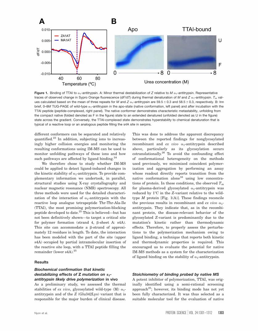
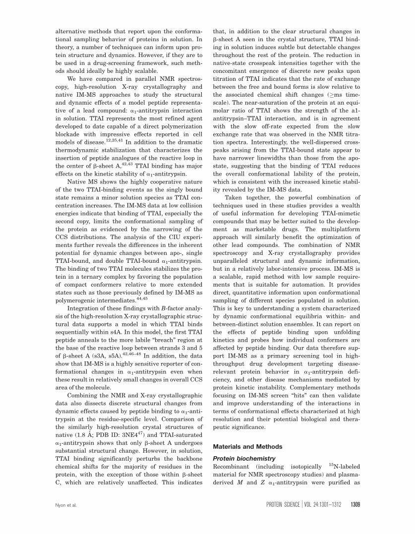
Figure 1. Binding of TTAI to a1-antitrypsin. A: Minor thermal destabilization of Z relative to M a1-antitrypsin. Representative
traces of observed change in Sypro Orange fluorescence (dF/dT) during thermal denaturation of M and Z a1-antitrypsin. Tm val-
ues calculated based on the mean of three repeats for M and Z a1-antitrypsin are 59.5 6 0.3 and 58.5 6 0.3, respectively. B: Inn
brief, 0–8M TUG-PAGE of wild-type a1-antitrypsin in the apo-state (native conformation, left panel) and after incubation with the
TTAI peptide (peptide-complexed, right panel). The native conformer demonstrates characteristic metastability, unfolding from
the compact native (folded denoted as F in the figure) state to an extended denatured (unfolded denoted as U in the figure)
state across the gradient. Conversely, the TTAI-complexed state demonstrates hyperstability to chemical denaturation that is
typical of a reactive loop or an analogous peptide filling the s4A site in serpins.
Nyon et al. PROTEIN SCIENCE VOL 24:1301—1312 1303
IM-MS techniques in identifying the modulators of
conformational stability.
In an initial experiment, wild-type a1-antitryp-
sin in the presence and absence of peptide was
probed by 0–8M of transverse urea gradient (TUG)-
PAGE [Fig. 1(B)]. The apoprotein unfolded at high
urea concentrations to give a slow-migrating
extended species as reported previously.27 The for-
mation of the a1-antitrypsin:peptide complex con-
ferred resistance to unfolding in conditions up to 8M
of urea and therefore ran as a compact species that
migrated similarly across the entire urea gradient.
By analogy with reactive loop-inserted a1-antitryp-
sin after the interaction with a serine protease,27
this stabilized conformation is highly consistent with
filling of the cryptic s4A.
To probe the interaction of TTAI with a1-anti-
trypsin further, the protein was incubated with
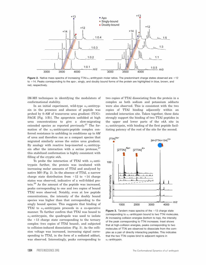
increasing molar amounts of TTAI and analyzed by
native MS (Fig. 2). In the absence of TTAI, a narrow
charge state distribution from 112 to 114 charge
states was observed, indicative of a well-folded pro-
tein.28 As the amount of the peptide was increased,
peaks corresponding to one and two copies of bound
TTAI were observed. Notably, even at low peptide
concentrations, the intensity of the doubly bound
species was higher than that corresponding to the
singly bound species. This suggests that binding of
TTAI to a1-antitrypsin proceeds in a co-operative
manner. To further confirm that TTAI was bound to
a1-antitrypsin, the quadrupole was used to isolate
the 113 charge state corresponding to the ternary
complex (two copies of TTAI bound), and subjected
to collision-induced dissociation (Fig. 3). As the colli-
sion voltage was increased, increasing signal corre-
sponding to TTAI, in the form of a sodiated adduct,
was observed. Interestingly, peaks corresponding to
two copies of TTAI dissociating from the protein in a
complex as both sodium and potassium adducts
were also observed. This is consistent with the two
copies of TTAI binding adjacently within an
extended interaction site. Taken together, these data
strongly support the binding of two TTAI peptides in
the upper and lower parts of the s4A site in
a1-antitrypsin, with binding of the first peptide facil-
itating patency of the rest of the site for the second.
Figure 2. Native mass spectra of increasing TTAI:a1-antitrypsin molar ratios. The predominant charge states observed are 112
to 114. Peaks corresponding to the apo-, singly, and doubly bound forms of the protein are highlighted in blue, brown, and
red, respectively.
Figure 3. Tandem mass spectra of the 113 charge state
corresponding to a1-antitrypsin bound to two TTAI molecules.
At increasing collision energies (bottom to top), the intensity
of the peak corresponding to TTAI increases. Inset shows
that at high-collision energies, peaks corresponding to two
molecules of TTAI are observed to dissociate from the com-
plex as a pair of directly interacting peptides. This indicates
that the two TTAI copies bind to adjacent regions in
a1-antitrypsin.
1304 PROTEINSCIENCE.ORG The Conformational Dynamics of a1-antitrypsin
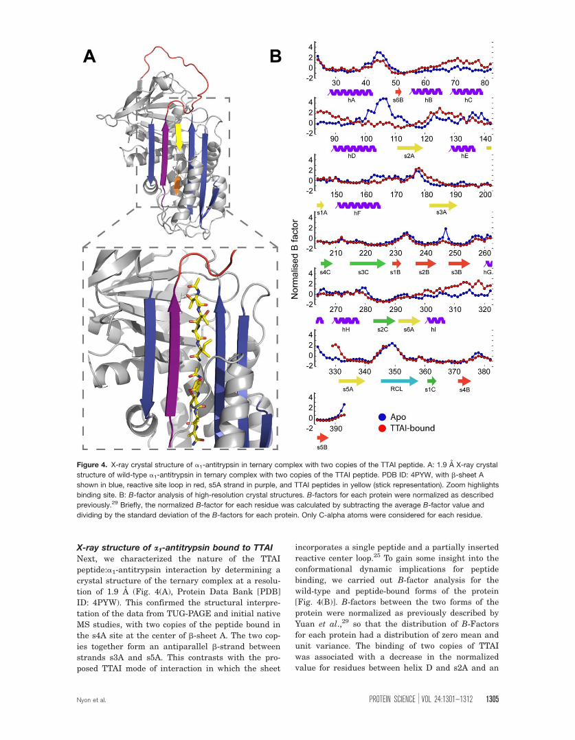
X-ray structure of a1-antitrypsin bound to TTAI
Next, we characterized the nature of the TTAI
peptide:a1-antitrypsin interaction by determining a
crystal structure of the ternary complex at a resolu-
tion of 1.9 A (Fig. 4(A), Protein Data Bank [PDB]
ID: 4PYW). This confirmed the structural interpre-
tation of the data from TUG-PAGE and initial native
MS studies, with two copies of the peptide bound in
the s4A site at the center of b-sheet A. The two cop-
ies together form an antiparallel b-strand between
strands s3A and s5A. This contrasts with the pro-
posed TTAI mode of interaction in which the sheet
incorporates a single peptide and a partially inserted
reactive center loop.25 To gain some insight into the
conformational dynamic implications for peptide
binding, we carried out B-factor analysis for the
wild-type and peptide-bound forms of the protein
[Fig. 4(B)]. B-factors between the two forms of the
protein were normalized as previously described by
Yuan et al.,29 so that the distribution of B-Factors
for each protein had a distribution of zero mean and
unit variance. The binding of two copies of TTAI
was associated with a decrease in the normalized
value for residues between helix D and s2A and an
Figure 4. X-ray crystal structure of a1-antitrypsin in ternary complex with two copies of the TTAI peptide. A: 1.9 A X-ray crystal
structure of wild-type a1-antitrypsin in ternary complex with two copies of the TTAI peptide. PDB ID: 4PYW, with b-sheet A
shown in blue, reactive site loop in red, s5A strand in purple, and TTAI peptides in yellow (stick representation). Zoom highlights
binding site. B: B-factor analysis of high-resolution crystal structures. B-factors for each protein were normalized as described
previously.29 Briefly, the normalized B-factor for each residue was calculated by subtracting the average B-factor value and
dividing by the standard deviation of the B-factors for each protein. Only C-alpha atoms were considered for each residue.
Nyon et al. PROTEIN SCIENCE VOL 24:1301—1312 1305
increase for residues between helix I and s5A
around the lower peptide insertion site. Away from
b-sheet A, RMSD comparisons of the crystallo-
graphic positions of backbone and side-chain atoms
indicated minimal differences (Supporting Informa-
tion Figs. 1 and 2).
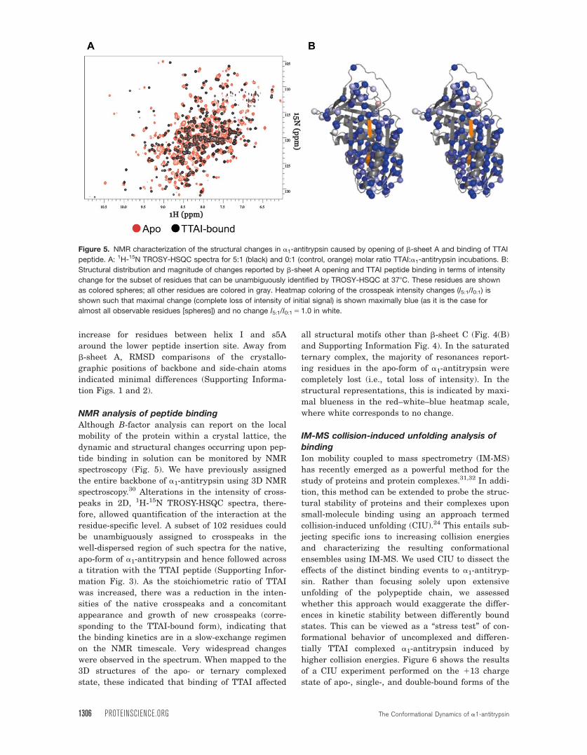
NMR analysis of peptide binding
Although B-factor analysis can report on the local
mobility of the protein within a crystal lattice, the
dynamic and structural changes occurring upon pep-
tide binding in solution can be monitored by NMR
spectroscopy (Fig. 5). We have previously assigned
the entire backbone of a1-antitrypsin using 3D NMR
spectroscopy.30 Alterations in the intensity of cross-
peaks in 2D, 1H-15N TROSY-HSQC spectra, there-
fore, allowed quantification of the interaction at the
residue-specific level. A subset of 102 residues could
be unambiguously assigned to crosspeaks in the
well-dispersed region of such spectra for the native,
apo-form of a1-antitrypsin and hence followed across
a titration with the TTAI peptide (Supporting Infor-
mation Fig. 3). As the stoichiometric ratio of TTAI
was increased, there was a reduction in the inten-
sities of the native crosspeaks and a concomitant
appearance and growth of new crosspeaks (corre-
sponding to the TTAI-bound form), indicating that
the binding kinetics are in a slow-exchange regimen
on the NMR timescale. Very widespread changes
were observed in the spectrum. When mapped to the
3D structures of the apo- or ternary complexed
state, these indicated that binding of TTAI affected
all structural motifs other than b-sheet C (Fig. 4(B)
and Supporting Information Fig. 4). In the saturated
ternary complex, the majority of resonances report-
ing residues in the apo-form of a1-antitrypsin were
completely lost (i.e., total loss of intensity). In the
structural representations, this is indicated by maxi-
mal blueness in the red–white–blue heatmap scale,
where white corresponds to no change.
IM-MS collision-induced unfolding analysis of
binding
Ion mobility coupled to mass spectrometry (IM-MS)
has recently emerged as a powerful method for the
study of proteins and protein complexes.31,32 In addi-
tion, this method can be extended to probe the struc-
tural stability of proteins and their complexes upon
small-molecule binding using an approach termed
collision-induced unfolding (CIU).24 This entails sub-
jecting specific ions to increasing collision energies
and characterizing the resulting conformational
ensembles using IM-MS. We used CIU to dissect the
effects of the distinct binding events to a1-antitryp-
sin. Rather than focusing solely upon extensive
unfolding of the polypeptide chain, we assessed
whether this approach would exaggerate the differ-
ences in kinetic stability between differently bound
states. This can be viewed as a “stress test” of con-
formational behavior of uncomplexed and differen-
tially TTAI complexed a1-antitrypsin induced by
higher collision energies. Figure 6 shows the results
of a CIU experiment performed on the 113 charge
state of apo-, single-, and double-bound forms of the
Figure 5. NMR characterization of the structural changes in a1-antitrypsin caused by opening of b-sheet A and binding of TTAI
peptide. A: 1H-15N TROSY-HSQC spectra for 5:1 (black) and 0:1 (control, orange) molar ratio TTAI:a1-antitrypsin incubations. B:
Structural distribution and magnitude of changes reported by b-sheet A opening and TTAI peptide binding in terms of intensity
change for the subset of residues that can be unambiguously identified by TROSY-HSQC at 378C. These residues are shown
as colored spheres; all other residues are colored in gray. Heatmap coloring of the crosspeak intensity changes (I5:1/I0:1) is
shown such that maximal change (complete loss of intensity of initial signal) is shown maximally blue (as it is the case for
almost all observable residues [spheres]) and no change I5:1/I0:1 5 1.0 in white.
1306 PROTEINSCIENCE.ORG The Conformational Dynamics of a1-antitrypsin
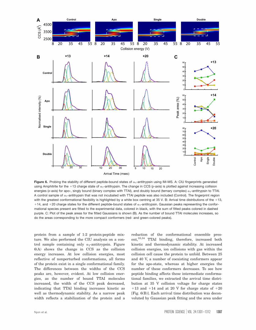
protein from a sample of 1:2 protein:peptide mix-
ture. We also performed the CIU analysis on a con-
trol sample containing only a1-antitrypsin. Figure
6(A) shows the change in CCS as the collision
energy increases. At low collision energies, most
reflective of nonperturbed conformations, all forms
of the protein exist in a single conformational family.
The differences between the widths of the CCS
peaks are, however, evident. At low collision ener-
gies, as the number of bound TTAI molecules
increased, the width of the CCS peak decreased,
indicating that TTAI binding increases kinetic as
well as thermodynamic stability. As a narrow peak
width reflects a stabilization of the protein and a
reduction of the conformational ensemble pres-
ent,33,34 TTAI binding, therefore, increased both
kinetic and thermodynamic stability. At increased
collision energies, ion collisions with gas within the
collision cell cause the protein to unfold. Between 25
and 40 V, a number of coexisting conformers appear
for the apo-state, whereas at higher energies the
number of these conformers decreases. To see how
peptide binding affects these intermediate conforma-
tional families, we extracted the arrival time distri-
bution at 35 V collision voltage for charge states
113 and 114 and at 20 V for charge state of 120
[Fig. 6(B)]. Each arrival time distribution was decon-
voluted by Gaussian peak fitting and the area under
Figure 6. Probing the stability of different peptide-bound states of a1-antitrypsin using IM-MS. A: CIU fingerprints generated
using Amphitrite for the 113 charge state of a1-antitrypsin. The change in CCS (y-axis) is plotted against increasing collision
energies (x-axis) for apo-, singly bound (binary complex with TTAI), and doubly bound (ternary complex) a1-antitrypsin to TTAI.
A control sample of a1-antitrypsin that was not incubated with TTAI peptide was also included (Control). The fingerprint region
with the greatest conformational flexibility is highlighted by a white box centring at 35 V. B: Arrival time distributions of the 113,
114, and 120 charge states for the different peptide-bound states of a1-antitrypsin. Gaussian peaks representing the confor-
mational species present are fitted to the experimental data, colored in black, with the sum of fitted peaks colored in dashed
purple. C: Plot of the peak areas for the fitted Gaussians is shown (B). As the number of bound TTAI molecules increases, so
do the areas corresponding to the more compact conformers (red- and green-colored peaks).
Nyon et al. PROTEIN SCIENCE VOL 24:1301—1312 1307
the curve for each of these Gaussians was plotted
[Fig. 6(C)]. We examined low charge states (113 and
114) as previous studies have shown that such
charge states are more reflective of structures found
in solution.28 We also examined a high charge state
(120) as an increase in charge predictably corre-
sponds to a more Coulombically stressed conforma-
tional ensemble. Reassuringly, no major differences
were observed between control a1-antitrypsin that
had not been incubated with TTAI, and apo-states of
the protein within the mixture of different binding
states in the presence of TTAI. This was true for all
charge states. Binding of a single TTAI molecule
resulted in a small increase in the population of more
compact conformers within the ensemble, with a con-
comitant reduction of the more extended ones. The
binding of the second TTAI peptide resulted in a
greater increase in the population of more compact
conformers relative to the more extended states. At
higher collision energies, the differences between
apo-, singly bound (binary complex with TTAI), and
doubly bound (ternary complex) became more pro-
nounced. A similar trend was observed for the high
charge state although the effect of binding a single
TTAI peptide is not as noticeable as for the low
charge states. This suggests that in higher charge
states the destabilizing effects of Coulombic repulsion
outweigh the kinetic stabilization caused by binding
of one but not two copies of the TTAI peptide.
Discussion
The structure and dynamics of proteins in solution
mediate their behavior in health and disease. These
can be related to energy landscapes of the available
conformational space determined by sequence and
environment35 and are well suited to characteriza-
tion by native IM-MS methods. Local and global
energy minima define thermodynamically metasta-
ble and stable states, respectively. Slope gradients
relate to the kinetics of transitions into these
states (folding) and between them (conformational
exchange). Pathogenic point mutations in translated
polypeptides, or pathological alterations in the envi-
ronment, can therefore affect either thermodynamic
or kinetic determinants of solution behavior or both.
Although thermodynamic effects may be more read-
ily quantified, kinetic destabilization has been
shown to be important for conformational disease
process.36
The previous studies of glycosylated a1-antitryp-
sin purified ex vivo from human plasma indicated
that the Z mutation caused a 98C reduction in Tm.
This change, together with the data from other
mutants assessed in the same way, suggested that
a1-antitrypsin polymerization tendency showed a
close inverse correlation with thermodynamic stabil-
ity.14 However, in native nonglycosylated a1-anti-
trypsin expressed in Pichia pastoris, the observed
thermodynamic stability was only mildly reduced by
the mutation (18C reduction in Tm) with kinetic
destabilization effects correlating better with poly-
merogenicity.17 This is consistent with the data from
the studies in cell models of a1-antitrypsin expres-
sion in health and disease. Mutations that
thermodynamically stabilize a1-antitrypsin by up to
108C (e.g., Thr114Phe by 158C, Gly117Phe by
1108C)37,38 reduce a1-antitrypsin polymerization
rates at physiological temperatures when introduced
on a wild-type background. However, they have no
or minimal effect on the polymerization/deficient
secretion phenotypes when introduced on the back-
ground of the Z mutation in cells.
Such data now indicate that the effects of the
common, severely pathogenic Z allele are mediated
more by kinetic rather than thermodynamic destabi-
lization.17,39 It follows that therapeutic strategies
targeted specifically to modulate conformational
sampling within the native state ensemble may be
beneficial in Z a1-antitrypsin deficiency. Such an
approach has more general potential.40 However, at
present, compound screens aimed at addressing
disease-relevant solution behavior are overwhelm-
ingly assessed by thermodynamic readouts, such as
change in thermal or chemical denaturation sensi-
tivity.12 It is therefore important to develop
Table I. Crystallographic Data Parameters
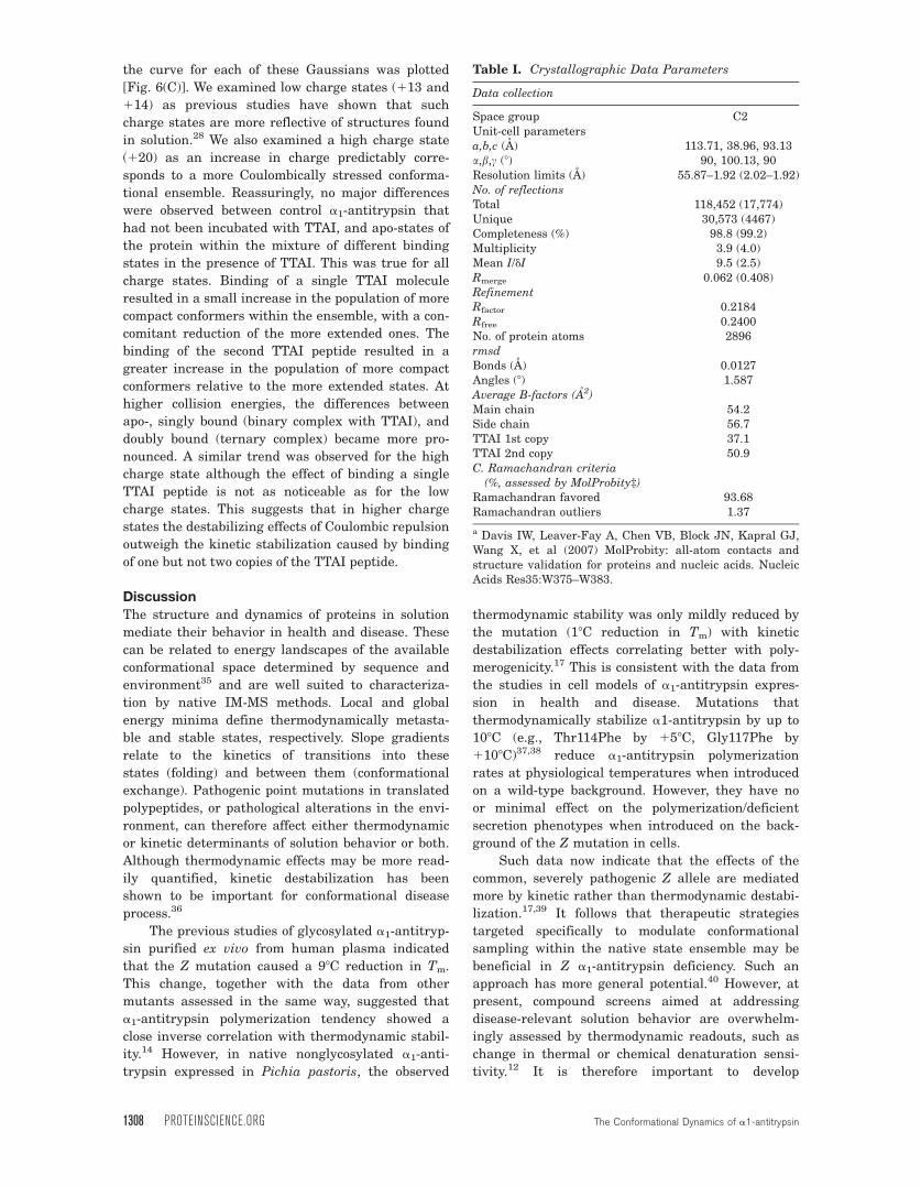
Data collection
Space group C2Unit-cell parametersa,b,c (A) 113.71, 38.96, 93.13a,b,c (8) 90, 100.13, 90Resolution limits (A) 55.87–1.92 (2.02–1.92)No. of reflectionsTotal 118,452 (17,774)Unique 30,573 (4467)Completeness (%) 98.8 (99.2)Multiplicity 3.9 (4.0)Mean I/dI 9.5 (2.5)Rmerge 0.062 (0.408)RefinementRfactor 0.2184Rfree 0.2400No. of protein atoms 2896rmsdBonds (A) 0.0127Angles (8) 1.587Average B-factors (A2)Main chain 54.2Side chain 56.7TTAI 1st copy 37.1TTAI 2nd copy 50.9C. Ramachandran criteria
(%, assessed by MolProbity‡)Ramachandran favored 93.68Ramachandran outliers 1.37
a Davis IW, Leaver-Fay A, Chen VB, Block JN, Kapral GJ,Wang X, et al (2007) MolProbity: all-atom contacts andstructure validation for proteins and nucleic acids. NucleicAcids Res35:W375–W383.
1308 PROTEINSCIENCE.ORG The Conformational Dynamics of a1-antitrypsin
alternative methods that report upon the conforma-
tional sampling behavior of proteins in solution. In
theory, a number of techniques can inform upon pro-
tein structure and dynamics. However, if they are to
be used in a drug-screening framework, such meth-
ods should ideally be highly scalable.
We have compared in parallel NMR spectros-
copy, high-resolution X-ray crystallography and
native IM-MS approaches to study the structural
and dynamic effects of a model peptide representa-
tive of a lead compound: a1-antitrypsin interaction
in solution. TTAI represents the most refined agent
developed to date capable of a direct polymerization
blockade with impressive effects reported in cell
models of disease.12,25,41 In addition to the dramatic
thermodynamic stabilization that characterizes the
insertion of peptide analogues of the reactive loop in
the center of b-sheet A,42,43 TTAI binding has major
effects on the kinetic stability of a1-antitrypsin.
Native MS shows the highly cooperative nature
of the two TTAI-binding events as the singly bound
state remains a minor solution species as TTAI con-
centration increases. The IM-MS data at low collision
energies indicate that binding of TTAI, especially the
second copy, limits the conformational sampling of
the protein as evidenced by the narrowing of the
CCS distributions. The analysis of the CIU experi-
ments further reveals the differences in the inherent
potential for dynamic changes between apo-, single
TTAI-bound, and double TTAI-bound a1-antitrypsin.
The binding of two TTAI molecules stabilizes the pro-
tein in a ternary complex by favoring the population
of compact conformers relative to more extended
states such as those previously defined by IM-MS as
polymerogenic intermediates.44,45
Integration of these findings with B-factor analy-
sis of the high-resolution X-ray crystallographic struc-
tural data supports a model in which TTAI binds
sequentially within s4A. In this model, the first TTAI
peptide anneals to the more labile “breach” region at
the base of the reactive loop between strands 3 and 5
of b-sheet A (s3A, s5A).42,46–48 In addition, the data
show that IM-MS is a highly sensitive reporter of con-
formational changes in a1-antitrypsin even when
these result in relatively small changes in overall CCS
area of the molecule.
Combining the NMR and X-ray crystallographic
data also dissects discrete structural changes from
dynamic effects caused by peptide binding to a1-anti-
trypsin at the residue-specific level. Comparison of
the similarly high-resolution crystal structures of
native (1.8 A; PDB ID: 3NE447) and TTAI-saturated
a1-antitrypsin shows that only b-sheet A undergoes
substantial structural change. However, in solution,
TTAI binding significantly perturbs the backbone
chemical shifts for the majority of residues in the
protein, with the exception of those within b-sheet
C, which are relatively unaffected. This indicates
that, in addition to the clear structural changes in
b-sheet A seen in the crystal structure, TTAI bind-
ing in solution induces subtle but detectable changes
throughout the rest of the protein. The reduction in
native-state crosspeak intensities together with the
concomitant emergence of discrete new peaks upon
titration of TTAI indicates that the rate of exchange
between the free and bound forms is slow relative to
the associated chemical shift changes (�ms time-
scale). The near-saturation of the protein at an equi-
molar ratio of TTAI shows the strength of the a1-
antitrypsin–TTAI interaction, and is in agreement
with the slow off-rate expected from the slow
exchange rate that was observed in the NMR titra-
tion spectra. Interestingly, the well-dispersed cross-
peaks arising from the TTAI-bound state appear to
have narrower linewidths than those from the apo-
state, suggesting that the binding of TTAI reduces
the overall conformational lability of the protein,
which is consistent with the increased kinetic stabil-
ity revealed by the IM-MS data.
Taken together, the powerful combination of
techniques used in these studies provides a wealth
of useful information for developing TTAI-mimetic
compounds that may be better suited to the develop-
ment as marketable drugs. The multiplatform
approach will similarly benefit the optimization of
other lead compounds. The combination of NMR
spectroscopy and X-ray crystallography provides
unparalleled structural and dynamic information,
but in a relatively labor-intensive process. IM-MS is
a scalable, rapid method with low sample require-
ments that is suitable for automation. It provides
direct, quantitative information upon conformational
sampling of different species populated in solution.
This is key to understanding a system characterized
by dynamic conformational equilibria within- and
between-distinct solution ensembles. It can report on
the effects of peptide binding upon unfolding
kinetics and probes how individual conformers are
affected by peptide binding. Our data therefore sup-
port IM-MS as a primary screening tool in high-
throughput drug development targeting disease-
relevant protein behavior in a1-antitrypsin defi-
ciency, and other disease mechanisms mediated by
protein kinetic instability. Complementary methods
focusing on IM-MS screen “hits” can then validate
and improve understanding of the interactions in
terms of conformational effects characterized at high
resolution and their potential biological and thera-
peutic significance.
Materials and Methods
Protein biochemistry
Recombinant (including isotopically 15N-labeled
material for NMR spectroscopy studies) and plasma-
derived M and Z a1-antitrypsin were purified as
Nyon et al. PROTEIN SCIENCE VOL 24:1301—1312 1309
described previously.37,44,49 Conformational homoge-
neity of the samples was confirmed by SDS-, nonde-
naturing, and TUG-PAGE and activity assay.50 TTAI
tetrapeptide, modified by an acetyl group at the N-
terminus and an amide group at the C-terminus,
was purchased from Pepceuticals, United Kingdom.
Tm values (mean from n 5 3 experiments) were
obtained by incubating 0.1 mg/mL of a1-antitrypsin
in a thermal ramp as described previously.51
X-ray crystallographyThe a1-antitrypsin:TTAI ternary complex was
formed by incubating purified recombinant a1-anti-
trypsin with TTAI at high concentration in a 2:1
molar ratio of 225 and 450 lM, respectively, for 30
min at 258C. Saturation of a1-antitrypsin with two
copies of bound TTAI was confirmed by native MS
studies. Crystals were grown by hanging drop vapor
diffusion in a buffer of 26% w/v PEG3350 and
10 mM of Bis-Tris (pH 6.5). The resulting rod-like
crystals were mounted in loops and cryocooled in
cryoprotectant (26% w/v PEG 3350; 10 mM Bis-Tris
[pH 6.5], 20% v/v glycerol). Synchrotron diffraction
data were collected on beamline 23.2 at the ESRF,
Grenoble, France (Table I). Data were processed
using iMOSFLM52 and SCALA53 and solved by
molecular replacement using PHASER.54 The coordi-
nates of the native a1-antitrypsin crystal structure
(PDB ID: 3NE447) were used as the search model to
solve the structure to a resolution of 1.92 A. The
structure of the ternary complex was deposited with
the Research Collaboratory for Structural Bioinfor-
matics (RCSB) PDB.
NMR spectroscopyPurified 15N-labeled a1-antitrypsin was prepared in
25 mM of Na2HPO4, 50 mM of NaCl, and 1 mM of
EDTA, pH 8.0 buffer containing 10% v/v D2O, and
1% v/v DSS. Alpha1-antitrypsin was incubated at
378C (urea, 0M) in the absence and presence of 10:1
molar equivalent of the tetrapeptide TTAI and NMR
spectra recorded on the equilibrium states. All NMR
data were recorded on a Bruker 700 Avance III spec-
trometer equipped with an HCN cryoprobe. 2D
TROSY 1H-15N HSQC spectra were processed and
analyzed using nmrPipe55 and CCPN56 software
packages. All spectra were referenced to DSS at 0.0
ppm, manually phased, and baseline corrected. Spec-
tra were analyzed using our previous assignment of
the protein backbone (Biological Magnetic Reso-
nance Data Bank accession number: 17804)30 by
changes in native crosspeak intensity relative to the
data on a1-antitrypsin in the absence of TTAI.
Native and IM-MS
In brief, 15 lM of a1-antitrypsin was incubated in
protein purification buffer (37 mM NaCl, 2.7 mM
KCl, 10 mM Na2HPO4, and 2 mM KH2PO4 [pH 7.4])
alone and in the presence of the TTAI tetrapeptide.
Peptide:protein molar ratios of 0.1:1, 0.2:1, 0.3:1,
0.4:1, 0.5:1, 1:1, 3:1, and 5:1 were used and incuba-
tion was carried out for 3 days at 37.58C.
Prior to MS experiments, samples were buffer
exchanged using Micro Bio-spin P-6 Gel Columns
(Biorad, United Kingdom) into 200 mM of ammonium
acetate (pH 8). Protein was further concentrated and
buffer exchanged using 10-kDa Amicon Ultra-0.5
Centrifugal Filters (Millipore, United Kingdom).
A first-generation Synapt traveling wave (T-
Wave) IM mass spectrometer (Waters, Manchester,
United Kingdom)57 was used for all native and IM
measurements. Proteins were introduced into the
mass spectrometer by means of nanoelectrospray
ionization using borosilicate glass capillaries pre-
pared in-house. Typical instrument parameters used
were as follows: capillary voltage, 1.1 kV; cone volt-
age, 55 V; extraction cone voltage, 4.0 V; tempera-
ture, 408C, trap CE, 6.0 V; transfer CE, 4.0 V; trap
pressure, 1.5 mbar; bias, 4.0 V; backing pressure, 4
mbar. Calibration was performed using 33 lM of
caesium iodide (Sigma-Aldrich), dissolved in high-
performance liquid chromatography-grade water and
acetonitrile, with formic acid (49:50:1, respectively).
For collision-induced dissociation experiments,
the quadrupole was used to isolate the charge state
of interest and fragmentation was performed in the
trap TWave of the Synapt by increasing the trap col-
lision voltage. For CIU, the same procedure was fol-
lowed but this time IM separation was performed as
well. For IM separations, the bias was increased to
19 V and the IM TWave velocity and wave height
were set to 300 m/s and 7 V, respectively.
Data were acquired using MassLynx v4.1
(Waters). Arrival time distributions were converted
to CCS by fitting to a power equation58 using the
software Amphitrite.59 A set of standards with
known CCS values60 were used and these were
Bovine Serum Albumin, b-lactoglobulin A, and con-
canavalin A, all purchased from Sigma. Amphitrite
was also used to plot CIU curves. ATD peak fitting
was performed using the program Fityk 1.2.9.61
Peaks were modeled as Gaussian and fitted to the
ATDs using the Levenberg–Marquardt algorithm.
References
1. Carrell RW, Gooptu B (1998) Conformational changesand disease—serpins, prions and Alzheimer’s. CurrOpin Struct Biol 8:799–809.
2. Stockley RA, Turner AM (2014) Alpha-1-antitrypsindeficiency: clinical variability, assessment, and treat-ment. Trends Mol Med 20:105–115.
3. Gooptu B, Dickens JA, Lomas DA (2014) The molecularand cellular pathology of a1-antitrypsin deficiency.Trends Mol Med 20:116–127.
4. Gooptu B, Lomas DA (2009) Conformational pathologyof the serpins: themes, variations and therapeuticstrategies. Annu Rev Biochem 78:147–176.
1310 PROTEINSCIENCE.ORG The Conformational Dynamics of a1-antitrypsin
5. Sharp HL, Bridges RA, Krivit W, Freier EF (1969) Cir-rhosis associated with alpha-1-antitrypsin deficiency: apreviously unrecognised inherited disorder. J Lab ClinMed 73:934–939.
6. Lomas DA, Evans DL, Finch JT, Carrell RW (1992)The mechanism of Z a1-antitrypsin accumulation inthe liver. Nature 357:605–607.
7. Laurell C-B, Eriksson S (1963) The electrophoretic a1-globulin pattern of serum in a1-antitrypsin deficiency.Scand J Clin Lab Invest 15:132–140.
8. Eriksson S (1964) Pulmonary emphysema and alpha1-antitrypsin deficiency. Acta Med Scand 175:197–205.
9. Gooptu B, Ekeowa UI, Lomas DA (2009) Mechanismsof emphysema in a1-antitrypsin deficiency: molecularand cellular insights. Eur Respir J 34:475–488.
10. Blanco I, Fern�andez E, Bustillo EF (2001) Alpha-1-antitrypsin PI phenotypes S and Z in Europe: an anal-ysis of the published surveys. Clin Genet 60:31–41.
11. Nyon MP, Gooptu B (2014) Therapeutic targeting ofmisfolding and conformational change in a1-antitrypsindeficiency. Future Med Chem 6:1047–1065.
12. Chang Y-P, Mahadeva R, Patschull AOM, Nobeli I,Ekeowa UI, McKay AR, Thalassinos K, Irving JA, HaqI, Nyon MP, Christodoulou J, Ord�o~nez A, Miranda E,Gooptu B (2011) Targeting serpins in high-throughputand structure-based drug design. Method Enzymol 501:139–175.
13. Whisstock JC, Bottomley SP (2006) Molecular gymnas-tics: serpin structure, folding and misfolding. CurrOpin Struct Biol 16:761–768.
14. Dafforn TR, Mahadeva R, Elliott PR, Sivasothy P,Lomas DA (1999) A kinetic mechanism for the poly-merisation of a1-antitrypsin. J Biol Chem 274:9548–9555.
15. Huntington JA, Read RJ, Carrell RW (2000) Structureof a serpin-protease complex shows inhibition by defor-mation. Nature 407:923–926.
16. Irving JA, Haq I, Dickens JA, Faull SV, Lomas DA(2014) Altered native stability is the dominant basisfor susceptibility of alpha1-antitrypsin mutants to poly-merization. Biochem J 460:103–115.
17. Knaupp AS, Levina V, Robertson AL, Pearce MC,Bottomley SP (2010) Kinetic instability of the serpin Za1-antitrypsin promotes aggregation. J Mol Biol 396:375–383.
18. Eichner T, Radford SE (2011) Understanding the com-plex mechanisms of 2-microglobulin amyloid assembly.FEBS J 278:3868–3883.
19. Eichner T, Kalverda AP, Thompson GS, Homans SW,Radford SE (2011) Conformational conversion duringamyloid formation at atomic resolution. Mol Cell 41:161–172.
20. Wojnowska M, Yan J, Sivalingam GN, Cryar A, Gor J,Thalassinos K, Djordjevic S (2013) Autophosphoryla-tion activity of a soluble hexameric histidine kinasecorrelates with the shift in protein conformationalequilibrium. Chem Biol 20:1411–1420.
21. Bohrer BC, Merenbloom SI, Koeniger SL, HilderbrandAE, Clemmer DE (2008) Biomolecule analysis by ionmobility spectrometry. Annu Rev Anal Chem 1:293–327.
22. Zhong Y, Hyung SJ, Ruotolo BT (2012) Ion mobility-mass spectrometry for structural proteomics. ExpertRev Proteomics 9:47–58.
23. Shi H, Clemmer DE (2014) Evidence for two new solu-tion states of ubiquitin by IMS-MS analysis. J PhysChem B 118:3498–3506.
24. Rabuck JN, Hyung SJ, Ko KS, Fox CC, Soellner MB,Ruotolo BT (2013) Activation state-selective kinase
inhibitor assay based on ion mobility-mass spectrome-try. Anal Chem 85:6995–7002.
25. Chang YP, Mahadeva R, Chang WS, Lin SC, Chu YH(2009) Small-molecule peptides inhibit Z alpha1-antitrypsin polymerization. J Cell Mol Med 13:2304–2236.
26. Verbanac KM, Heath EC (1983) Biosynthesis and proc-essing of rat alpha 1-antitrypsin. Arch Biochem Bio-phys 223:149–157.
27. Mast AE, Enghild JJ, Salvesen G (1992) Conformationof the reactive site loop of a1-proteinase inhibitorprobed by limited proteolysis. Biochemistry 31:2720–2728.
28. Scarff CA, Thalassinos K, Hilton GR, Scrivens JH(2008) Travelling wave ion mobility mass spectrometrystudies of protein structure: biological significance andcomparison with X-ray crystallography and nuclearmagnetic resonance spectroscopy measurements. RapidCommun Mass Spectrom 22:3297–3304.
29. Yuan Z, Zhao J, Wang Z-X (2003) Flexibility analysis ofenzyme active sites by crystallographic temperaturefactors. Protein Eng 16:109–114.
30. Nyon MP, Kirkpatrick J, Cabrita LD, Christodoulou J,Gooptu B (2012) 1H, 15N and 13C backbone resonanceassignments of the archetypal serpin a1-antitrypsin.Biomol NMR Assign 6:153–156.
31. Thalassinos K, Pandurangan AP, Xu M, Alber F, TopfM (2013) Conformational states of macromolecularassemblies explored by integrative structure calcula-tion. Structure 21:1500–1508.
32. Benesch JL, Ruotolo BT (2011) Mass spectrometry:come of age for structural and dynamical biology. CurrOpin Struct Biol 21:641–649.
33. Hilton GR, Thalassinos K, Grabenauer M, Sanghera N,Slade SE, Wyttenbach T, Robinson PJ, Pinheiro TJ,Bowers MT, Scrivens JH (2010) Structural analysis ofprion proteins by means of drift cell and travelingwave ion mobility mass spectrometry. J Am Soc MassSpectrom 21:845–854.
34. Zhou M, Morgner N, Barrera NP, Politis A, IsaacsonSC, Matak-Vinkovic D, Murata T, Bernal RA, Stock D,Robinson CV (2011) Mass spectrometry of intact V-typeATPases reveals bound lipids and the effects of nucleo-tide binding. Science 334:380–385.
35. Dill KA, Chan HS (1997) From levinthal to pathwaysto funnels. Nat Struct Biol 4:10–19.
36. Karamanos TK, Kalverda AP, Thompson GS, RadfordSE. (2014) Visualization of transient protein-proteininteractions that promote or inhibit amyloid assembly.Mol Cell 55:212–226.
37. Parfrey H, Mahadeva R, Ravenhill N, Zhou A, DaffornTR, Foreman RC, Lomas DA (2003) Targeting a surfacecavity of a1-antitrypsin to prevent conformational dis-ease. J Biol Chem 278:33060–33066.
38. Gooptu B, Miranda E, Nobeli I, Mallya M, Purkiss A,Leigh Brown SC, Summers C, Phillips RL, Lomas DA,Barrett TE (2009) Crystallographic and cellular charac-terisation of two mechanisms stablising the native foldof a1-antitrypsin: implications for disease and drugdesign. J Mol Biol 387:857–868.
39. Hughes VA, Meklemburg R, Bottomley SP, WintrodePL (2014) The Z mutation alters the global structuraldynamics of a1-antitrypsin. PLoS One 9:e102617.
40. Holdom MD, Davies AM, Nettleship JE, Bagby SC,Dhaliwal B, Girardi E, Hunt J, Gould HJ, Beavil AJ,McDonnell JM, Owens RJ, Sutton BJ (2011) Conforma-tional changes in IgE contribute to its uniquely slowdissociation rate from receptor FceRI. Nat Struct MolBiol 18:571–576.
Nyon et al. PROTEIN SCIENCE VOL 24:1301—1312 1311
41. Chang YP, Mahadeva R, Chang WSW, Shukla A,Dafforn TR, Chu YH (2006) Identification of a 4-merpeptide inhibitor that effectively blocks the polymeriza-tion of pathogenic Z a1-antitrypsin. Am J Respir CellMol Biol. 35:540–548.
42. Zhou A, Stein PE, Huntington JA, Sivasothy P, LomasDA, Carrell RW (2004) How small peptides blockand reverse serpin polymerization. J Mol Biol 342:931–941.
43. Skinner R, Chang W-SW, Jin L, Pei X, Huntington JA,Abrahams J-P, Carrell RW, Lomas DA (1998) Implica-tions for function and therapy of a 2.9A structure ofbinary-complexed antithrombin. J Mol Biol 283:9–14.
44. Nyon MP, Segu L, Cabrita LD, L�evy GR, KirkpatrickJ, Roussel BD, Patschull AOM, Barrett TE, EkeowaUI, Kerr R, Waudby CA, Kalsheker N, Hill M,Thalassinos K, Lomas DA, Christodoulou J, Gooptu B(2012) Structural dynamics associated with intermedi-ate formation in an archetypal conformational disease.Structure 20:504–512.
45. Ekeowa UI, Freeke J, Miranda E, Gooptu B, Bush MF,Perez J, Teckman JH, Robinson CV, Lomas DA (2010)Defining the mechanism of polymerization in theserpinopathies. Proc Natl Acad Sci USA 107:17146–17151.
46. Whisstock JC, Skinner R, Lesk AM (1998) An atlas ofserpin conformations. Trends Biochem Sci 23:63–67.
47. Fitton HL, Pike RN, Carrell RW, Chang W-SW (1997)Mechanisms of antithrombin polymerisation and hepa-rin activation probed by insertion of synthetic reactiveloop peptides. Biol Chem 378:1059–1063.
48. Patschull AOM, Segu L, Nyon MP, Lomas DA, NobeliI, Barrett TE, Gooptu B (2011) Therapeutic target sitevariability in a1-antitrypsin characterized at high reso-lution. Acta Cryst F 67:1492–1497.
49. Tan L, Perez J, Mela M, Miranda E, Burling KA,Rouhani FN, DeMeo DL, Haq I, Irving JA, Ord�o~nez A,Dickens JA, Brantly M, Marciniak SJ, Alexander GJ,Gooptu B, Lomas DA (2015) Characterising the associ-ation of latency with a1-antitrypsin polymerisationusing a novel monoclonal antibody. Int J Biochem CellBiol 58:81–91.
50. Dafforn TR, Pike RN, Bottomley SP (2004) Physicalcharacterization of serpin conformations. Methods 32:150–158.
51. Haq I, Irving JA, Faull SV, Dickens JA, Ord�o~nez A,Belorgey D, Gooptu B, Lomas DA (2013) Reactivecentre loop mutants of a-1-antitrypsin reveal position-specific effects on intermediate formation along thepolymerization pathway. Biosci Rep 33:499–511.
52. Leslie AGW (1992Recent changes to the MOSFLMpackage for processing film and image data, in JointCCP4 and ESF-EACMB Newsletter on Protein Crystal-lography. Warrington, UK: SERC, Daresbury Laboratory.
53. Evans P (2006) Scaling and assessment of data quality.Acta Cryst D Biol Crystallogr 62:72–82.
54. McCoy AJ, Grosse-Kunstleve RW, Adams PD, WinnMD, Storoni LC, Read RJ (2007) Phaser crystallo-graphic software. J Appl Cryst 40:658–674.
55. Delaglio F, Grzesiek S, Vuister G, Zhu G, Pfeifer J,Bax A (1995) NMRPipe: a multidimensional spectralprocessing system based on UNIX pipes. J BiomolNMR 6:277–293.
56. Fogh R, Ionides J, Ulrich E, Boucher W, Vranken W,Linge JP, Habeck M, Rieping W, Bhat TN, WestbrookJ, Henrick K, Gilliland G, Berman H, Thornton J,Nilges M, Markley J, Laue E (2002) The CCPN project:an interim report on a data model for the NMR com-munity. Nat Struct Biol 9:416–418.
57. Pringle SD, Giles K, Wildgoose JL, Williams JP, SladeSE, Thalassinos K, Bateman RH, Bowers MT, ScrivensJH (2007) An investigation of the mobility separationof some peptide and protein ions using a new hybridquadrupole/travelling wave IMS/oa-ToF instrument.Int J Mass Spectrom 261:1–12.
58. Thalassinos K, Grabenauer M, Slade SE, Hilton GR,Bowers MT, Scrivens JH (2009) Characterization ofphosphorylated peptides using traveling Wave-basedand drift cell ion mobility mass spectrometry. AnalChem 81:248–254.
59. Sivalingam GN, Yan J, Sahota H, Thalassinos K (2013)Amphitrite: a program for processing travelling waveion mobility mass spectrometry data. Int J Mass Spec-trom 345–347:54–62.
60. Bush MF, Hall Z, Giles K, Hoyes J, Robinson CV, RuotoloBT (2010) Collision cross-sections of proteins and theircomplexes: a calibration framework and database forgas-phase structural biology. Anal Chem 82:9557–9565.
61. Wojdyr M (2010) Fityk: a general-purpose peak fittingprogram. J Appl Crystallogr 43:1126–1128.
1312 PROTEINSCIENCE.ORG The Conformational Dynamics of a1-antitrypsin