Amyloid-β metabolism in Niemann-Pick C disease … NPC revised.pdf · 2012-09-19 · Amyloid-β...
32
1 Amyloid-β metabolism in Niemann-Pick C disease models and patients Niklas Mattsson* 1 , Maria Olsson 1 , Mikael K. Gustavsson 1 , Marko Kosicek 2 , Martina Malnar 2 , Jan-Eric Månsson 1 , Maria Blomqvist 1 , Johan Gobom 1 , Ulf Andreasson 1 , Gunnar Brinkmalm 1 , Charles Vite 3 , Silva Hecimovic 2 , Caroline Hastings 4 , Kaj Blennow 1 , Henrik Zetterberg 1 , Erik Portelius 1 1 Clinial Neurochemistry Laboratory, Institute of Neuroscience and Physiology, Department of Psychiatry and Neurochemistry, the Sahlgrenska Academy, University of Gothenburg, Sweden 2 Division of Molecular Medicine, Rudjer Boskovic Institute, Zagreb, Croatia 3 School of Veterinary Medicine, University of Pennsylvania, USA 4 Children’s Hospital & Research Center Oakland, Oakland, California, USA *Correspondence to: Niklas Mattsson, Clinical Neurochemistry Laboratory, V-huset, Sahlgrenska University Hospital/Mölndal, SE-431 20, Sweden. E-mail: [email protected]. Phone: +46706393851.
Transcript of Amyloid-β metabolism in Niemann-Pick C disease … NPC revised.pdf · 2012-09-19 · Amyloid-β...
1
Amyloid-β metabolism in Niemann-Pick C disease models and patients
Niklas Mattsson*1, Maria Olsson1, Mikael K. Gustavsson1, Marko Kosicek2, Martina Malnar2, Jan-Eric
Månsson1, Maria Blomqvist1, Johan Gobom1, Ulf Andreasson1, Gunnar Brinkmalm1, Charles Vite3, Silva
Hecimovic2, Caroline Hastings4, Kaj Blennow1, Henrik Zetterberg1, Erik Portelius1
1Clinial Neurochemistry Laboratory, Institute of Neuroscience and Physiology, Department of Psychiatry and
Neurochemistry, the Sahlgrenska Academy, University of Gothenburg, Sweden
2Division of Molecular Medicine, Rudjer Boskovic Institute, Zagreb, Croatia
3School of Veterinary Medicine, University of Pennsylvania, USA
4Children’s Hospital & Research Center Oakland, Oakland, California, USA
*Correspondence to: Niklas Mattsson, Clinical Neurochemistry Laboratory, V-huset, Sahlgrenska University
Hospital/Mölndal, SE-431 20, Sweden. E-mail: [email protected]. Phone: +46706393851.

2
Abstract
Niemann-Pick type C (NPC) is a progressive neurodegenerative lysosomal disease with altered cellular lipid
trafficking. The metabolism of amyloid-β (Aβ) - previously mainly studied in Alzheimer’s disease - has been
suggested to be altered in NPC. Here we aimed to perform a detailed characterization of metabolic products
from the amyloid precursor protein (APP) in NPC models and patients. We used multiple analytical
technologies, including immunoassays and immunoprecipitation followed by mass spectrometry (IP-MS) to
characterize Aβ peptides and soluble APP fragments (sAPP-α/β) in cell media from pharmacologically
(U18666A) and genetically (NPC1-/-) induced NPC cell models, and cerebrospinal fluid (CSF) from NPC cats
and human patients. The pattern of Aβ peptides and sAPP-α/β fragments in cell media was differently affected
by NPC-phenotype induced by U18666A treatment and by NPC1-/- genotype. U18666A treatment increased the
secreted media levels of sAPP-α, AβX-40 and AβX-42 and reduced the levels of sAPP-β, Aβ1-40 and Aβ1-42,
while IP-MS showed increased relative levels of Aβ5-38 and Aβ5-40 in response to treatment. NPC1-/- cells had
reduced media levels of sAPP-α and Aβ1-16, and increased levels of sAPP-β. NPC cats had altered CSF
distribution of Aβ peptides compared with normal cats. Cats treated with the potential disease-modifying
compound 2-hydroxypropyl-β-cyclodextrin had increased relative levels of short Aβ peptides including Aβ1-16
compared with untreated cats. NPC patients receiving β-cyclodextrin had reduced levels over time of CSF Aβ1-
42, AβX-38, AβX-40, AβX-42 and sAPP-β, as well as reduced levels of the axonal damage markers tau and
phosphorylated tau. We conclude that NPC models have altered Aβ metabolism, but with differences across
experimental systems, suggesting that NPC1-loss of function, such as in NPC1-/- cells, or NPC1-dysfunction,
seen in NPC patients and cats as well as in U18666A-treated cells, may cause subtle but different effects on
APP degradation pathways. The preliminary findings from NPC cats suggest that treatment with cyclodextrin
may have an impact on APP processing pathways. CSF Aβ, sAPP and tau biomarkers were dynamically altered
over time in human NPC patients.
Keywords: Niemann-Pick type C; amyloid-β; amyloid precursor protein; biomarker; cerebrospinal fluid.

3
Introduction
Niemann-Pick type C (NPC) is a rare autosomal recessive disease caused by mutations in either of the two
genes encoding for the lysosome-associated lipid trafficking proteins NPC1 and NPC2 (Swardfager et al. 2010).
The mutations cause intracellular accumulation and altered distribution of different lipid species. The major
clinical feature is progressive neurological deterioration, with onset typically in childhood. Treatment for NPC
is limited, but the glucosylceramide synthase inhibitor miglustat (Zavesca, Actelion Inc.) may have an effect on
amelioration of neurological symptoms (Patterson et al. 2007; Pineda et al. 2009). Recently, the cholesterol
modulating compound β-cyclodextrin was proposed for NPC treatment (Rosenbaum and Maxfield 2011; Peake
and Vance 2012).
NPC may also have late onset in adulthood and is often confused with more common neurological or psychiatric
diseases, such as psychosis, depression, or other neurodegenerative diseases (Patterson et al. 2012).
Interestingly, NPC shares several pathologic features with Alzheimer’s disease (AD), including neurofibrillary
tangles (Auer et al. 1995), trafficking abnormalities in endosomes and lysosomes (Nixon et al. 2008), disease
exacerbation by the APOE ε4 allele (Saito et al. 2002) and cellular lipid dysregulation (Grimm et al. 2007).
Also, NPC1 polymorphisms are associated with late-onset AD (Erickson et al. 2008), and AD patients have
increased expression of NPC1 in hippocampus and frontal cortex (Kagedal et al. 2010). Notably, in NPC there
are indications of abnormal metabolism of the peptide amyloid-β (Aβ) (Jin et al. 2004), which has been
proposed to drive AD pathogenesis (Selkoe 1991; Hardy and Higgins 1992). AD patients have extracellular
deposits of Aβ and reduced cerebrospinal fluid (CSF) levels of the 42 amino acid isoform Aβ1-42. In contrast,
NPC patients accumulate intracellular Aβ (Jin et al. 2004) and may have elevated CSF Aβ levels (Mattsson et al.
2011a). Aβ is formed by proteolysis from the amyloid precursor protein (APP) through complicated enzymatic
pathways that depend both on the cellular lipid environment and on vesicular trafficking within the cell,
ultimately giving rise to a large number of different Aβ peptides and soluble APP (sAPP) fragments (Portelius
et al. 2011a). The altered lipid homeostasis and the disturbed vesicular trafficking seen in NPC may be expected
to affect APP metabolism, which is dependent on both the cellular lipid topography (Holmes et al. 2012) and the
endosomal vesicular system (Cirrito et al. 2008). Previous studies have indeed found that cells lacking the
NPC1-protein have reduced surface levels of APP, increased release of sAPP-β (Malnar et al. 2010; Kosicek et
al. 2010) and intracellular accumulation of Aβ peptides (Malnar et al. 2010; Yamazaki et al. 2001). NPC mice
accumulate Aβ peptides in their brains (Olson and Humpel 2010; Burns et al. 2003; Boland et al. ; Yamazaki et
al. 2001), and have increased brain activity and levels of Aβ generating enzymes (Kodam et al. 2010). Also,
APPxPS1 transgenic mice (an established Alzheimer’s disease model) have a more rapid accumulation of brain
Aβ if they are NPC1-heterozygous than if they are NPC1+/+ (Borbon and Erickson 2011).
These studies point to disturbed APP processing pathways in NPC, but detailed data on APP degradation
products is needed to understand precisely how NPC changes APP metabolism. Importantly, a careful analysis
of this may give general clues to links between lipid metabolism, vesicular trafficking and APP processing,
which may be important to understand mechanisms in other diseases, such as AD. We therefore undertook an
investigation with the aim to measure a large number of different Aβ and sAPP-α/β species, using multiple
orthogonal and complementing technologies. To assess the robustness of the findings, we included both
pharmacological and genetical cell models, the major large animal NPC model, which is a cat strain carrying a

4
missense mutation in the NPC1 gene (2864G-C), resulting in a phenotype that is biochemically,
neuropathologically and symptomatically similar to the human disease (Somers et al. 2003; Vite et al. 2008),
and CSF from human NPC patients. The results differed across the systems, emphasizing the need for
carefulness when translating findings between models and human patients.
Experimental procedures
U18666A cell model
Human neuroblastoma SH-SY5Y cells (Biedler et al. 1978) over-expressing wild type human APP695
(APP695wt) were cultivated in Dulbecco’s modified Eagle’s medium/F-12 (Invitrogen) supplemented with 10%
fetal bovine serum, 2 mM L-glutamine and 1 % penicillin-streptomycin solution. Cells were treated with the
amphiphile U18666A (3-β-[2-(diethylamino)ethoxy]androst-5-en-17-one) at 3 µg/mL cell media for 24 hours to
induce the NPC cholesterol storage phenotype. U18666A blocks cellular cholesterol transport and is a well-
established method to emulate the NPC phenotype in cell experiments (Runz et al. 2002; Lange et al. 2000)
(Roff et al. 1991). Filipin staining was used to confirm the presence of cholesterol storage. Cells were also
treated with the β-secretase (β-site aspartyl cleaving enzyme 1, BACE1) inhibitor β-secretase inhibitor IV
(Calbiochem, Merck, compound 3 in (Stachel et al. 2004)), the enzyme cathepsin B inhibitor Z-FA-FMK (BD
Biosciences, San Jose, CA, USA) or DMSO.
NPC1-null cell model
Since U18666A may influence APP processing enzymes directly (Crestini et al. 2006; Sidera et al. 2005), it is
difficult to determine how much of its effects that are related to NPC1 inhibition. We therefore tested Chinese
Hamster Ovary wild type cells (CHOwt) and CHO NPC1-null cells (CHO NPC1-/-) (all originally provided by
Dr. Daniel Ory to SH) in which NPC1 gene was deleted. These were used and prepared essentially as described
previously (Kosicek et al. 2010; Malnar et al. 2010). In short, cells were maintained in Dulbecco’s modified
Eagle’s medium / F-12 supplemented with 10% FBS, 2 mM L-glutamine and antibiotics/antimycotic solution
(100 U/ml penicillin, 100 mg/ml streptomycin and 0.25 mg/ml amphotericin B). For transient expression, cells
were transfected using Lipofectamine LTX (Invitrogen) according to the supplier’s instructions. APP695wt,
APPswe and C99 6myc-tagged constructs were generated using pCS2+6MT vector (Hecimovic et al. 2004).
Twenty four hours after transfection medium was removed, fresh medium was added, incubated further for 24 h
and collected for analysis.
NPC1 cats
To investigate if the NPC-dependent effects seen in cell experiments were present also in mammals, we
performed experiments on cats. As opposed to CHO NPC1-/- cells which lack the NPC1 gene, NPC1 cats carry a
5
spontaneous disease causing mutation in the NPC1 gene. Control and NPC1 cats were raised in the animal
colony at the School of Veterinary Medicine at the University of Pennsylvania under NIH and USDA guidelines
for the care and use of animals in research, as described previously (Ward et al. 2010). We also included cats
treated with 2-hydroxypropyl-β-cyclodextrin (called cyclodextrin below).
NPC patients
Finally, we assessed APP degradation products in human NPC patients by CSF examination. We have
previously reported increased levels of CSF AβX-38, AβX-40, AβX-42 and Aβ1-42 in NPC patients compared
to controls, with no major differences during one year of follow-up (Mattsson et al. 2011a, b) (for AβX-
peptides, quantification was done by a technique with low specificity for the N-terminal amino acid of the Aβ
sequence). Here we analyzed serial CSF samples from two NPC1 patients before and during treatment with
cyclodextrin. The samples were obtained in conjunction with treatment with intravenous or intrathecal injections
of the compound (under approval for compassionate use Investigation New Drugs from the Food and Drug
Administration, IND104,114 and IND104,116). The patients were on simultaneous off-label use of miglustat,
which is registered for NPC in the European Union. For contrast values, CSF samples collected by lumbar
puncture from 9 age-matched patients (Table 1) were obtained from clinical routine samples at Clinical
Neurochemistry Laboratory, Sahlgrenska University Hospital, Mölndal, Sweden. Contrast samples were
selected from patients with normal CSF cell counts, albumin CSF to serum ratio, and no signs of intrathecal
immunoglobulin production. All CSF samples were centrifuged at 2,000 x g at 4°C for 10 min and stored at -
80°C pending analysis.
Fluorescent bead-based assays
The xMAP assay INNO-BIA AlzBio3 (Innogenetics, Ghent, Belgium) was used for quantifications of Aβ1-42,
T-tau, and P-tau, as explained previously (Olsson et al. 2005). The xMAP assay INNO-BIA Aβ forms
(Innogenetics) was used for quantifications of Aβ1-40 and Aβ1-42 (format A) and AβX-40 and AβX-42 (format
B), as explained previously (Hansson et al. 2010). Both format A and B use the monoclonal antibodies 21F12
and 2G3, which specifically bind Aβ peptides ending at Ala42 and Val40, respectively, as capture antibodies. In
format A, 3D6 (Aβ N-terminal neoepitope epitope starting at Asp1) was used as detection antibody, providing
specific quantifications of Aβ1-40/42 isoforms. In format B, 4G8 (epitope within Aβ18-22) was used as
detection antibody, providing quantification of AβX-40/42 isoforms.
Electrochemiluminescense assays
The MSD Human/Rodent Abeta Triplex assay (Meso Scale Discovery, Gaithersburg, MD, USA) was used for
quantifications of AβX-38, AβX-40 and AβX-42, and the MSD sAPPα/sAPPβ Multiplex Assay for
quantifications of sAPP-α and sAPP-β, as explained previously (Mattsson et al. 2011a).
6
Immunoprecipitation and mass spectrometry
Aβ peptides were analyzed by immunoprecipitation and mass spectrometry (IP-MS) by a method previously
developed at our laboratory (Portelius et al. 2007). In short, anti-Aβ antibodies coupled to magnetic beads were
used for IP. After elution, Aβ isoforms were analyzed by mass spectrometry on an UltraFlextreme matrix-
assisted laser-desorption/ionization time-of-flight/time-of-flight (MALDI TOF/TOF) instrument or an AutoFlex
MALDI TOF (Bruker Daltonics, Bremen, Germany). An in-house developed MATLAB (Mathworks Inc.
Natick, MA, USA) program was used for relative quantifications of Aβ isoforms in the spectra. For each peak
the sum of the intensities for the three strongest isotopic signals were calculated and normalized against the sum
for all the Aβ peaks in the spectrum followed by averaging of duplicates. This method allows for relative
quantification of different Aβ isoforms.
Liquid chromatography and tandem mass spectrometry (LC-MS/MS)
Aβ isoform identities were confirmed by liquid chromatography (LC) combined with high resolution tandem
mass spectrometry (MS/MS) (Portelius et al. 2007). LC-MS/MS analysis was performed on an Ettan MDLC
nanoflow chromatographic system (GE Healthcare) using HotSep Kromasil C4 columns (G&T Septech)
coupled to a Thermo LTQ FT Ultra electrospray ionization hybrid linear quadrupole ion trap/Fourier transform
ion cyclotron resonance (ESI-LQIT/FTICR) mass spectrometer (Thermo Fisher Scientific). All spectra were
acquired in FTICR mode and collision induced dissociation (CID) as well as electron capture dissociation
(ECD) was used to obtain fragment ion data.
Ethics
All subjects or care-givers gave informed and written consent. The cat study was approved by the University of
Pennsylvania Institutional Animal Care and Use Committee. The human study was approved by the local
Institutional Review Boards at the treating hospitals and was performed in accordance with the ethical standards
laid down in the 1964 Declaration of Helsinki.
Results
Aβ and sAPP-α/β profile in U18666A treated SH-SY5Y cells
U18666A treatment induced an NPC cholesterol storage phenotype in SH-SY5Y cells with increased
concentrations of sAPP-α, AβX-40 and AβX-42 and reduced concentrations of sAPP-β, Aβ1-40 and Aβ1-42 in
cell media (experiment repeated twice, Figure 1). The treatment also increased the AβX-42 to AβX-40 ratio
(data not shown), similar to what was previously seen in NPC patients’ CSF (Mattsson et al. 2011a). Treatment
with a BACE1 inhibitor reduced sAPP-β, Aβ1-40 and Aβ1-42 levels (Supplementary figure 1), confirming an
efficient BACE1-inhibition, but did not counter-act the U18666A elevations of AβX-40 and AβX-42, indicating
that these originated from BACE1-independent pathways. Inhibition of the enzyme cathepsin B, which has been
suggested as an alternative Aβ producing enzyme (Hook et al. 2005) reduced the concentrations of Aβ1-40,
Aβ1-42, AβX-40 and AβX-42 in media from both untreated and U18666A treated cells, which made it difficult

7
to determine if this enzyme was involved in any specific APP degradation pathway (Supplementary figure 2).
However, we noted that cathepsin B inhibition reduced AβX-40 and AβX-42 levels in U18666A-treated cells
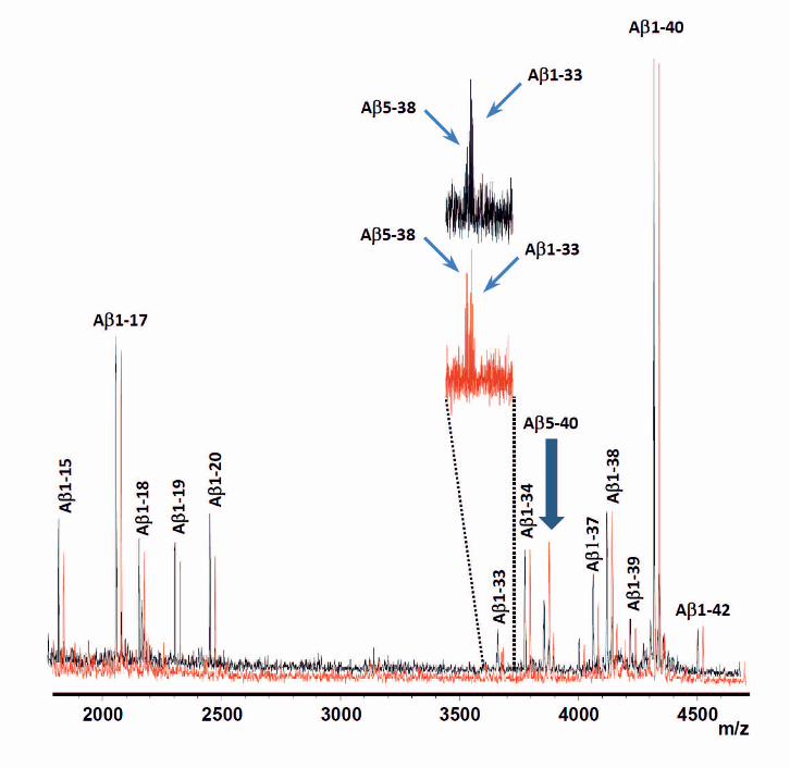
almost to the levels as in vehicle (DMSO)-treated cells. IP-MADLI-TOF MS analyses showed increased levels
of Aβ species starting at position 5 (Aβ5-38 and Aβ5-40) in the U18666A treated cells (Figure 2), which are
peptides known to be formed without BACE1-processing (Mattsson et al. 2012b; Takeda et al. 2004; Portelius
et al. 2011b). Such N-truncated Aβ species may account for at least part of the increased concentrations of AβX-
40/42 detected by immunoassays after U18666A treatment.
Aβ and sAPP-α/β profile in NPC1-/- cells
In contrast to U18666A-treatment, the NPC1-/- genotype did not have major effect on media levels of Aβ1-40,
Aβ1-42, AβX-40 and AβX-42 (Supplementary figure 3), but seemed to reduce sAPP-α levels (at least in
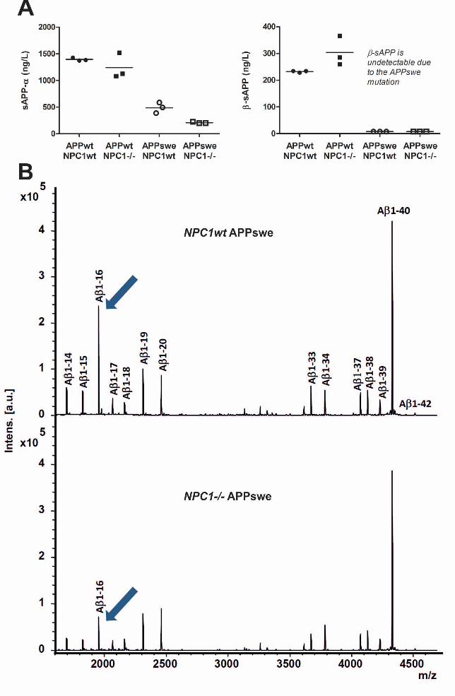
APPswe transfected cells) and increase sAPP-β levels (Figure 3, panel A). Notably, the sAPP-β fragment was
not detected from APPswe media which is expected due to the swe mutation (Mullan et al. 1992) (Figure 3,
panel A). Interestingly, IP-MS showed that media from NPC1-/- cells had reduced relative levels of Aβ1-16
(Figure 3, panel B). In combination with reduced sAPP-α this argues for a shift away from α-secretase
pathways, since both the formation of sAPP-α and Aβ1-16 requires α-secretase processing (Portelius et al.
2011b). In cells only expressing endogenous APP, the NPC1-/- genotype reduced AβX-40 levels (Figure 4, panel
A). Similarly, in cells transfected with C99 the NPC1-/- genotype caused a slight reduction of AβX-38, AβX-40
and AβX-42 (Figure 4). IP-MALDI-TOFMS unexpectedly showed a major difference in the overall Aβ peptide
pattern for C99 transfected cells compared to other cells, with elevated relative Aβ1-41 signals and reduced
relative Aβ1-40 and Aβ1-42 signals, and the NPC1-/- genotype increased the relative Aβ1-41 signal even more
(Supplementary Figure 4).
Aβ profiling in the CSF of NPC1 cat
We analyzed Aβ species in NPC1 cat CSF samples by IP-MALDI-TOFMS. While the peak profile of Aβ
peptides in cat CSF resembled that seen in human CSF, we noted a +14 Da shift of all Aβ species compared to
the human variant (Figure 5, panel a). By MS/MS analysis of m/z 4342.175, using MALDI-TOF/TOF, and
comparing the fragment ion pattern to that of the human Aβ1-40 analog we assigned this mass difference to an
amino acid substitution in the cat Aβ sequence, with an Asp7 → Glu replacement compared to the human
sequence (Figure 5, panel b). In combination with homology with human Aβ1-40 it could be established that the
cat Aβ1-40 sequence is DAEFRHESGYEVHHQKLVFFAEDVGSNKGAIIGLMVGGVV. In parallel with this
experiment, the cat Aβ sequence was further verified using ECD-MS/MS as described recently (Brinkmalm et
al. 2012). The protein sequence data will appear in the UniProt Knowledgebase under the accession number
P86906. The amino acid difference at position 7 renders 6E10 non-functional in cat experiments as reflected by
no signals corresponding to Aβ when the 6E10 was used in the IP. To include coverage of short C-terminal
truncated Aβ peptides, we therefor used the antibodies 82E1 (N-terminal specific) and 4G8. There were some
differences in the relative Aβ peptide distribution between NPC1 cats and wild type cats, with lower relative

8
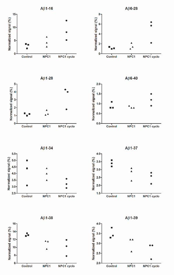
levels of Aβ1-37, Aβ1-38 and Aβ1-39 in NPC1 cats compared to controls. Some of the cats on cyclodextrin had
increased relative levels of Aβ1-16, Aβ6-28 and Aβ1-28 compared to the other groups (Figure 6, Supplementary
fig 5).
Aβ, sAPP-α/β and T-tau/P-tau profile in NPC patient CSF
Finally, we examined serial CSF samples in two NPC patients, twin girls. Measurements on the first two sample
rounds have been reported previously (Mattsson et al. 2011b, a), but for this study the original samples were re-
run together with all follow-up samples. See Table 1 for baseline values and Figures 7-8 for changes over time.
As indicated in the figures, the patients started oral miglustat treatment after the first sampling (the first
sampling is the baseline value, prior to miglustat or cyclodextrin treatment) and intravenous (IV) cyclodextrin
treatment after the second sampling. The patients initiated therapy with low dose (100 mg/kg) IV cyclodextrin
and the dose was increased with every drug administration until they reached 2500 mg/kg. The IV cyclodextrin
treatment continued until the 4th sampling when the patients received every 2 week intrathecal (IT) cyclodextrin
(175 mg) in addition to continuing the IV dosing at a stable level of 2500 mg/kg every 2 weeks. After an initial
12 week trial period of IT treatment the FDA placed a clinical hold on the IT treatment until the safety data
could be assessed (the IV cyclodextrin treatment and the miglustat treatment were continuous). Following a 6
weeks intermission, the IT dosing was re-initiated and subsequently the IT dosing has been doubled to 375 mg.
The patients have not had any deleterious effects of the drugs, either IV or IT. There have been no grade 3 or 4
toxicities and no apparent dose limitation to date. Additionally, the patients have been physically well, with
stability of many symptoms (developmental delay, ataxia), improvements (improved hearing threshold, better
swallowing), and have shown some progression typically of NPC (difficult control of seizures and gelastic
cataplexy). Overall, the patients clinical progression in this pilot study appears to be delayed and some
immediate, albeit short lived, effects are apparent after drug administration (increased alertness and energy).
The CSF levels of Aβ1-42, AβX-38, AβX-40, AβX-42 and sAPP-β decreased over time in both patients,
suggesting that miglustat and cyclodextrin treatment may have effects on APP and Aβ metabolism. IP-MS
analyses (data not shown) revealed no major shifts in the Aβ peptide pattern distribution in the patients
compared to a contrast group (the contrast group is described in Table 1).
In addition to APP and Aβ markers we analyzed the axonal damage markers T-tau and P-tau (Table 1 and
Figure 8), both of which decreased over time. Noticeably, P-tau started to decrease later, after introduction of
cyclodextrin treatment. The decrease in T-tau and P-tau during treatment might suggest a beneficial effect on the
neuronal degeneration, but this has to be examined further in a larger series of patients.
Discussion
Here we report on APP degradation products across multiple NPC models and in human patient CSF. In sum,
pharmacologically or genetically induced NPC phenotype have altered patterns of APP degradation products,
but the specific results differ between models, which may be a consequence of complex interactions between
9
APP metabolism and NPC-induced pathways. The changes over time in CSF Aβ, sAPP, and tau biomarkers
may support the use of these biomarkers to study the disease process in NPC studies and drug trials.
First, we noted that U18666A treatment shifted the APP processing away from the β-secretase dependent
pathways with the result that the release of both sAPP-α and Aβ5-X isoforms increased. The latter have
previously been shown to be BACE1-independent (Mattsson et al. 2012b) and likely linked to α-secretase
activity (Takeda et al. 2004).
Several previous studies have examined Aβ metabolism after U18666A treatment, but the results are difficult to
compare due to differences in which cell model that was used and/or methods used for APP and Aβ
quantification (e.g. not discriminating between Aβ1-40/42 and AβX-40/42). Runz et al. found that U18666A
treatment reduced the release of AβX-40, AβX-42, and cellular levels of β-CTF in SH-SY5Y APP695-
transfected cells, arguing for reduced β-secretase processing (Runz et al. 2002). Likewise, Davis found that
U18666A increased the release of sAPP-α, concluding that U18666A treatment likely blocks the APP re-
endocytosis that is necessary for β-secretase processing in the endocytic pathway in neurons (Davis 2008). In
contrast, Yamazaki et al. found no effect of U18666A treatment on secretion of Aβ1-40 or Aβ1-42 from CHO
cells, although the treatment lead to intracellular accumulation of Aβ (especially Aβ42) in late endosomes
(Yamazaki et al. 2001). Jin et al. found that U18666A led to accumulation of AβX-42 and CTFs in APP695
transfected primary mouse cortical neurons, but not in C99 transfected cells, suggesting that the treatment
increased the activity of other secretases than γ-secretase (Jin et al. 2004). Few studies have explored systems
with endogenous APP expression, but Koh et al. found that U18666A treatment reduced the release of Aβ40 and
Aβ42 while the intracellular levels increased in mouse cortical neurons only expressing endogenous APP (Koh
et al. 2006).
U18666A has several effects, including transcriptional up-regulation of the γ-secretase components presenilin-1
and presenilin-2 (Crestini et al. 2006) and altered glycosylation of BACE1 (Sidera et al. 2005), which makes it
difficult to determine if the effects on APP metabolism due to U18666A treatment are directly related to NPC1
inhibition or not. We therefore performed analyses on CHO-NPC1-/- cells that were transiently transfected with
either APPwt or APPswe construct. In our experiments on NPC1-/- cells, we found the opposite result from
U18666A treatment, with increased β-secretase processing and reduced α-secretase processing. This adds to
previous data showing that these cells have reduced cell surface APP levels, increased CTF levels in lipid rafts,
and increased release of sAPP-β (Malnar et al. 2010; Kosicek et al. 2010). In addition, in accordance with
previous findings (Yamazaki et al. 2001; Malnar et al. 2010) we observed no major changes in secreted Aβ
species in NPC1-/- cells compared to NPCwt cells. However, in future studies it would be interesting to analyze
intracellular Aβ by IP-MS since intracellular Aβ has been shown to accumulate in several NPC model cells
(Malnar et al. 2010; Yamazaki et al. 2001). Unexpectedly, transfection with C99 increased the relative release of
Aβ1-41 and reduced the relative release of Aβ1-40 and Aβ1-42. Also, although the NPC1-/- genotype had no
effect on secreted Aβ1-40/42 and AβX-38/40/42 in APPswe and APPwt transfected cells, it reduced the release
of AβX-38, AβX-40 and AβX-42 in C99 transfected cells, and further increased the relative release of Aβ1-41.
In cats, we found that the Aβ sequence differs from human and dog Aβ at amino acid position Aβ7, although the
general CSF Aβ peptide distribution was very similar to what is seen in humans and dogs (Portelius et al. 2006;
10
Portelius et al. 2010; Mattsson et al. 2012b). To our knowledge, this is the first investigation of APP and Aβ
metabolism in the NPC1 cat. We found that NPC1-cats had slightly reduced relative CSF levels of Aβ1-37,
Aβ1-38, and Aβ1-39 compared to controls. Previous NPC1 animal studies have shown that NPC1 mice
accumulate β-CTF, Aβ40, and Aβ42 in their brains (Olson and Humpel 2010; Burns et al. 2003; Boland et al. ;
Yamazaki et al. 2001), but have unchanged levels of APP, sAPP, PS-1, and BACE protein (Burns et al. 2003).
Also, NPC1 mice brain homogenates have slightly increased γ-secretase activity (Burns et al. 2003). In addition,
a recent study by Kodam et al. (Kodam et al. 2010) identified increased β-secretase activity along with increased
levels of APP, BACE1 and all four components of the γ-secretase complex in NPC mice cerebellum and
hippocampus compared to controls.
In the CSF of the two NPC patients studied, we found a decrease over time in Aβ1-42, AβX-38, AβX-40, AβX-
42, and sAPP-β (to about 50 % of baseline levels), which may indicate a continuous loss of functional APP-
processing synapses or neurons as the disease progresses. This is consistent with a previous study, where CSF
AβX-38, AβX-40, AβX-42, and sAPP-β levels were lower in more severely than in less severely affected NPC
patients (Mattsson et al. 2011a). Extending these observations to other neurodegenerative diseases, such as AD,
patients who are severely affected have lower CSF AβX-40, sAPP-α and sAPP-β levels (Rosen et al. 2012), and
CSF AβX-40 was recently even found to decline over time in AD patients (Mattsson et al. 2012a). We also
found that CSF levels of T-tau and P-tau decreased in the patients (with the baseline levels determined prior to
miglustat or cyclodextrin therapy). From this study, it cannot be determined if this reflects the natural course of
the disease, or if it represents an effect of miglustat and/or cyclodextrin therapy on the intensity of the axonal
degenerative process, which is a proposed interpretation for similar tau changes in AD treatment studies
(Blennow et al. 2010; Blennow et al. 2012). Studies with extensive longitudinal CSF sampling in
neurodegenerative diseases are still rare. As shown here, such studies may be useful to track changes in CNS
metabolism over time.
Cyclodextrin has been proposed as a treatment directed against the lipid alterations in NPC. Lipid metabolism
has a complex relationship with APP and Aβ metabolism (Grimm et al. 2007). The α-secretase pathway, which
releases the N-terminal ectodomain sAPP-α and prevents Aβ formation, occurs mainly outside cholesterol rich
lipid raft membrane domains. In contrast, BACE1 and γ-secretase, which releases Aβ peptides, are most active
within lipid rafts. Consequently, cholesterol depletion inhibits Aβ formation (Simons et al. 1998), while
cholesterol enrichment may reduce sAPP-α secretion (Bodovitz and Klein 1996) and increase Aβ deposition
(Refolo et al. 2000). In cats, cyclodextrin increased the relative CSF levels of Aβ1-16, which is generated by
combined cleavage from α- and β-secretase (Portelius et al. 2011b), as well as the levels of Aβ6-28 and Aβ1-28,
indicating a shift away from the classical amyloidogenic pathway towards α-secretase dependent pathways,
perhaps as a consequence of cellular cholesterol depletion. We could not replicate this finding in human NPC
CSF using IP-MS. One limitation to consider, in addition to the very small sample number in this pilot study, is
that the patients were in an advanced stage of disease when treatment started, while the cats were treated from
birth.
The main findings of this study is that (1) pharmacologically and genetically induced NPC1 models had signs of
altered APP processing, but with differences between models, with U18666A treatment shifting APP processing
away from β-secretase dependent pathways and NPC1-/- genotype instead increasing β-secretase processing,
11
which may partly explain previous differences in results between studies; (2) NPC1-loss of function, such as in
NPC1-/- cells, may have subtle but different effects on APP processing and Aβ formation compared to NPC1-
dysfunction seen in NPC patients and cats as well as in U18666A treated cells; (3) NPC cats had altered CSF
distribution of Aβ peptides compared with normal cats and cats treated with cyclodextrin had increased relative
levels of short Aβ peptides compared with untreated cats; and (4) NPC patients receiving treatment with
miglustat, IV and IT β-cyclodextrin decreased in CSF Aβ, sAPP and tau measurements over time, suggesting
that these biomarkers may be useful tools to monitor the disease process and treatment effect. Importantly,
although cell models may give support for pathogenetic mechanisms, careful animal studies and patient studies
are needed to fully understand the mechanisms of disease in NPC.
Conflict of interest
The authors declare that they have no conflict of interest.
Acknowledgments
We would like to thank Åsa Källén, Monica Christiansson, Sara Hullberg and Dzemila Secic for excellent
technical assistance. We would also like to thank Chris Hempel for her support of this work. This study was
supported by grants from Sahlgrenska University Hospital, Sahlgrenska Academy, the Lundbeck Foundation,
Stiftelsen Psykiatriska Forskningsfonden, Stiftelsen Gamla Tjänarinnor, Uppsala Universitets Medicinska
Fakultet stiftelse för psykiatrisk och neurologisk forskning, the Swedish Brain Fund, Göteborgs läkaresällskap,
Thuréus stiftelse, Pfannenstills stiftelse, Demensfonden, Magn. Bergvalls stiftelse, Gun och Bertil Stohnes
stiftelse, Sweden and Ministry of Science, Education and Sports of the Republic of Croatia (098-0982522-2525
to S.H.).

12
Table 1. CSF biomarkers at baseline in NPC patients and contrast group
Patient 1 Patient 2 Contrast group (N=9)
Age (years) 2 2 3.7 (1.6-11)
Sex F F 7 M: 2 F
AβX-38 (ng/L) 1143 1827 651 (100-1517)
AβX-40 (ng/L) 10257 17243 7315 (868-13733)
AβX-42 (ng/L) 1247 2026 601 (53-1396)
Aβ1-42 (ng/L) 552 667 236 (84-408)
sAPP-α (ng/mL) 467 794 313 (110-781)
sAPP-β (ng/mL) 213 326 102 (31-285)
T-tau (ng/L) 1399 1981 62 (40-135)
P-tau (ng/L) 121 133 35 (24-61)
Data for the contrast group is median (range). Regarding AβX-38/40/42 peptides, the X refers to the fact that
quantification was done by a technique with low specificity for the N-terminal amino acid of the Aβ sequence
13
Figure legends
Fig. 1 Cells treated to emulate NPC phenotype have altered release of Aβ- and sAPP-peptides. SH-SY5Y
APP695wt cells were treated with DMSO, U18666A, or untreated (blank). Cell media concentrations of sAPP-α
(A), sAPP-β (B), Aβ1-40 (C), Aβ1-42 (D), AβX-40 (E and G) and AβX-42 (F and H), were determined using
fluorescent bead-based assay (FORM; C-F) and/or electrochemiluminescent based assay (MSD; A, B, G and H).
Each data point represents individual cell cultures. Lines are means. Two data points are missing from the
U18666A treatment group in panels C-D due to technical error in measurements.
Fig. 2 Cells treated to emulate NPC phenotype have increased relative release of Aβ-peptides starting at
position Aβ5. The spectra of secreted Aβ peptides in U18666A-treated SH-SY5Y APP695wt cells were
analyzed by immunoprecipitation and mass spectrometry. IP-MALDI-TOF MS showed increased relative
signals of Aβ5-38 and Aβ5-40 in U18666A treated cells (red).
Fig. 3 NPC1-/- cells have reduced relative release of Aβ1-16. NPC1wt and NPC1-/- CHO cells were
transiently transfected with either APPwt or APPsw construct. Cell media concentrations of sAPP-α and sAPP-β
were determined using an electrochemiluminescense assay, while Aβ species were analyzed by IP-MALDI-MS
TOF. Each data point represents individual cell cultures. In parallel with decreased sAPP-α levels in NPC1-/-
cells (A) we observed reduced relative signal of Aβ1-16 in the same cells (B). Note that in APPsw-transfected
cells we were unable to analyze sAPP-β, which is expected due to the swe mutation.
Fig. 4 Effect of NPC1-/- on release of AβX-38, AβX-40 and AβX-42. AβX-40 (A), AβX-38 (B) and AβX-42
(C) were analyzed in untransfected (APPendo) and either APPsw or C99-transfected (A-C) NPC1wt and NPC1-/-
CHO cells by an electrochemiluminescent based assay (MSD). Each data point represents individual cell
cultures. In untransfected cells expressing only endogenous APP, the NPC1-/- genotype reduced AβX-40 levels
(A). Similarly, in cells transfected with C99, the NPC1-/- genotype caused a slight reduction of AβX-38, AβX-40
and AβX-42 (A-C).
Fig. 5 The cat Aβ sequence. IP-MALDI-TOF/TOF tandem mass spectrum of cat Aβ identifies an amino acid
substitution at position 7 compared to the human analog. (a) Analysis in MS mode. A mass shift of +14 Da is
observed for all Aβ peptides in cat CSF (top panel) compared to human (bottom panel). (b) MS/MS analysis of
Aβ1-40 from cat (top panel) and human (bottom panel). For human Aβ1-40, containing Arg at position 5 and
Asp at position 7 and 23, MS/MS analysis of singly charged ions is expected to be dominated by charge-remote
fragmentation, which is confirmed by the strong b7, b23 signals. In the MS/MS spectrum of the cat Aβ1-40, b23
is strong, but b7 is significantly less intense. This observation together with the 14 Da mass difference indicates
that Asp-7 is replaced by Glu in cat Aβ1-40.
Fig. 6 The effects of NPC and cyclodextrin treatment on CSF Aβ peptides in cats. Aβ peptides were
determined by immunoprecipitation and mass spectrometry in CSF from control cats and cyclodextrin
treated/untreated NPC1-cats. Untreated NPC1-cats (middle panel) had slightly reduced relative CSF levels of
Aβ1-37, Aβ1-38, and Aβ1-39 compared to controls (top panel). Cyclodextrin treated NPC cat (bottom panel)
show increased relative levels of Aβ1-16, Aβ6-28 and Aβ1-28 compared to the other groups.
14
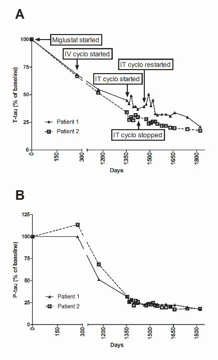
Fig. 7 Longitudinal data on CSF Aβ and sAPP biomarkers in two NPC patients. Biomarkers determined by
fluorescent bead-based and electrochemiluminescent assays. X-axes show days from baseline. The patients
started oral miglustat treatment after the first sampling and intravenous (IV) cyclodextrin (cyclo) treatment after
the second sampling. The IV cyclodextrin treatment continued until the 4th sampling when the patients received
intrathecal (IT) cyclodextrin in addition to continuing the IV cyclodextrin dosing. After 12 week trial period IT
treatment was stopped for 6 weeks before it was restarted (the IV cyclodextrin treatment and the miglustat
treatment were continuous). CSF levels of Aβ1-42 (A), AβX-42 (B), AβX-38 (C), AβX-40 (D) and sAPP-β (F)
were decreased over time to 50% of the baseline in the two examined NPC patients. Meanwhile, sAPP-α levels
were only slightly changed (E).
Fig. 8 Longitudinal data on CSF T-tau and P-tau in two NPC patients. T-tau and P-tau were determined in
the CSF of treated NPC patients using fluorescent bead-based assay. X-axes show days from baseline. The
patients started oral miglustat treatment after the first sampling and intravenous (IV) cyclodextrin (cyclo)
treatment after the second sampling. The IV cyclodextrin treatment continued until the 4th sampling when the
patients received intrathecal (IT) cyclodextrin in addition to continuing the IV cyclodextrin dosing. After 12
week trial period IT treatment was stopped for 6 weeks before it was restarted (the IV cyclodextrin treatment
and the miglustat treatment were continuous). CSF levels of T-tau (A) and P-tau (B) were decreased over time
to the baseline levels in the two examined NPC patients.
15
Supplementary fig. 1 Effects of BACE1-inhibition on NPC phenotype cells. SH-SY5Y APP695wt cells were
treated with DMSO, BACE1 inhibition with β-secretase inhibitor IV (Calbiochem, Merck), and/or U18666A.
Cell media concentrations of Aβ1-40 (A), Aβ1-42 (B), AβX-40 (C) and AβX-42 (D) were determined using
fluorescent bead-based assay. Each data point represents individual cell cultures. Treatment with a BACE1
inhibitor reduced Aβ1-40 and Aβ1-42 levels.
Supplementary fig. 2 Effects of cathepsin B-inhibition on NPC phenotype cells. SH-SY5Y APP695wt cells
were treated with DMSO, cathepsin B inhibition with Z-FA-FMK (BD Biosciences, San Jose, CA, USA),
and/or U18666A. Cell media concentrations of Aβ1-40 (A), Aβ1-42 (B), AβX-40 (C) and AβX-42 (D) were
determined using fluorescent bead-based assay. Each data point represents individual cell cultures. Cathepsin B
inhibition reduced AβX-40 and AβX-42 levels in U18666A-treated cells almost to the levels as in vehicle
(DMSO)-treated cells.
Supplementary fig. 3 Effects of NPC1-/- on release of Aβ peptides. NPC1wt and NPC1-/- CHO cells were
transiently transfected with either APPwt or APPsw construct. Cell media concentrations of Aβ1-40, Aβ1-42,
AβX-40 and AβX-42 were determined using fluorescent bead-based assay (FORM) and/or
electrochemiluminescent based assay (MSD). Each data point represents individual cell cultures. NPC1-/-
genotype did not have a major effect on media levels of Aβ1-40, Aβ1-42, AβX-40 and AβX-42.
Supplementary fig. 4 Release of Aβ peptides from C99 transfected cells. NPC1wt and NPC1-/- CHO cells
were transiently transfected with either APPswe or C99 construct. Signals of Aβ1-40, Aβ1-41 and Aβ1-42 were
determined in the cell media with IP-MALDI-TOFMS. Each data point represents individual cell cultures. IP-
MALDI-TOFMS analysis showed elevated relative Aβ1-41 signals and reduced relative Aβ1-40 and Aβ1-42
signals in C99 transfected cells compared to APPswe transfected cells. NPC1-/- genotype increased the relative
Aβ1-41 signal even more.
Supplementary fig. 5 Aβ peptides in cat CSF. Aβ1-16, Aβ6-28, Aβ1-28, Aβ6-40, Aβ1-34, Aβ1-37, Aβ1-38
and Aβ1-39 peptides were determined by IP-MALDI-TOFMS in CSF from control and cyclodextrin
treated/untreated NPC1-cats. Each data point represents individual animals. Some of the cats treated with
cyclodextrin showed increased relative levels of Aβ1-16, Aβ6-28 and Aβ1-28 compared to the other groups.

16
References
Auer IA, Schmidt ML, Lee VM, Curry B, Suzuki K, Shin RW, Pentchev PG, Carstea ED, Trojanowski JQ (1995) Paired helical filament tau (PHFtau) in Niemann-Pick type C disease is similar to PHFtau in Alzheimer's disease. Acta Neuropathol 90 (6):547-551.
Biedler JL, Roffler-Tarlov S, Schachner M, Freedman LS (1978) Multiple neurotransmitter synthesis by human neuroblastoma cell lines and clones. Cancer Res 38 (11 Pt 1):3751-3757.
Blennow K, Hampel H, Weiner M, Zetterberg H (2010) Cerebrospinal fluid and plasma biomarkers in Alzheimer disease. Nat Rev Neurol 6 (3):131-144.
Blennow K, Zetterberg H, Rinne JO, Salloway S, Wei J, Black R, Grundman M, Liu E, for the AABI (2012) Effect of Immunotherapy With Bapineuzumab on Cerebrospinal Fluid Biomarker Levels in Patients With Mild to Moderate Alzheimer Disease. Arch Neurol.
Bodovitz S, Klein WL (1996) Cholesterol modulates alpha-secretase cleavage of amyloid precursor protein. J Biol Chem 271 (8):4436-4440.
Boland B, Smith DA, Mooney D, Jung SS, Walsh DM, Platt FM Macroautophagy is not directly involved in the metabolism of amyloid precursor protein. J Biol Chem 285 (48):37415-37426.
Borbon IA, Erickson RP (2011) Interactions of Npc1 and amyloid accumulation/deposition in the APP/PS1 mouse model of Alzheimer's. Journal of applied genetics 52 (2):213-218.
Brinkmalm G, Portelius E, Ohrfelt A, Mattsson N, Persson R, Gustavsson MK, Vite CH, Gobom J, Mansson JE, Nilsson J, Halim A, Larson G, Ruetschi U, Zetterberg H, Blennow K, Brinkmalm A (2012) An online nano-LC-ESI-FTICR-MS method for comprehensive characterization of endogenous fragments from amyloid beta and amyloid precursor protein in human and cat cerebrospinal fluid. J Mass Spectrom 47 (5):591-603.
Burns M, Gaynor K, Olm V, Mercken M, LaFrancois J, Wang L, Mathews PM, Noble W, Matsuoka Y, Duff K (2003) Presenilin redistribution associated with aberrant cholesterol transport enhances beta-amyloid production in vivo. J Neurosci 23 (13):5645-5649.
Cirrito JR, Kang JE, Lee J, Stewart FR, Verges DK, Silverio LM, Bu G, Mennerick S, Holtzman DM (2008) Endocytosis is required for synaptic activity-dependent release of amyloid-beta in vivo. Neuron 58 (1):42-51.
Crestini A, Napolitano M, Piscopo P, Confaloni A, Bravo E (2006) Changes in cholesterol metabolism are associated with PS1 and PS2 gene regulation in SK-N-BE. J Mol Neurosci 30 (3):311-322.
Davis W, Jr. (2008) The cholesterol transport inhibitor U18666a regulates amyloid precursor protein metabolism and trafficking in N2aAPP "Swedish" cells. Curr Alzheimer Res 5 (5):448-456.
Erickson RP, Larson-Thome K, Weberg L, Szybinska A, Mossakowska M, Styczynska M, Barcikowska M, Kuznicki J (2008) Variation in NPC1, the gene encoding Niemann-Pick C1, a protein involved in intracellular cholesterol transport, is associated with Alzheimer disease and/or aging in the Polish population. Neurosci Lett 447 (2-3):153-157.
Grimm MO, Grimm HS, Hartmann T (2007) Amyloid beta as a regulator of lipid homeostasis. Trends Mol Med 13 (8):337-344.
Hansson O, Zetterberg H, Vanmechelen E, Vanderstichele H, Andreasson U, Londos E, Wallin A, Minthon L, Blennow K (2010) Evaluation of plasma Abeta(40) and Abeta(42) as predictors of conversion to Alzheimer's disease in patients with mild cognitive impairment. Neurobiol Aging 31 (3):357-367.
Hardy JA, Higgins GA (1992) Alzheimer's disease: the amyloid cascade hypothesis. Science 256 (5054):184-185.
Hecimovic S, Wang J, Dolios G, Martinez M, Wang R, Goate AM (2004) Mutations in APP have independent effects on Abeta and CTFgamma generation. Neurobiol Dis 17 (2):205-218.
Holmes O, Paturi S, Ye W, Wolfe MS, Selkoe DJ (2012) Effects of membrane lipids on the activity and processivity of purified gamma-secretase. Biochemistry 51 (17):3565-3575.
Hook V, Toneff T, Bogyo M, Greenbaum D, Medzihradszky KF, Neveu J, Lane W, Hook G, Reisine T (2005) Inhibition of cathepsin B reduces beta-amyloid production in regulated secretory

17
vesicles of neuronal chromaffin cells: evidence for cathepsin B as a candidate beta-secretase of Alzheimer's disease. Biol Chem 386 (9):931-940.
Jin LW, Shie FS, Maezawa I, Vincent I, Bird T (2004) Intracellular accumulation of amyloidogenic fragments of amyloid-beta precursor protein in neurons with Niemann-Pick type C defects is associated with endosomal abnormalities. Am J Pathol 164 (3):975-985.
Kagedal K, Kim WS, Appelqvist H, Chan S, Cheng D, Agholme L, Barnham K, McCann H, Halliday G, Garner B (2010) Increased expression of the lysosomal cholesterol transporter NPC1 in Alzheimer's disease. Biochim Biophys Acta 1801 (8):831-838.
Kodam A, Maulik M, Peake K, Amritraj A, Vetrivel KS, Thinakaran G, Vance JE, Kar S (2010) Altered levels and distribution of amyloid precursor protein and its processing enzymes in Niemann-Pick type C1-deficient mouse brains. Glia 58 (11):1267-1281.
Koh CH, Whiteman M, Li QX, Halliwell B, Jenner AM, Wong BS, Laughton KM, Wenk M, Masters CL, Beart PM, Bernard O, Cheung NS (2006) Chronic exposure to U18666A is associated with oxidative stress in cultured murine cortical neurons. J Neurochem 98 (4):1278-1289.
Kosicek M, Malnar M, Goate A, Hecimovic S (2010) Cholesterol accumulation in Niemann Pick type C (NPC) model cells causes a shift in APP localization to lipid rafts. Biochem Biophys Res Commun 393 (3):404-409.
Lange Y, Ye J, Rigney M, Steck T (2000) Cholesterol movement in Niemann-Pick type C cells and in cells treated with amphiphiles. J Biol Chem 275 (23):17468-17475.
Malnar M, Kosicek M, Mitterreiter S, Omerbasic D, Lichtenthaler SF, Goate A, Hecimovic S (2010) Niemann-Pick type C cells show cholesterol dependent decrease of APP expression at the cell surface and its increased processing through the beta-secretase pathway. Biochim Biophys Acta 1802 (7-8):682-691.
Mattsson N, Portelius E, Rolstad S, Gustavsson M, Andreasson U, Stridsberg M, Wallin A, Blennow K, Zetterberg H (2012a) Longitudinal Cerebrospinal Fluid Biomarkers over Four Years in Mild Cognitive Impairment. J Alzheimers Dis 30 (4):767-778.
Mattsson N, Rajendran L, Zetterberg H, Gustavsson M, Andreasson U, Olsson M, Brinkmalm G, Lundkvist J, Jacobson LH, Perrot L, Neumann U, Borghys H, Mercken M, Dhuyvetter D, Jeppsson F, Blennow K, Portelius E (2012b) BACE1 Inhibition Induces a Specific Cerebrospinal Fluid beta-Amyloid Pattern That Identifies Drug Effects in the Central Nervous System. PLoS One 7 (2):e31084.
Mattsson N, Zetterberg H, Bianconi S, Yanjanin NM, Fu R, Mansson JE, Porter FD, Blennow K (2011a) Gamma-secretase-dependent amyloid-beta is increased in Niemann-Pick type C: a cross-sectional study. Neurology 76 (4):366-372.
Mattsson N, Zetterberg H, Bianconi S, Yanjanin NM, Fu R, Mansson JE, Porter FD, Blennow K (2011b) Miglustat treatment may reduce cerebrospinal fluid levels of the axonal degeneration marker tau in Niemann-Pick type C. JIMD Reports Accepted for publication.
Mullan M, Crawford F, Axelman K, Houlden H, Lilius L, Winblad B, Lannfelt L (1992) A pathogenic mutation for probable Alzheimer's disease in the APP gene at the N-terminus of beta-amyloid. Nat Genet 1 (5):345-347.
Nixon RA, Yang DS, Lee JH (2008) Neurodegenerative lysosomal disorders: a continuum from development to late age. Autophagy 4 (5):590-599.
Olson L, Humpel C (2010) Growth factors and cytokines/chemokines as surrogate biomarkers in cerebrospinal fluid and blood for diagnosing Alzheimer's disease and mild cognitive impairment. Exp Gerontol 45 (1):41-46.
Olsson A, Vanderstichele H, Andreasen N, De Meyer G, Wallin A, Holmberg B, Rosengren L, Vanmechelen E, Blennow K (2005) Simultaneous measurement of beta-amyloid(1-42), total tau, and phosphorylated tau (Thr181) in cerebrospinal fluid by the xMAP technology. Clin Chem 51 (2):336-345.
Patterson MC, Hendriksz CJ, Walterfang M, Sedel F, Vanier MT, Wijburg F, on behalf of the NPCGWG (2012) Recommendations for the diagnosis and management of Niemann-Pick disease type C: An update. Mol Genet Metab.

18
Patterson MC, Vecchio D, Prady H, Abel L, Wraith JE (2007) Miglustat for treatment of Niemann-Pick C disease: a randomised controlled study. Lancet Neurol 6 (9):765-772.
Peake KB, Vance JE (2012) Normalization of cholesterol homeostasis by 2-hydroxypropyl-beta-cyclodextrin in neurons and glia from Niemann-Pick C1-deficient mice. J Biol Chem.
Pineda M, Wraith JE, Mengel E, Sedel F, Hwu WL, Rohrbach M, Bembi B, Walterfang M, Korenke GC, Marquardt T, Luzy C, Giorgino R, Patterson MC (2009) Miglustat in patients with Niemann-Pick disease Type C (NP-C): a multicenter observational retrospective cohort study. Mol Genet Metab 98 (3):243-249.
Portelius E, Mattsson N, Andreasson U, Blennow K, Zetterberg H (2011a) Novel abeta isoforms in Alzheimer's disease - their role in diagnosis and treatment. Curr Pharm Des 17 (25):2594-2602.
Portelius E, Price E, Brinkmalm G, Stiteler M, Olsson M, Persson R, Westman-Brinkmalm A, Zetterberg H, Simon AJ, Blennow K (2011b) A novel pathway for amyloid precursor protein processing. Neurobiol Aging 32 (6):1090-1098.
Portelius E, Tran AJ, Andreasson U, Persson R, Brinkmalm G, Zetterberg H, Blennow K, Westman-Brinkmalm A (2007) Characterization of amyloid beta peptides in cerebrospinal fluid by an automated immunoprecipitation procedure followed by mass spectrometry. J Proteome Res 6 (11):4433-4439.
Portelius E, Van Broeck B, Andreasson U, Gustavsson MK, Mercken M, Zetterberg H, Borghys H, Blennow K (2010) Acute effect on the Abeta isoform pattern in CSF in response to gamma-secretase modulator and inhibitor treatment in dogs. J Alzheimers Dis 21 (3):1005-1012.
Portelius E, Westman-Brinkmalm A, Zetterberg H, Blennow K (2006) Determination of beta-Amyloid Peptide Signatures in Cerebrospinal Fluid Using Immunoprecipitation-Mass Spectrometry. J Proteome Res 5 (4):1010-1016.
Refolo LM, Malester B, LaFrancois J, Bryant-Thomas T, Wang R, Tint GS, Sambamurti K, Duff K, Pappolla MA (2000) Hypercholesterolemia accelerates the Alzheimer's amyloid pathology in a transgenic mouse model. Neurobiol Dis 7 (4):321-331.
Roff CF, Goldin E, Comly ME, Cooney A, Brown A, Vanier MT, Miller SP, Brady RO, Pentchev PG (1991) Type C Niemann-Pick disease: use of hydrophobic amines to study defective cholesterol transport. Developmental neuroscience 13 (4-5):315-319.
Rosen C, Andreasson U, Mattsson N, Marcusson J, Minthon L, Andreasen N, Blennow K, Zetterberg H (2012) Cerebrospinal fluid profiles of amyloid beta-related biomarkers in Alzheimer's disease. Neuromolecular Med 14 (1):65-73.
Rosenbaum AI, Maxfield FR (2011) Niemann-Pick type C disease: molecular mechanisms and potential therapeutic approaches. J Neurochem 116 (5):789-795.
Runz H, Rietdorf J, Tomic I, de Bernard M, Beyreuther K, Pepperkok R, Hartmann T (2002) Inhibition of intracellular cholesterol transport alters presenilin localization and amyloid precursor protein processing in neuronal cells. J Neurosci 22 (5):1679-1689.
Saito Y, Suzuki K, Nanba E, Yamamoto T, Ohno K, Murayama S (2002) Niemann-Pick type C disease: accelerated neurofibrillary tangle formation and amyloid beta deposition associated with apolipoprotein E epsilon 4 homozygosity. Ann Neurol 52 (3):351-355.
Selkoe DJ (1991) The molecular pathology of Alzheimer's disease. Neuron 6 (4):487-498. Sidera C, Parsons R, Austen B (2005) The regulation of beta-secretase by cholesterol and statins in
Alzheimer's disease. J Neurol Sci 229-230:269-273. Simons M, Keller P, De Strooper B, Beyreuther K, Dotti CG, Simons K (1998) Cholesterol depletion
inhibits the generation of beta-amyloid in hippocampal neurons. Proc Natl Acad Sci U S A 95 (11):6460-6464.
Somers KL, Royals MA, Carstea ED, Rafi MA, Wenger DA, Thrall MA (2003) Mutation analysis of feline Niemann-Pick C1 disease. Mol Genet Metab 79 (2):99-103.
Stachel SJ, Coburn CA, Steele TG, Jones KG, Loutzenhiser EF, Gregro AR, Rajapakse HA, Lai MT, Crouthamel MC, Xu M, Tugusheva K, Lineberger JE, Pietrak BL, Espeseth AS, Shi XP, Chen-

19
Dodson E, Holloway MK, Munshi S, Simon AJ, Kuo L, Vacca JP (2004) Structure-based design of potent and selective cell-permeable inhibitors of human beta-secretase (BACE-1). J Med Chem 47 (26):6447-6450.
Swardfager W, Lanctot K, Rothenburg L, Wong A, Cappell J, Herrmann N (2010) A meta-analysis of cytokines in Alzheimer's disease. Biol Psychiatry 68 (10):930-941.
Takeda K, Araki W, Akiyama H, Tabira T (2004) Amino-truncated amyloid beta-peptide (Abeta5-40/42) produced from caspase-cleaved amyloid precursor protein is deposited in Alzheimer's disease brain. FASEB J 18 (14):1755-1757.
Ward S, O'Donnell P, Fernandez S, Vite CH (2010) 2-hydroxypropyl-beta-cyclodextrin raises hearing threshold in normal cats and in cats with Niemann-Pick type C disease. Pediatr Res 68 (1):52-56.
Vite CH, Ding W, Bryan C, O'Donnell P, Cullen K, Aleman D, Haskins ME, Van Winkle T (2008) Clinical, electrophysiological, and serum biochemical measures of progressive neurological and hepatic dysfunction in feline Niemann-Pick type C disease. Pediatr Res 64 (5):544-549.
Yamazaki T, Chang TY, Haass C, Ihara Y (2001) Accumulation and aggregation of amyloid beta-protein in late endosomes of Niemann-pick type C cells. J Biol Chem 276 (6):4454-4460.