Altered Wnt and NF-κB Signaling in Facet Joint ... · China (Tian et al. 2014). Lumbar...
9
Tohoku J. Exp. Med., 2018, 245, 69-77 69 Received February 26, 2018; revised and accepted May 9, 2018. Published online May 26, 2018; doi: 10.1620/tjem.245.69. *These authors contributed equally to the work. Correspondence: Zhi-Ming Cui, Department of Spine Surgery, The Second Affiliated Hospital of Nantong University, 6 Hai’er Alley North, Chongchuan District, Nantong, Jiangsu 226001, China. e-mail: czmspine@126.com Altered Wnt and NF-κB Signaling in Facet Joint Osteoarthritis: Insights from RNA Deep Sequencing Chu Chen, 1, * Guo-Feng Bao, 1, * Guanhua Xu, 1 Yuyu Sun 1 and Zhi-Ming Cui 1 1 Department of Spine Surgery, The Second Affiliated Hospital of Nantong University, Nantong, Jiangsu, China Facet joint osteoarthritis is common lumbar osteoarthritis characterized by facet joint cartilage degeneration. However, the molecular basis of facet joint osteoarthritis remains largely undetermined. In the current study, we collected facet joint tissue samples from 10 control patients and 48 patients with facet joint osteoarthritis (20 patients with moderate degeneration and 28 with severe degeneration). The control patients underwent internal fixation of the lumbar spine due to vertebral fracture. RNA deep sequencing was performed, and Bioinformatic tools were applied. Among top 30 enriched signaling pathways, we focused on two inflammation-related signaling pathways, Wnt and NF-κ B signaling pathways. Subsequently, using the quantitative RT-PCR analysis, we confirmed that in Wnt signaling pathway, the mRNA levels of Dickkopf WNT Signaling Pathway Inhibitor 2 (DKK2), Sex-determining Region Y-box 17 (SOX17), MYC, Cyclin D1, Calcium/Calmodulin Dependent Protein Kinase II Alpha (CAMK2A), and Wnt Family Member 11 and 5 were increased in facet joint osteoarthritis, while the mRNA levels of WNT Inhibitory Factor 1, Casein Kinase 1 Alpha 1, Transcription Factor 7/Lymphoid Enhancer Binding Factor 1 (TCF7/LEF1), and VANGL Planar Cell Polarity Protein 2 were decreased. In NF- κ B signaling pathway, the mRNA levels of C-C Motif Chemokine Ligand 4 (CCL4) and C-C Motif Chemokine Ligand 4 Like 2 (CCL4L2) were increased, while the mRNA levels of BCL2 Related Protein A1 were decreased. These results suggest that Wnt and NF- κ B signaling may be altered in the process of facet joint cartilage degeneration. The present study will expand our understanding of the molecular bases underlying facet joint osteoarthritis. Keywords: bioinformatic analysis; facet joint osteoarthritis; NF-κB signaling pathway; RNA-seq; Wnt signaling pathway Tohoku J. Exp. Med., 2018 May, 245 (1), 69-77. © 2018 Tohoku University Medical Press Introduction Osteoarthritis is a worldwide joint disease that affects about 10% of men and 18% of women aged over 60 years (Woolf and Pfleger 2003). Osteoarthritis is characterized by low bone mass, tissue degeneration, and cartilage loss. It causes pain, joint stiffness, function loss, disability, and leads to severe economic and social burden (Katz 2006; Das and Farooqi 2008; Glynjones et al. 2015). Osteoarthritis affects joints at different sites, including vertebral column, shoulder, hip, knee, ankle, and temporomandibular joint (Vieira et al. 2015). Lumbar osteoarthritis, an osteoarthritis disease caused by lumbar degeneration, is very common in China (Tian et al. 2014). Lumbar osteoarthritis can mainly be classified to facet joint osteoarthritis, facet syndrome, facet joint disease, hypertrophic facet joint arthritis, and degenerative facet joint disease. Facet joint osteoarthritis is a universal form of lumbar osteoarthritis that can be caused by genetic factors and many environmental factors, such as age, occupation, injury, physical activity, and physical loading (Battie et al. 2002; Riihimäki and Viikari-Juntura 2008). The features, symptoms, and diagnostic images of facet joint osteoarthri- tis have been investigated (Gellhorn et al. 2013). Recently, biomarkers of facet joint osteoarthritis have also been dis- covered (Nakamura et al. 2016). Although much knowl- edge about facet cartilage degeneration has been gained, the pathological changes of facet cartilage degeneration, espe- cially the cellular and molecular changes, remain largely undetermined. Thus, it is urgent to obtain a global view of facet joint osteoarthritis from the cellular and molecular aspect. RNA deep sequencing (RNA-seq) is a widely used high-throughput genotyping method that measures the expression levels of thousands of coding and non-coding genes with high signal-to-noise ratio, high dynamic range,
Transcript of Altered Wnt and NF-κB Signaling in Facet Joint ... · China (Tian et al. 2014). Lumbar...
Sequencing Analysis of Facet Joint Osteoarthritis 69Tohoku J. Exp. Med., 2018, 245, 69-77
69
Received February 26, 2018; revised and accepted May 9, 2018. Published online May 26, 2018; doi: 10.1620/tjem.245.69.*These authors contributed equally to the work.Correspondence: Zhi-Ming Cui, Department of Spine Surgery, The Second Affiliated Hospital of Nantong University, 6 Hai’er Alley
North, Chongchuan District, Nantong, Jiangsu 226001, China.e-mail: [email protected]
Altered Wnt and NF-κB Signaling in Facet Joint Osteoarthritis: Insights from RNA Deep Sequencing
Chu Chen,1,* Guo-Feng Bao,1,* Guanhua Xu,1 Yuyu Sun1 and Zhi-Ming Cui1
1Department of Spine Surgery, The Second Affiliated Hospital of Nantong University, Nantong, Jiangsu, China
Facet joint osteoarthritis is common lumbar osteoarthritis characterized by facet joint cartilage degeneration. However, the molecular basis of facet joint osteoarthritis remains largely undetermined. In the current study, we collected facet joint tissue samples from 10 control patients and 48 patients with facet joint osteoarthritis (20 patients with moderate degeneration and 28 with severe degeneration). The control patients underwent internal fixation of the lumbar spine due to vertebral fracture. RNA deep sequencing was performed, and Bioinformatic tools were applied. Among top 30 enriched signaling pathways, we focused on two inflammation-related signaling pathways, Wnt and NF-κB signaling pathways. Subsequently, using the quantitative RT-PCR analysis, we confirmed that in Wnt signaling pathway, the mRNA levels of Dickkopf WNT Signaling Pathway Inhibitor 2 (DKK2), Sex-determining Region Y-box 17 (SOX17), MYC, Cyclin D1, Calcium/Calmodulin Dependent Protein Kinase II Alpha (CAMK2A), and Wnt Family Member 11 and 5 were increased in facet joint osteoarthritis, while the mRNA levels of WNT Inhibitory Factor 1, Casein Kinase 1 Alpha 1, Transcription Factor 7/Lymphoid Enhancer Binding Factor 1 (TCF7/LEF1), and VANGL Planar Cell Polarity Protein 2 were decreased. In NF-κB signaling pathway, the mRNA levels of C-C Motif Chemokine Ligand 4 (CCL4) and C-C Motif Chemokine Ligand 4 Like 2 (CCL4L2) were increased, while the mRNA levels of BCL2 Related Protein A1 were decreased. These results suggest that Wnt and NF-κB signaling may be altered in the process of facet joint cartilage degeneration. The present study will expand our understanding of the molecular bases underlying facet joint osteoarthritis.
Keywords: bioinformatic analysis; facet joint osteoarthritis; NF-κB signaling pathway; RNA-seq; Wnt signaling pathwayTohoku J. Exp. Med., 2018 May, 245 (1), 69-77. © 2018 Tohoku University Medical Press
IntroductionOsteoarthritis is a worldwide joint disease that affects
about 10% of men and 18% of women aged over 60 years (Woolf and Pfleger 2003). Osteoarthritis is characterized by low bone mass, tissue degeneration, and cartilage loss. It causes pain, joint stiffness, function loss, disability, and leads to severe economic and social burden (Katz 2006; Das and Farooqi 2008; Glynjones et al. 2015). Osteoarthritis affects joints at different sites, including vertebral column, shoulder, hip, knee, ankle, and temporomandibular joint (Vieira et al. 2015). Lumbar osteoarthritis, an osteoarthritis disease caused by lumbar degeneration, is very common in China (Tian et al. 2014). Lumbar osteoarthritis can mainly be classified to facet joint osteoarthritis, facet syndrome, facet joint disease, hypertrophic facet joint arthritis, and degenerative facet joint disease.
Facet joint osteoarthritis is a universal form of lumbar
osteoarthritis that can be caused by genetic factors and many environmental factors, such as age, occupation, injury, physical activity, and physical loading (Battie et al. 2002; Riihimäki and Viikari-Juntura 2008). The features, symptoms, and diagnostic images of facet joint osteoarthri-tis have been investigated (Gellhorn et al. 2013). Recently, biomarkers of facet joint osteoarthritis have also been dis-covered (Nakamura et al. 2016). Although much knowl-edge about facet cartilage degeneration has been gained, the pathological changes of facet cartilage degeneration, espe-cially the cellular and molecular changes, remain largely undetermined. Thus, it is urgent to obtain a global view of facet joint osteoarthritis from the cellular and molecular aspect.
RNA deep sequencing (RNA-seq) is a widely used high-throughput genotyping method that measures the expression levels of thousands of coding and non-coding genes with high signal-to-noise ratio, high dynamic range,
C. Chen et al.70
high reliability, and accuracy (Mortazavi et al. 2008; Nagalakshmi et al. 2010; Chen et al. 2017). Considering that there remains great unknown about facet joint osteoar-thritis, in the current study, we collected samples from healthy controls and patients with facet cartilage degenera-tion and performed RNA-seq. We made comparisons between the expression levels of genes in the control group and the facet joint osteoarthritis group, identified signifi-cantly differentially expressed genes, and investigated enriched signaling pathways of these differentially expressed genes by using Kyoto Enrichment of Genes and Genomes (KEGG). Two enriched signaling pathways that were highly related with inflammation, Wnt signaling path-way and nuclear factor kappa-light-chain-enhancer of acti-vated B cells (NF-κB) signaling pathway, were studied in detail.
Materials and MethodsPatient information
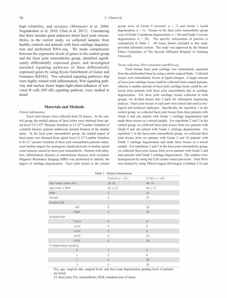
Facet joint tissues were collected from 58 donors. In the con-trol group, the medial aspects of facet joints were obtained from spi-nal level T12 (12th Thoracic Vertebra) to L2 (2nd Lumbar Vertebra) of vertebral fracture patients underwent internal fixation of the lumbar spine. In the facet joint osteoarthritis group, the medial aspect of facet joints was obtained from spinal level L3 (3rd Lumbar Vertebra) to S1 (1st sacrum Vertebra) of facet joint osteoarthritis patients under-went lumbar surgery for neurogenic claudication due to lumbar spinal canal stenosis caused by facet joint osteoarthritis. Patients with infec-tion, inflammatory diseases or autoimmune diseases were excluded. Magnetic Resonance Imaging (MRI) was performed to identify the degree of cartilage degeneration. Facet joint tissues in the control
group were of Grade 0 (normal; n = 7) and Grade 1 (mild degeneration; n = 3). Tissues in the facet joint osteoarthritis group were of Grade 2 (moderate degeneration; n = 20) and Grade 3 (severe degeneration; n = 28). The specific information of patients is summarized in Table 1. All tissue donors included in this study provided informed consent. The study was approved by the Human Ethics Committee of The Second Affiliated Hospital of Nantong University.
Tissue collection, RNA extraction and RNA-seqFresh human facet joint cartilage was immediately separated
from the subchondral bone by using a sterile surgical blade. Collected tissues were immediately frozen in liquid nitrogen. A larger amount of facet joint cartilage tissue could be collected from control patients, whereas a smaller amount of facet joint cartilage tissue could be col-lected from patients with facet joint osteoarthritis due to cartilage degeneration. For facet joint cartilage tissues collected in both groups, we divided tissues into 3 parts for subsequent sequencing analysis. Facet joint tissues in each part were mixed and used as bio-logical and technical triplicates. Specifically, for repetition 1 in the control group, we collected facet joint tissues from three patients with Grade 0 and one patient with Grade 1 cartilage degeneration and made these tissues as a mixed sample. For repetitions 2 and 3 in the control group, we collected facet joint tissues from two patients with Grade 0 and one patient with Grade 1 cartilage degeneration. For repetition 1 in the facet joint osteoarthritis group, we collected facet joint tissues from six patients with Grade 2 and 10 patients with Grade 3 cartilage degeneration and made these tissues as a mixed sample. For repetitions 2 and 3 in the facet joint osteoarthritis group, we collected facet joint tissues from seven patients with Grade 2 and nine patients with Grade 3 cartilage degeneration. The samples were homogenized by using the Cell crusher tissue pulverizer. Total RNA was isolated by using TRIzol reagent (Invitrogen, Carlsbad, CA) and
Control ( n = 10 ) FJ OA ( n = 48 )
Age range ( years old ) 18-35 46-79
Age mean ± SEM 25 ± 1.2 64 ± 1.7
Male 5 21
Female 5 27
Surgical side
Left 6 22
Right 4 26
Surgical level
T12/L1 6 0
L1/L2 4 0
L3/L4 0 6
L4/L5 0 23
L5/S1 0 19
FJ degeneration grading
0 7 0
1 3 0
2 0 20
3 0 28
Sex, age, surgical side, surgical level, and facet joint degeneration grading level of patients are listed.FJ, facet joint; OA, osteoarthritis; SEM, standard error of mean.
Table 1. Patient information.
Sequencing Analysis of Facet Joint Osteoarthritis 71
purified by using RNeasy Mini kit (Qiagen, Valencia, CA). High quality RNA samples were achieved. For RNA in the control group, the OD260/280 was 1.76, RNA concentration was 62.51 ng/μl, and RNA Integrity Number (RIN) was 6.2. For RNA in the facet joint osteoarthritis group, the OD260/280 was 1.80, RNA concentration was 34.50 ng/μl, and RIN was 6.2.
RNA-seq was performed by using Illumina Hiseq X10 plat-form. Obtained primary sequencing data were subjected to quality control by using perl scripts. Clean reads were obtained by removing reads with adapters, reads that contains more than 5% unknown bases, and low quality reads. Reference genome and gene model annotation files were directly downloaded from the genome website. The reference genome index was built by using Bowtie (v2.1.0) (Langmead and Salzberg 2012) and paired-end clean reads were
aligned to the reference genome using TopHat (v2.1.1) (Kim et al. 2013).
Bioinformatic analysisGene expression levels were calculated by fragments per kilo-
base of exon per million fragments mapped (FPKM). The expression level of each gene in the facet joint osteoarthritis group was compared with the level in the control group. Gene that obtains a log2Ratio ≤ –1 or ≥ 1 and a q-value ≤ 0.05 was assigned as significantly differ-entially expressed. Differentially expressed genes underwent KEGG analysis to classify correlating gene sets into their respective path-ways.
Primer names Primer ( 5' to 3' ) SequenceWIF1 Forward TGCGGTGCCCATGGA
Reverse CTGCCACGAACCCAGBP3 Forward GGAACTGGATTCGCAACATAGA
Reverse GACCCTTGGAAGTTGGCACAGCCSNK1A1 Forward CTTCTTGTCTGTAAGCCAGC
Reverse TCTTATGTCTTCACAGGTAAGCTCF7 Forward CTACTCCGCCTTCAATCTGC
Reverse AGAGAGTTGTGGGACGCTGTLEF1 Forward AAGGAACACTGACATCAATT
Reverse TTTGGAACTTGGCTCTTGWNT11 Forward CTCACCGGATTACTGCACACAGAAC
Reverse CCATTCATTAACTCTTTCACCATCCWNT5B Forward GAACAACAGCTTGAGCCAGAATA
Reverse GAGGAACGGGAACCTTAACAGACCAMK2A Forward TGAGAGCACCAACACCACCATCG
Reverse TGTCATTCCAGGGTCGCACATCTTCPLCB2 Forward GAGCAAATCGCCAAGATGAT
Reverse CCTTGTCTGTGGTGACCTTGDKK2 Forward TGGAAGATACTGCCACAGTCC
Reverse ACCATGGTTGCGATCTCTATGSOX17 Forward GCCGAGCCAAAGCGG
Reverse GTCAACGCCTTCCAAGACTTGMYC Forward TCTCCACTCACCAGCACAACTACG
Reverse ATCTGCTTCAGGACCCTCCND1 Forward CCCAACAACTTCCTCTCCT
Reverse TCCAGAAGGGCTTCAATCTGVANGL2 Forward AGGTGAATGAGTGCCTGGAC
Reverse ATTTGTGCCTCCCAGTATGCMMP7 Forward AGCAGCTATGCAGCTGGCCGT
Reverse GCCCTGAGCCTGTTCCCACTGCCCL4 Forward AGCTGTGGTATTCCAGACCAA
Reverse TCAAGGTCATCCACGTACTCCCCL4L2 Forward AGAAAACCTCTTTGCCACCA
Reverse GCAGACTTGCTTGCCTCTTTCXCL8 Forward AATGAAAAGATGAGGGTGCAT
Reverse GCTTGTGTGCTCTGCTGTCTBCL2A1 Forward GGATAAGGCAAAACGGAGGCTG
Reverse CAGTATTGCTTCAGGAGAGATAGCTNFSF13B Forward TTCCATGGCTTCTCAGCTTT
Reverse GTCCCATGGCGTAGGTCTTACXCL12 Forward TACAGATGCCCATGCCGA
Reverse CTGAAGGGCACAGTTTGGAGTNFSF14 Forward TCAGCTGCTCTGGCATGGAGAGTG
Reverse TGCTGGGTTGGCCTGGTGAGATCGThe primers used in this study are listed.
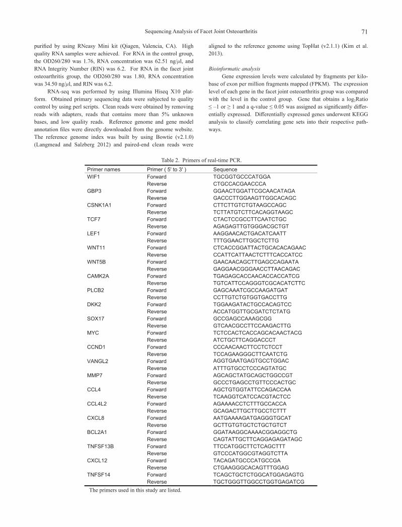
Table 2. Primers of real-time PCR.
C. Chen et al.72
qRT-PCRTotal RNA was isolated from cartilage tissues by using TRIzol
reagent. Isolated RNA was reverse-transcribed and qPCR was per-formed with an AB Step One Plus Real-Time PCR system. The qPCR conditions were as follows: 95°C for 2 minutes, 40 cycles of 95°C for 15 seconds and 60°C for 30 seconds. The primers used in this study are listed in Table 2.
Statistical analysisAll data were expressed as mean ± standard error. Student’s
t-test was used to compare differences between the control groups and the facet joint osteoarthritis group. All statistical analyses were per-formed with SPSS for Windows 11.0.1 (SPSS Inc., Chicago, IL) and all significance levels were set at p-value < 0.05.
ResultsOverview of the transcriptome changes in facet joint osteo-arthritis
Three replicated samples from the healthy control group or facet joint osteoarthritis group were collected and
underwent RNA isolation and subsequent RNA-seq. The four nitrogen-containing nucleobases (adenine [A], thymine [T], guanine [G], and cytosine [C]) were randomly distrib-uted, and RNA-seq reads were mapped to the reference genome. These parameters indicated that we achieved high quality outcomes.
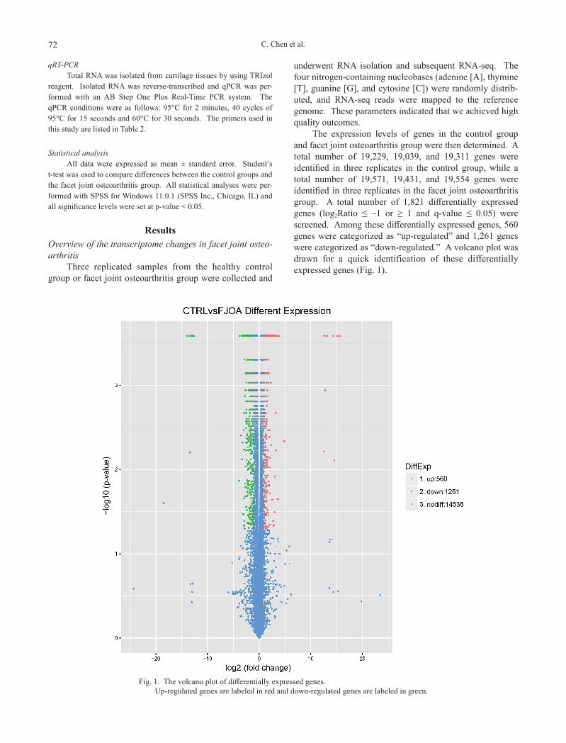
The expression levels of genes in the control group and facet joint osteoarthritis group were then determined. A total number of 19,229, 19,039, and 19,311 genes were identified in three replicates in the control group, while a total number of 19,571, 19,431, and 19,554 genes were identified in three replicates in the facet joint osteoarthritis group. A total number of 1,821 differentially expressed genes (log2Ratio ≤ –1 or ≥ 1 and q-value ≤ 0.05) were screened. Among these differentially expressed genes, 560 genes were categorized as “up-regulated” and 1,261 genes were categorized as “down-regulated.” A volcano plot was drawn for a quick identification of these differentially expressed genes (Fig. 1).
Fig. 1. The volcano plot of differentially expressed genes. Up-regulated genes are labeled in red and down-regulated genes are labeled in green.
Sequencing Analysis of Facet Joint Osteoarthritis 73
Significantly enriched signaling pathways in facet joint osteoarthritis
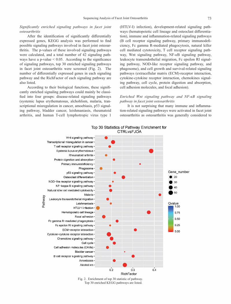
After the identification of significantly differentially expressed genes, KEGG analysis was performed to find possible signaling pathways involved in facet joint osteoar-thritis. The p-values of these involved signaling pathways were calculated, and a total number of 42 signaling path-ways have a p-value < 0.05. According to the significance of signaling pathways, top 30 enriched signaling pathways in facet joint osteoarthritis were screened (Fig. 2). The number of differentially expressed genes in each signaling pathway and the RichFactor of each signaling pathway are also listed.
According to their biological functions, these signifi-cantly enriched signaling pathways could mainly be classi-fied into four groups: disease-related signaling pathways (systemic lupus erythematosus, alchohlism, malaria, tran-scriptional misregulation in cancer, amoebiasis, p53 signal-ing pathway, bladder cancer, leishmaniasis, rheumatoid arthritis, and human T-cell lymphotropic virus type 1
(HTLV-I) infection), development-related signaling path-ways (hematopoietic cell lineage and osteoclast differentia-tion), immune and inflammation-related signaling pathways (B cell receptor signaling pathway, primary immunodefi-ciency, Fc gamma R-mediated phagocytosis, natural killer cell mediated cytotoxicity, T cell receptor signaling path-way, Wnt signaling pathway, NF-κB signaling pathway, leukocyte transendothelial migration, Fc epsilon RI signal-ing pathway, NOD-like receptor signaling pathway, and phagosome), and cell growth and survival-related signaling pathways (extracellular matrix (ECM)-receptor interaction, cytokine-cytokine receptor interaction, chemokines signal-ing pathway, cell cycle, protein digestion and absorption, cell adhesion molecules, and focal adhesion).
Enriched Wnt signaling pathway and NF-κB signaling pathway in facet joint osteoarthritis
It is not surprising that many immune and inflamma-tion-related signaling pathways were activated in facet joint osteoarthritis as osteoarthritis was generally considered to
Fig. 2. Enrichment of top 30 statistic of pathway. Top 30 enriched KEGG pathways are listed.
C. Chen et al.74
be highly associated with immune and inflammation. Two immune and inflammation-related signaling pathways, Wnt signaling pathway and NF-κB signaling pathway, have pre-viously been identified to be involved in osteoarthritis (Kobayashi et al. 2013; Rigoglou and Papavassiliou 2013; Takamatsu et al. 2014; Funck-Brentano et al. 2015). However, whether these two signaling pathways are also activated in facet joint osteoarthritis has not been deter-mined yet. Here, Wnt signaling pathway and NF-κB sig-naling pathway were investigated in detail to identify their
involvement in facet joint osteoarthritis.Based on the KEGG Orthology online database (http://
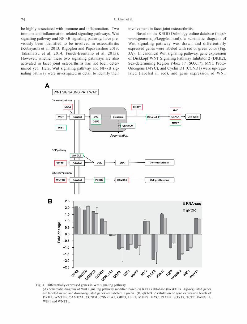
www.genome.jp/kegg/ko.html), a schematic diagram of Wnt signaling pathway was drawn and differentially expressed genes were labeled with red or green color (Fig. 3A). In canonical Wnt signaling pathway, gene expression of Dickkopf WNT Signaling Pathway Inhibitor 2 (DKK2), Sex-determining Region Y-box 17 (SOX17), MYC Proto-Oncogene (MYC), and Cyclin D1 (CCND1) were up-regu-lated (labeled in red), and gene expression of WNT
Fig. 3. Differentially expressed genes in Wnt signaling pathway. (A) Schematic diagram of Wnt signaling pathway modified based on KEGG database (ko04310). Up-regulated genes
are labeled in red and down-regulated genes are labeled in green. (B) qRT-PCR validation of gene expression levels of DKK2, WNT5B, CAMK2A, CCND1, CSNK1A1, GBP3, LEF1, MMP7, MYC, PLCB2, SOX17, TCF7, VANGL2, WIF1 and WNT11.
Sequencing Analysis of Facet Joint Osteoarthritis 75
Inhibitory Factor 1 (WIF1), Guanylate Binding Protein 3 (GBP3), Casein Kinase 1 Alpha 1 (CSNK1A1), Matrix Metallopeptidase 7 (MMP7) and Transcription Factor 7/Lymphoid Enhancer Binding Factor 1 (TCF7/LEF1) were down-regulated (labeled in green). In planar cell polarity (PCP) signaling pathway, Wnt Family Member 11 (WNT11) was up-regulated and VANGL Planar Cell Polarity Protein 2 (VANGL2) was down-regulated. In Wnt/Ca2+ signaling pathway, Wnt Family Member 5 (WNT5) and Calcium/Calmodulin Dependent Protein Kinase II Alpha (CAMK2A) were up-regulated and Phospholipase C
Beta 2 (PLCB2) was down-regulated. The expression lev-els of representative genes were further validated by qRT-PCR. Outcomes from qRT-PCR showed that consistent with the RNA-seq data, the mRNA levels of DKK2, SOX17, MYC, CCND1, CAMK2A, WNT11 and WNT5 were increased in facet joint osteoarthritis, while the mRNA levels of WIF1, CSNK1A1, TCF7/LEF1, and VANGL2 were decreased (Fig. 3B).
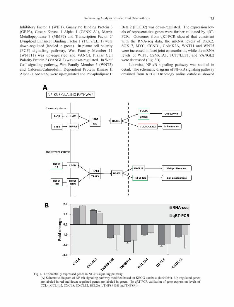
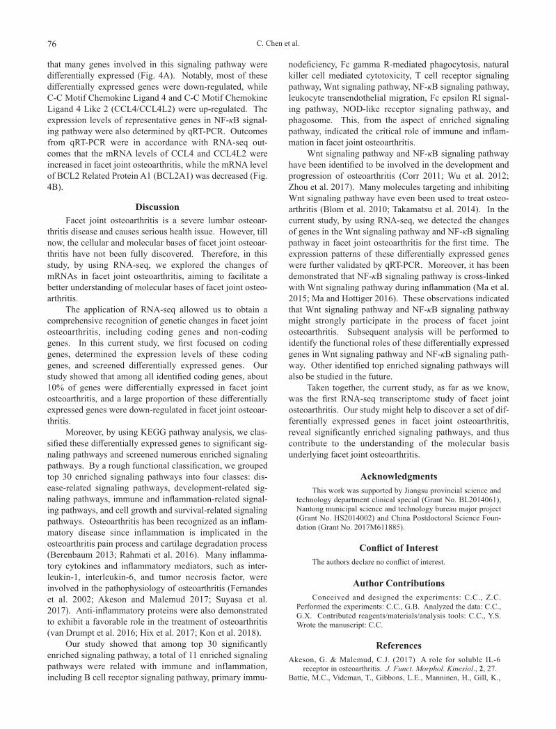
Likewise, NF-κB signaling pathway was studied in detail. The schematic diagram of NF-κB signaling pathway obtained from KEGG Orthology online database showed
Fig. 4. Differentially expressed genes in NF-κB signaling pathway. (A) Schematic diagram of NF-κB signaling pathway modified based on KEGG database (ko04064). Up-regulated genes
are labeled in red and down-regulated genes are labeled in green. (B) qRT-PCR validation of gene expression levels of CCL4, CCL4L2, CXCL8, CXCL12, BCL2A1, TNFSF13B and TNFSF14.
C. Chen et al.76
that many genes involved in this signaling pathway were differentially expressed (Fig. 4A). Notably, most of these differentially expressed genes were down-regulated, while C-C Motif Chemokine Ligand 4 and C-C Motif Chemokine Ligand 4 Like 2 (CCL4/CCL4L2) were up-regulated. The expression levels of representative genes in NF-κB signal-ing pathway were also determined by qRT-PCR. Outcomes from qRT-PCR were in accordance with RNA-seq out-comes that the mRNA levels of CCL4 and CCL4L2 were increased in facet joint osteoarthritis, while the mRNA level of BCL2 Related Protein A1 (BCL2A1) was decreased (Fig. 4B).
DiscussionFacet joint osteoarthritis is a severe lumbar osteoar-
thritis disease and causes serious health issue. However, till now, the cellular and molecular bases of facet joint osteoar-thritis have not been fully discovered. Therefore, in this study, by using RNA-seq, we explored the changes of mRNAs in facet joint osteoarthritis, aiming to facilitate a better understanding of molecular bases of facet joint osteo-arthritis.
The application of RNA-seq allowed us to obtain a comprehensive recognition of genetic changes in facet joint osteoarthritis, including coding genes and non-coding genes. In this current study, we first focused on coding genes, determined the expression levels of these coding genes, and screened differentially expressed genes. Our study showed that among all identified coding genes, about 10% of genes were differentially expressed in facet joint osteoarthritis, and a large proportion of these differentially expressed genes were down-regulated in facet joint osteoar-thritis.
Moreover, by using KEGG pathway analysis, we clas-sified these differentially expressed genes to significant sig-naling pathways and screened numerous enriched signaling pathways. By a rough functional classification, we grouped top 30 enriched signaling pathways into four classes: dis-ease-related signaling pathways, development-related sig-naling pathways, immune and inflammation-related signal-ing pathways, and cell growth and survival-related signaling pathways. Osteoarthritis has been recognized as an inflam-matory disease since inflammation is implicated in the osteoarthritis pain process and cartilage degradation process (Berenbaum 2013; Rahmati et al. 2016). Many inflamma-tory cytokines and inflammatory mediators, such as inter-leukin-1, interleukin-6, and tumor necrosis factor, were involved in the pathophysiology of osteoarthritis (Fernandes et al. 2002; Akeson and Malemud 2017; Suyasa et al. 2017). Anti-inflammatory proteins were also demonstrated to exhibit a favorable role in the treatment of osteoarthritis (van Drumpt et al. 2016; Hix et al. 2017; Kon et al. 2018).
Our study showed that among top 30 significantly enriched signaling pathway, a total of 11 enriched signaling pathways were related with immune and inflammation, including B cell receptor signaling pathway, primary immu-
nodeficiency, Fc gamma R-mediated phagocytosis, natural killer cell mediated cytotoxicity, T cell receptor signaling pathway, Wnt signaling pathway, NF-κB signaling pathway, leukocyte transendothelial migration, Fc epsilon RI signal-ing pathway, NOD-like receptor signaling pathway, and phagosome. This, from the aspect of enriched signaling pathway, indicated the critical role of immune and inflam-mation in facet joint osteoarthritis.
Wnt signaling pathway and NF-κB signaling pathway have been identified to be involved in the development and progression of osteoarthritis (Corr 2011; Wu et al. 2012; Zhou et al. 2017). Many molecules targeting and inhibiting Wnt signaling pathway have even been used to treat osteo-arthritis (Blom et al. 2010; Takamatsu et al. 2014). In the current study, by using RNA-seq, we detected the changes of genes in the Wnt signaling pathway and NF-κB signaling pathway in facet joint osteoarthritis for the first time. The expression patterns of these differentially expressed genes were further validated by qRT-PCR. Moreover, it has been demonstrated that NF-κB signaling pathway is cross-linked with Wnt signaling pathway during inflammation (Ma et al. 2015; Ma and Hottiger 2016). These observations indicated that Wnt signaling pathway and NF-κB signaling pathway might strongly participate in the process of facet joint osteoarthritis. Subsequent analysis will be performed to identify the functional roles of these differentially expressed genes in Wnt signaling pathway and NF-κB signaling path-way. Other identified top enriched signaling pathways will also be studied in the future.
Taken together, the current study, as far as we know, was the first RNA-seq transcriptome study of facet joint osteoarthritis. Our study might help to discover a set of dif-ferentially expressed genes in facet joint osteoarthritis, reveal significantly enriched signaling pathways, and thus contribute to the understanding of the molecular basis underlying facet joint osteoarthritis.
AcknowledgmentsThis work was supported by Jiangsu provincial science and
technology department clinical special (Grant No. BL2014061), Nantong municipal science and technology bureau major project (Grant No. HS2014002) and China Postdoctoral Science Foun-dation (Grant No. 2017M611885).
Conflict of InterestThe authors declare no conflict of interest.
Author ContributionsConceived and designed the experiments: C.C., Z.C.
Performed the experiments: C.C., G.B. Analyzed the data: C.C., G.X. Contributed reagents/materials/analysis tools: C.C., Y.S. Wrote the manuscript: C.C.
ReferencesAkeson, G. & Malemud, C.J. (2017) A role for soluble IL-6
receptor in osteoarthritis. J. Funct. Morphol. Kinesiol., 2, 27.Battie, M.C., Videman, T., Gibbons, L.E., Manninen, H., Gill, K.,
Sequencing Analysis of Facet Joint Osteoarthritis 77
Pope, M. & Kaprio, J. (2002) Occupational driving and lumbar disc degeneration: a case-control study. Lancet, 360, 1369-1374.
Berenbaum, F. (2013) Osteoarthritis as an inflammatory disease (osteoarthritis is not osteoarthrosis!). Osteoarthritis Cartilage, 21, 16-21.
Blom, A.B., van Lent, P.L., van der Kraan, P.M. & van den Berg, W.B. (2010) To seek shelter from the WNT in osteoarthritis? WNT-signaling as a target for osteoarthritis therapy. Curr. Drug Targets, 11, 620-629.
Chen, G., Shi, T. & Shi, L. (2017) Characterizing and annotating the genome using RNA-seq data. Sci. China Life Sci., 60, 116-125.
Corr, M. (2011) Wnt-beta-catenin signaling in the pathogenesis of osteoarthritis. Nat. Clin. Pract. Rheumatol., 4, 550-556.
Das, S.K. & Farooqi, A. (2008) Osteoarthritis. Best Pract. Res. Clin. Rheumatol., 22, 657-675.
Fernandes, J.C., Martel-Pelletier, J. & Pelletier, J.P. (2002) The role of cytokines in osteoarthritis pathophysiology. Biorhe-ology, 39, 237-246.
Funck-Brentano, T., Bouaziz, W., Marty, C., Geoffroy, V., Hay, E. & Cohen-Solal, M. (2015) Dkk-1-mediated inhibition of Wnt signaling in bone ameliorates osteoarthritis in mice. Arthritis. Rheumatol., 66, 3028-3039.
Gellhorn, A.C., Katz, J.N. & Suri, P. (2013) Osteoarthritis of the spine: the facet joints. Nat. Rev. Rheumatol., 9, 216-224.
Glynjones, S., Palmer, A.J., Agricola, R., Price, A.J., Vincent, T.L., Weinans, H. & Carr, A.J. (2015) Osteoarthritis. Lancet, 386, 376-387.
Hix, J., Klaassen, M., Foreman, R., Cullen, E., Toler, K., King, W. & Woodell-May, J. (2017) An autologous anti-inflammatory protein solution yielded a favorable safety profile and signifi-cant pain relief in an open-label pilot study of patients with osteoarthritis. Biores. Open Access, 6, 151-158.
Katz, J.N. (2006) Lumbar disc disorders and low-back pain: socio-economic factors and consequences. J. Bone Joint Surg. Am., 88 Suppl 2, 21-24.
Kim, D., Pertea, G., Trapnell, C., Pimentel, H., Kelley, R. & Salzberg, S.L. (2013) TopHat2: accurate alignment of tran-scriptomes in the presence of insertions, deletions and gene fusions. Genome Biol., 14, R36.
Kobayashi, H., Hirata, M., Saito, T., Itoh, S., Chung, U.I. & Kawaguchi, H. (2013) Transcriptional induction of ADAMTS5 protein by nuclear factor-κB (NF-κB) family member RelA/p65 in chondrocytes during osteoarthritis development. J. Biol. Chem., 288, 28620-28629.
Kon, E., Engebretsen, L., Verdonk, P., Nehrer, S. & Filardo, G. (2018) Clinical outcomes of knee osteoarthritis treated with an autologous protein solution injection: a 1-year pilot double-blinded randomized controlled trial. Am. J. Sports Med., 46, 171-180.
Langmead, B. & Salzberg, S.L. (2012) Fast gapped-read align-ment with Bowtie 2. Nat. Methods, 9, 357-359.
Ma, B., Fey, M. & Hottiger, M.O. (2015) WNT/β-catenin signaling
inhibits CBP-mediated RelA acetylation and expression of proinflammatory NF-kappaB target genes. J. Cell Sci., 128, 2430-2436.
Ma, B. & Hottiger, M.O. (2016) Crosstalk between WNT/β-catenin and NF-kappaB signaling pathway during inflamma-tion. Front. Immunol., 7, 378.
Mortazavi, A., Williams, B.A., McCue, K., Schaeffer, L. & Wold, B. (2008) Mapping and quantifying mammalian transcrip-tomes by RNA-Seq. Nat. Methods, 5, 621-628.
Nagalakshmi, U., Waern, K. & Snyder, M. (2010) RNA-Seq: a method for comprehensive transcriptome analysis. Curr. Protoc. Mol. Biol., 89, 4.11.1-4.11.13.
Nakamura, A., Rampersaud, Y.R., Sharma, A., Lewis, S.J., Wu, B., Datta, P., Sundararajan, K., Endisha, H., Rossomacha, E., Rockel, J.S., Jurisica, I. & Kapoor, M. (2016) Identification of microRNA-181a-5p and microRNA-4454 as mediators of facet cartilage degeneration. JCI Insight, 1, e86820.
Rahmati, M., Mobasheri, A. & Mozafari, M. (2016) Inflammatory mediators in osteoarthritis: a critical review of the state-of-the-art, current prospects, and future challenges. Bone, 85, 81-90.
Rigoglou, S. & Papavassiliou, A.G. (2013) The NF-κB signalling pathway in osteoarthritis. Int. J. Biochem. Cell Biol., 45, 2580-2584.
Riihimäki, H. & Viikari-Juntura, E. (2008) Back and Limb Disor-ders, BMJ Publishing Group, UK.
Suyasa, I.K., Kawiyana, I.K., Bakta, I.M. & Widiana, I.G. (2017) Interleukin-6 and ratio of plasma interleukin-6/interleukin-10 as risk factors of symptomatic lumbar osteoarthritis. World J. Orthop., 8, 149-155.
Takamatsu, A., Ohkawara, B., Ito, M., Masuda, A., Sakai, T., Ishiguro, N. & Ohno, K. (2014) Verapamil protects against cartilage degradation in osteoarthritis by inhibiting Wnt/beta-catenin signaling. PLoS One, 9, e92699.
Tian, W., Lv, Y., Liu, Y., Xiao, B. & Han, X. (2014) The high prevalence of symptomatic degenerative lumbar osteoarthritis in Chinese adults: a population-based study. Spine, 39, 1301-1310.
van Drumpt, R.A., van der Weegen, W., King, W., Toler, K. & Macenski, M.M. (2016) Safety and treatment effectiveness of a single autologous protein solution injection in patients with knee osteoarthritis. Biores. Open Access, 5, 261-268.
Vieira, S., Dibai-Filho, A.V., Brandino, H.E., Ferreira, V.T. & Scheicher, M.E. (2015) Abdominal muscle strength is related to the quality of life among older adults with lumbar osteoar-thritis. J. Bodyw. Mov. Ther., 19, 273-277.
Woolf, A.D. & Pfleger, B. (2003) Burden of major musculoskeletal conditions. Bull. World Health Organ., 81, 646-656.
Wu, L., Huang, X., Li, L., Huang, H., Xu, R. & Luyten, W. (2012) Insights on biology and pathology of HIF-1alpha/-2alpha, TGFβ/BMP, Wnt/β-catenin, and NF-kappaB pathways in osteoarthritis. Curr. Pharm. Des., 18, 3293-3312.
Zhou, Y., Wang, T., Hamilton, J.L. & Chen, D. (2017) Wnt/β-catenin signaling in osteoarthritis and in other forms of arthritis. Curr. Rheumatol. Rep., 19, 53.