AICT 2010-Athens Interventional Cardiovascular Therapeutics XI 8-9 OCTOBER 2010 Divani Caravel...
40
AICT 2010-Athens Interventional Cardiovascular Therapeutics XI 8-9 OCTOBER 2010 Divani Caravel Hotel, Αthens Euromedica – Kyanous Stavros, Euromedica – Kyanous Stavros, Thessaloniki. Thessaloniki. C. Graidis, D. Dimitriadis, A. Ntatsios, V. Karasavvides MULTIVESSEL PCI. IN DRUG-ELUTING STENT RESTENOSIS DUE TO STENT FRACTURE, TREATED WITH REPEAT DES IMPLANTATION
-
Upload
karin-miller -
Category
Documents
-
view
224 -
download
0
Transcript of AICT 2010-Athens Interventional Cardiovascular Therapeutics XI 8-9 OCTOBER 2010 Divani Caravel...

AICT 2010-Athens Interventional Cardiovascular Therapeutics XI
8-9 OCTOBER 2010
Divani Caravel Hotel, Αthens
Euromedica – Kyanous Stavros, Euromedica – Kyanous Stavros, Thessaloniki.Thessaloniki.
C. Graidis, D. Dimitriadis, A. Ntatsios, V. Karasavvides
MULTIVESSEL PCI. IN DRUG-ELUTING STENT RESTENOSIS DUE TO STENT FRACTURE, TREATED
WITH REPEAT DES IMPLANTATION

Criteria for case selection:
DES Restenosis - Stent Fracture
IVUS
Venture catheter
Rotablator
Bifurcation
Dissection
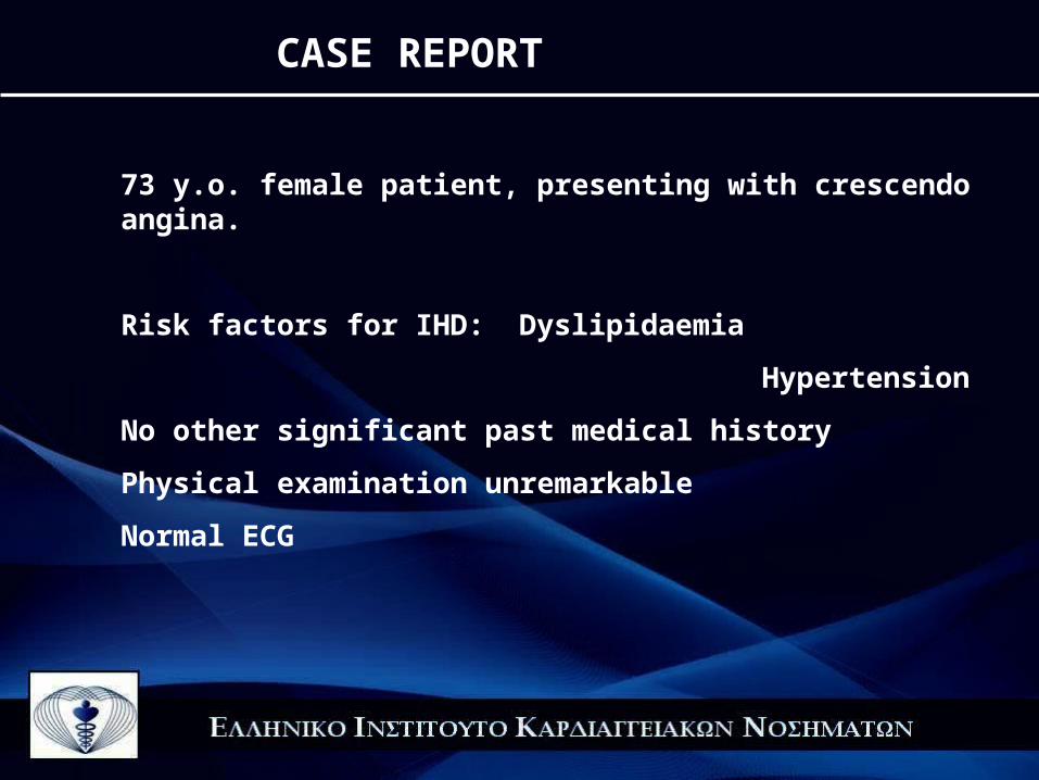
73 y.o. female patient, presenting with crescendo angina.
Risk factors for IHD: Dyslipidaemia
Hypertension
No other significant past medical history
Physical examination unremarkable
Normal ECG
CASE REPORT
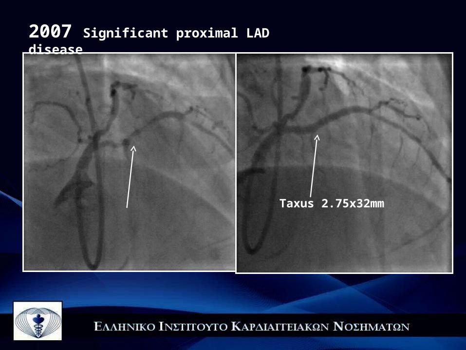
Taxus 2.75x32mm
2007 Significant proximal LAD disease
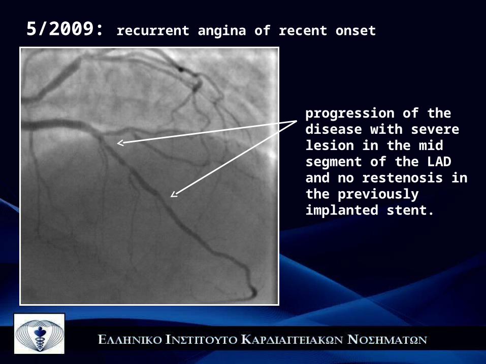
5/2009: recurrent angina of recent onset
progression of the disease with severe lesion in the mid segment of the LAD and no restenosis in the previously implanted stent.
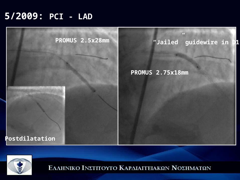
5/2009: PCI - LAD
PROMUS 2.5x28mm
PROMUS 2.75x18mm
Postdilatation
“Jailed” guidewire in D1

5/2009: PCI - LAD FINAL RESULT
Stent length 46mm

5/2009
Severe lesion in the intermediate branch and the circumflex artery.

Promus 2.5x12mm
DISSECTION !!!!
5/2009: PCI-INTERMEDIATE BRANCH
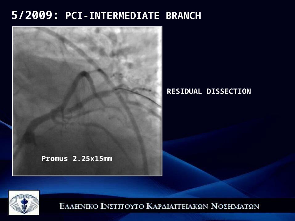
Promus 2.25x15mm
RESIDUAL DISSECTION
5/2009: PCI-INTERMEDIATE BRANCH

Promus 2.25x15mm
5/2009: PCI-INTERMEDIATE BRANCH

Promus 2.5x12mm, 2.25x15mm, 2,25x15mm
5/2009: PCI-INTERMEDIATE BRANCH FINAL RESULT

12/2009: recurrent chest pain!!!
Severe stenosis of the intermediate branch within the stented segment, with stent strut fracture near the distal overlapping site.
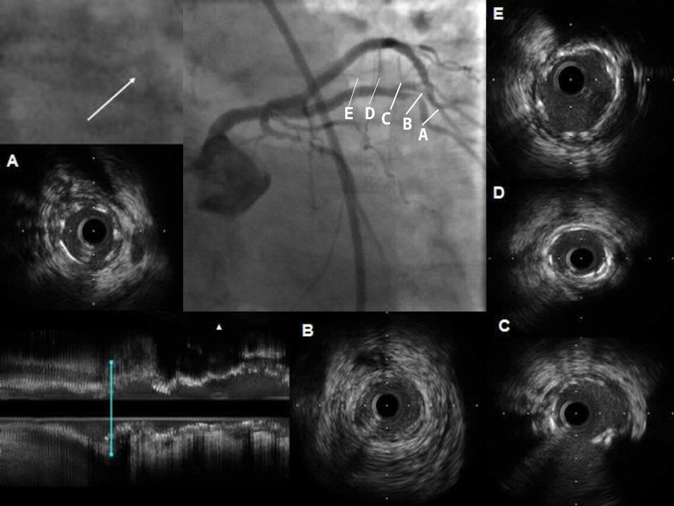
ABCDE
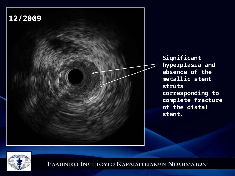
Significant hyperplasia and absence of the metallic stent struts corresponding to complete fracture of the distal stent.
12/2009

Promus Element 2.25x16 mm
12/2009
FINAL RESULT

Promus Element 2.25x12mm
Due to severe tortuosity of the proximal segment of the Cx, the Venture wire control catheter was used, in order to facilitate the passage of the guide-wire.
12/2009: PCI-Cx
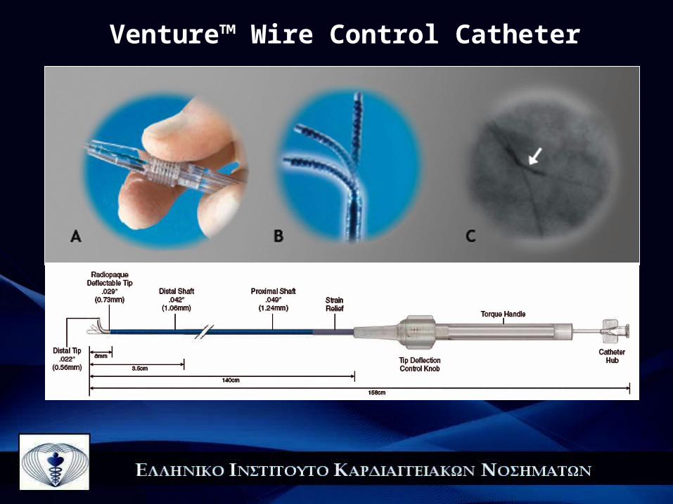
Venture™ Wire Control Catheter

RCA severely calcified with angulation at its proximal segment. Significant stenosis at the bifurcation with an acute marginal branch.
12/2009: PCI-RCA
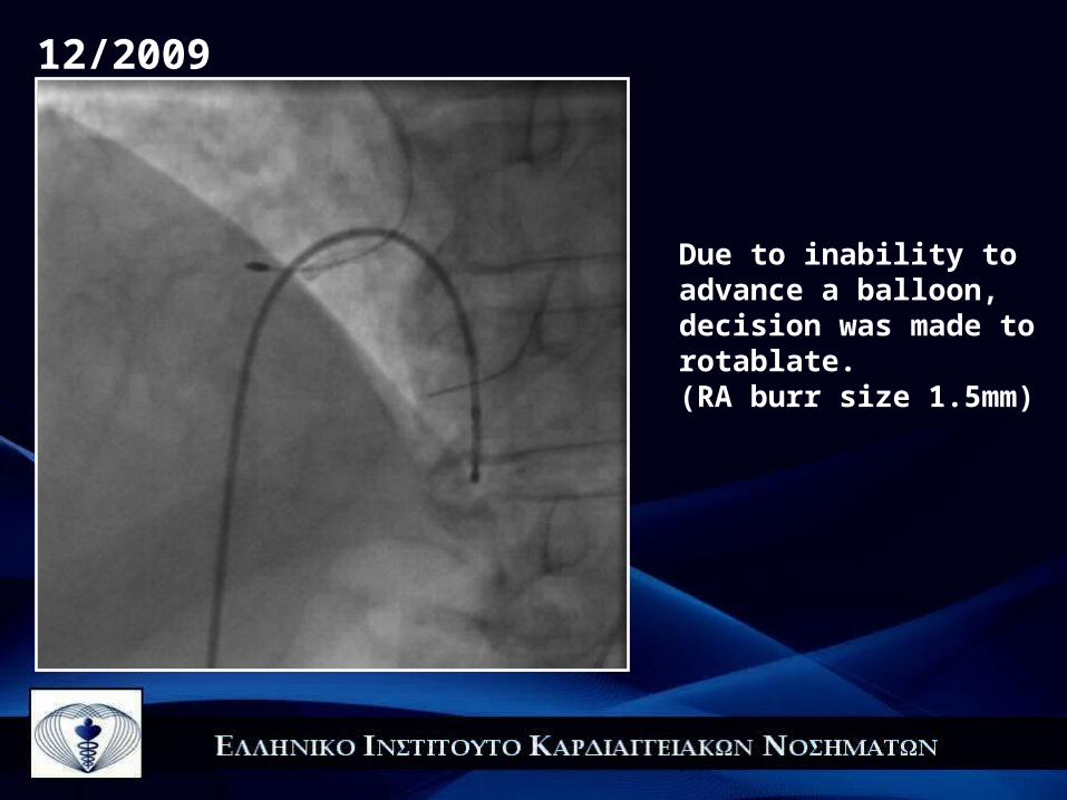
Due to inability to advance a balloon, decision was made to rotablate.(RA burr size 1.5mm)
12/2009
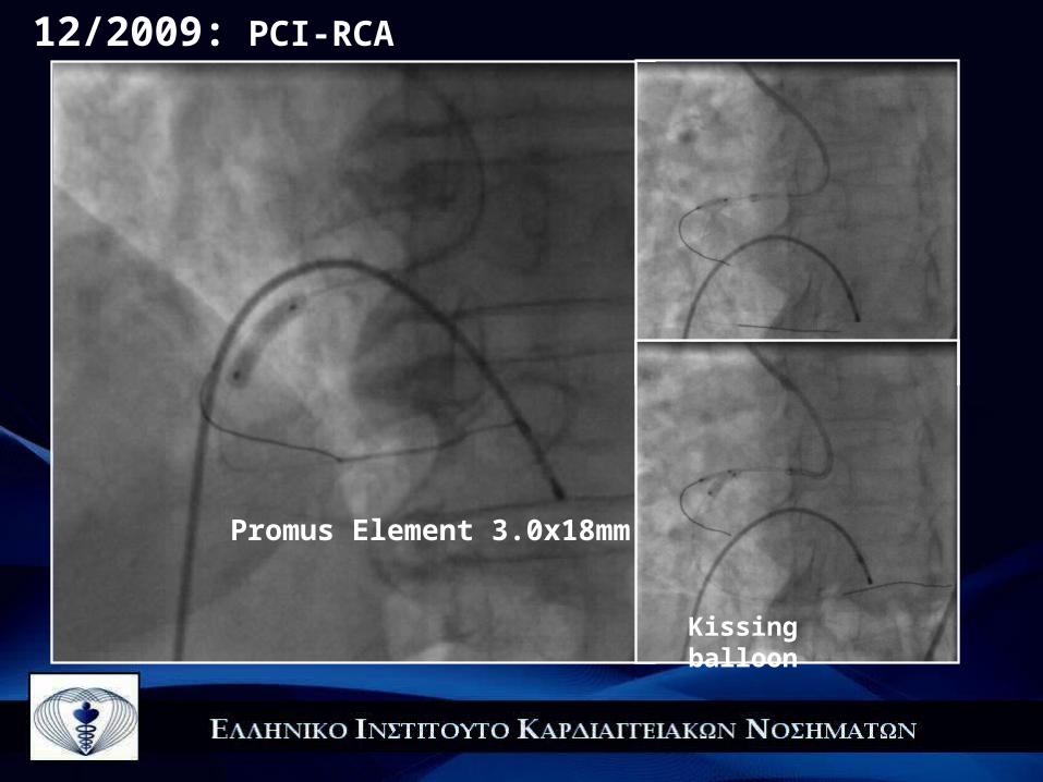
Promus Element 3.0x18mm
Kissing balloon
12/2009: PCI-RCA

12/2009: PCI-RCA FINAL RESULT
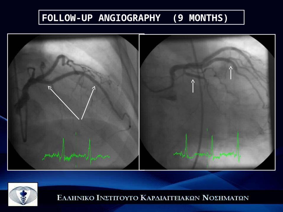
FOLLOW-UP ANGIOGRAPHY (9 MONTHS)
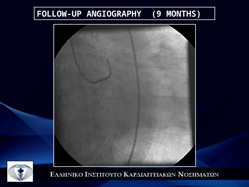
FOLLOW-UP ANGIOGRAPHY (9 MONTHS)

FOLLOW-UP ANGIOGRAPHY (9 MONTHS)
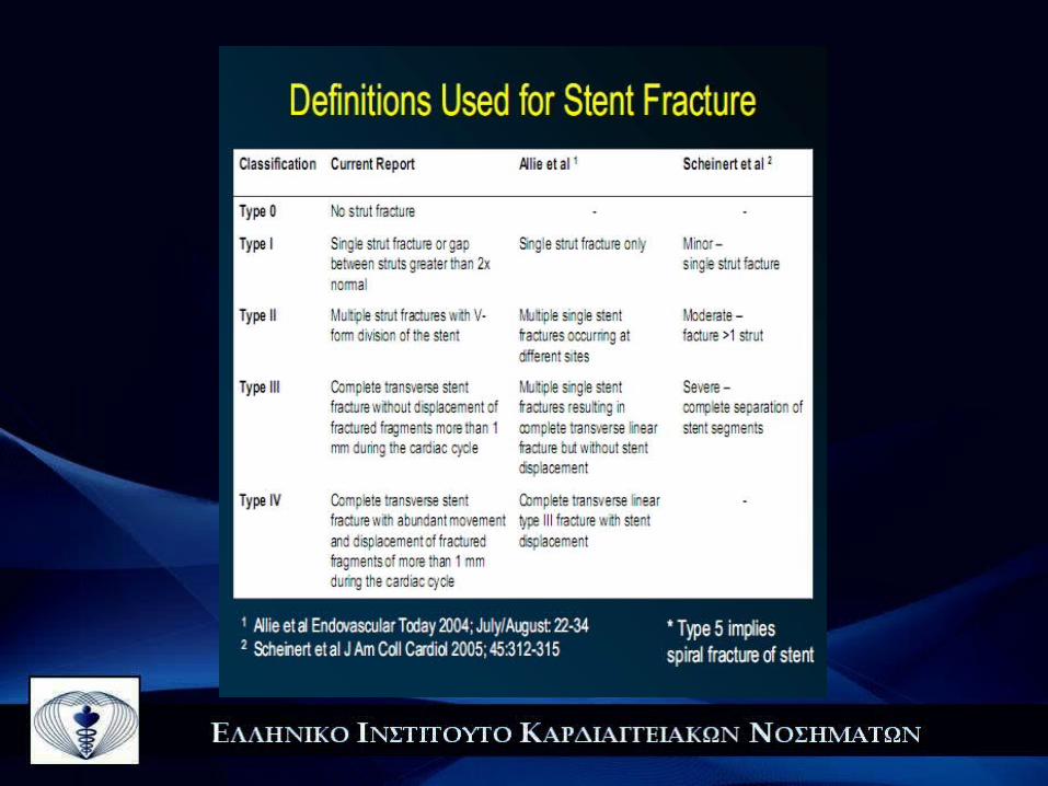
Coronary stent fracture is a rare but potentially serious complication
The reported incidence of SF in different studies ranges between 0.8% and 7.7%, mainly due to variations - in the rates of angiographic follow-up - use of IVUS - definition of stent fracture and - differences in the types of stents used
IVUS was more sensitive than fluoroscopy in detecting stent strut fractures in multiple studies
Since IVUS evaluation is not routinely performed during follow-up,
stent fracture is likely to be underdiagnosed
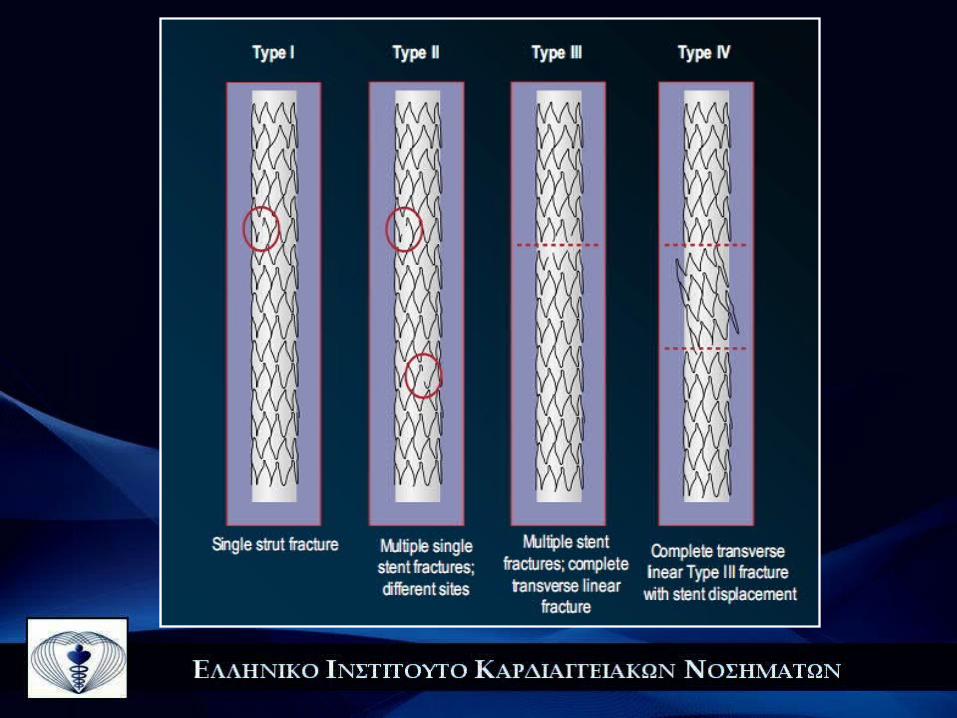

Factors predisposing to (drug-eluting) Stent Fracture
Anatomic Procedural Stent design Cardiac motion Long stent Closed cell
Vessel angulation Overlapping stent
Right coronary artery High pressure
Saphenous vein graft Overexpansion

The change in vessel angulation after stent implantation is also associated with the occurrence of SF.
Stent placement often straightens out the vessel; however, the
vessel continues to return to its original shape. The force to return the vessel to its original shape may be stronger if the change in vessel angulation after stent implantation is increased.
Longer stents are subjected to higher radial forces compared to shorter ones, and they may be prone to fracture, especially when placed in tortuous vessels or calcified lesions.

Most studies reported overlapping stents as a significant risk factor for SF, with an average rate of over 60% associated with overlapping stented lesions
The majority of stent fractures occurred within 10 mm from areas of increased rigidity caused by strut overlap.

The closed-cell design of SES may be more prone to fracture when sheer forces are beyond its flexibility.
The closed-cell design of the Cypher stent is more rigid, resulting in more straightening of coronary vessels compared to the opencell Taxus design. However, it also must be taken into account that the Cypher stent is more radio-opaque and stent fracture may be easier to diagnose by angiography.
The majority of DES fracture events have been reported in SESs
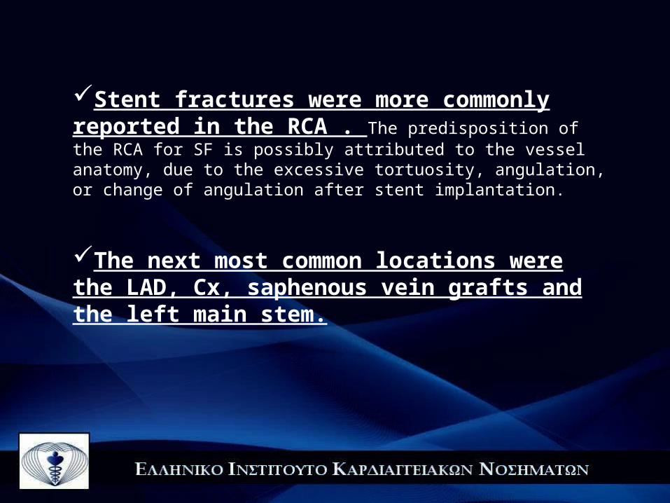
Stent fractures were more commonly reported in the RCA . The predisposition of the RCA for SF is possibly attributed to the vessel anatomy, due to the excessive tortuosity, angulation, or change of angulation after stent implantation.
The next most common locations were the LAD, Cx, saphenous vein grafts and the left main stem.

Clinical Implications
The complications observed with DES fracture include :
in-stent restenosis and target lesion revascularization,
stent thrombosis
myocardial infarction and sudden death
Patients may remain asymptomatic

While most of the stent fractures have been discovered during follow-up angiography six months or more post-implantation, SF cases have been reported from immediately after to several years after the index procedure
The length of time that a vessel wall is exposed to fractured stent struts may be important and possibly determine clinical outcomes. An early fracture affects local drug delivery and can lead to focal neointimal hyperplasia.
On the other hand, since significant restenosis was not observed in a significant number of fracture sites, it may be presumed that in these cases, stent strut fracture occurred long enough after implantation so that the drug effect was not compromised .

Management strategies
The management of stent fractures remains controversial.
If focal in-stent restenosis occurs, it seems reasonable to repeat percutaneous revascularisation with a short stent.
With repeat stenting there is a possibility of recurrence of SF whereas, on the other hand, the effect of balloon angioplasty on disrupted DESs is unknown.
The use of paclitaxel-eluting stents or other newly developed DESs might be an appropriate selection since their platform is more flexible.

In our case, it seems that the use of three, overlapping stents along with the presence of angulation and increased vessel movememt contributed to the fracture. Despite the fact that each of them had relatively short length, the fracture occurred very close to the distal overlapping site, at a point where there was a vessel angulation.
Point of distal overlapping

A careful examination of the vessel anatomy, careful selection of the stent length and type and a proper deployment technique is required for special coronary situations in order to reduce the occurrence of this phenomenon.

ΕΥΧΑΡΙΣΤΩ!ΕΥΧΑΡΙΣΤΩ!