Activation of the Human Immunodeficiency Virus Long ... · Varicella-zoster virus (VZV)1 is an...
16
Published in : Journal of biological chemistry (1998), vol. 273, iss. 22, pp. 13636-13644 Status : Postprint (Author’s version) Activation of the Human Immunodeficiency Virus Long Terminal Repeat by Varicella-zoster Virus IE4 Protein Requires Nuclear Factor-κB and Involves Both the Amino-terminal and the Carboxyl- terminal Cysteine-rich Region Defechereux-Thibaut de Maisieres P [1] , Baudoux-Tebache L [1] , Merville M-P [2] , Rentier B [1] , Bours V [2] Piette J [1] [1] Laboratory of Fundamental Virology and Immunology, Institute of Pathology, University of Liege, B-4000 Liege, Belgium [2] Laboratory of Medical Chemistry and Medical Oncology, Institute of Pathology, University of Liege, B-4000 Liege, Belgium Abstract : Varicella-zoster virus open reading frame 4-encoded protein (IE4) possesses transactivating properties for varicella-zoster virus genes as well as for those of heterologous viruses such as the human immunodeficiency virus type 1 (HIV-1). Mechanisms of HIV-1 LTR (long terminal repeat) transactivation were investigated in HeLa cells transiently transfected with an IE4 expression plasmid and a CAT reporter gene under the control of the HIV-1 LTR. These results demonstrated that IE4- mediated transactivation of the HIV-1 LTR in HeLa cells required transcription factor κB (NF-κB). Using the gel retardation assay, it was shown that transfection of the IE4 expression vector in HeLa cells was not associated with induction of NF-κB under the p50-p65 het-erodimeric form and that no direct binding of IE4 to the κB sites could be detected. Both Western blot and immunofluorescence analyses suggested that the ability of IE4 to activate transcription through κB motives was not connected with its capacity to override the inhibitory activities of IκB-a or p105. Finally, in vitro protein-protein interactions involving IE4 and basal transcription factors such as TATA-binding protein and transcription factor IIB were carried out. A direct interaction between IE4 and TATA-binding protein or transcription factor IIB components of the basal complex of transcription was evidenced, as well as binding to the p50 and p65 NF-κB subunits. Mutagenesis analysis of IE4 indicated that the COOH-terminal cysteine-rich and ar-ginine-rich regions (residues 82-182) were critical for transactivation, whereas the first 81 amino acids appeared dispensable. Moreover, the arginine-rich region is required for the in vitro binding activity, whereas the COOH-terminal end did not appear essential. Varicella-zoster virus (VZV) 1 is an α-herpesvirus, which causes two distinct diseases in man: chicken pox and shingles. Shortly after entry into the infected cells, VZV genes are expressed in a temporal cascade. The immediate-early genes are expressed first; these stimulate early gene expression, providing most of the proteins necessary for viral DNA replication. After DNA synthesis has occurred, genes of the late class, which mainly encode structural proteins, are expressed. This orderly pattern of expression has been proposed mainly by comparison with herpes simplex virus type 1 (HSV-1) (1), another α-herpesvirus that has been intensively studied. The use of transient transfection assays has clearly shown that several VZV proteins, i.e. those encoded by ORFs (open reading frames) 4, 10, 29, 61, 62, and 63, possess regulatory properties (2–8). Three of these polypeptides, encoded by ORFs 4, 62, and 63, are expressed during the immediate-early phase of lytic infection (9–11), and are thus referred to as IE4, IE62, and IE63. Hence, VZV immediate-early proteins contribute to the control of the viral cycle progression as in other herpesviruses. The IE4 protein is a transactivator of gene expression whose regulatory properties are not yet fully understood (2, 4, 11–14). IE4 stimulates VZV gene expression regardless of the cell type envisaged, i.e. monkey fibroblasts or human T lymphocytes (2, 4, 11, 12). It also appears that IE4 is capable of heterologous transactivation (11–13). The available data suggest that IE4 could exert its functions through transcriptional and post-transcriptional mechanisms (11–13). Evidence for transcriptionally mediated activation was provided by the characterization of critical cis-elements necessary for the IE4 inducibility of heterologous viral promoters (13). The presence of a TATA box alone is apparently not sufficient to convey transactivation by IE4, because transactivation of a minimal promoter containing only a TATA box is not possible (13). Two different factor-binding sites, the Sp1 and nuclear factor κB (NF-κB)-respon-sive elements, have been proposed as critical for IE4-mediated transactivation (13).
Transcript of Activation of the Human Immunodeficiency Virus Long ... · Varicella-zoster virus (VZV)1 is an...
Published in : Journal of biological chemistry (1998), vol. 273, iss. 22, pp. 13636-13644Status : Postprint (Author’s version)
Activation of the Human Immunodeficiency Virus Long TerminalRepeat by Varicella-zoster Virus IE4 Protein Requires NuclearFactor-κB and Involves Both the Amino-terminal and the Carboxyl-terminal Cysteine-rich Region
Defechereux-Thibaut de Maisieres P[1], Baudoux-Tebache L[1], Merville M-P[2], Rentier B[1], BoursV[2] Piette J[1]
[1]Laboratory of Fundamental Virology and Immunology, Institute of Pathology, University of Liege,B-4000 Liege, Belgium[2] Laboratory of Medical Chemistry and Medical Oncology, Institute of Pathology, University of Liege,B-4000 Liege, Belgium
Abstract : Varicella-zoster virus open reading frame 4-encoded protein (IE4) possesses transactivatingproperties for varicella-zoster virus genes as well as for those of heterologous viruses such as thehuman immunodeficiency virus type 1 (HIV-1). Mechanisms of HIV-1 LTR (long terminal repeat)transactivation were investigated in HeLa cells transiently transfected with an IE4 expression plasmidand a CAT reporter gene under the control of the HIV-1 LTR. These results demonstrated that IE4-mediated transactivation of the HIV-1 LTR in HeLa cells required transcription factor κB (NF-κB).Using the gel retardation assay, it was shown that transfection of the IE4 expression vector in HeLacells was not associated with induction of NF-κB under the p50-p65 het-erodimeric form and that nodirect binding of IE4 to the κB sites could be detected. Both Western blot and immunofluorescenceanalyses suggested that the ability of IE4 to activate transcription through κB motives was notconnected with its capacity to override the inhibitory activities of IκB-a or p105. Finally, in vitroprotein-protein interactions involving IE4 and basal transcription factors such as TATA-binding proteinand transcription factor IIB were carried out. A direct interaction between IE4 and TATA-bindingprotein or transcription factor IIB components of the basal complex of transcription was evidenced, aswell as binding to the p50 and p65 NF-κB subunits. Mutagenesis analysis of IE4 indicated that theCOOH-terminal cysteine-rich and ar-ginine-rich regions (residues 82-182) were critical fortransactivation, whereas the first 81 amino acids appeared dispensable. Moreover, the arginine-richregion is required for the in vitro binding activity, whereas the COOH-terminal end did not appearessential.
Varicella-zoster virus (VZV)1 is an α-herpesvirus, which causes two distinct diseases in man: chickenpox and shingles. Shortly after entry into the infected cells, VZV genes are expressed in a temporalcascade. The immediate-early genes are expressed first; these stimulate early gene expression,providing most of the proteins necessary for viral DNA replication. After DNA synthesis has occurred,genes of the late class, which mainly encode structural proteins, are expressed. This orderly pattern ofexpression has been proposed mainly by comparison with herpes simplex virus type 1 (HSV-1) (1),another α-herpesvirus that has been intensively studied. The use of transient transfection assays hasclearly shown that several VZV proteins, i.e. those encoded by ORFs (open reading frames) 4, 10, 29,61, 62, and 63, possess regulatory properties (2–8). Three of these polypeptides, encoded by ORFs 4,62, and 63, are expressed during the immediate-early phase of lytic infection (9–11), and are thusreferred to as IE4, IE62, and IE63. Hence, VZV immediate-early proteins contribute to the control ofthe viral cycle progression as in other herpesviruses.The IE4 protein is a transactivator of gene expression whose regulatory properties are not yet fullyunderstood (2, 4, 11–14). IE4 stimulates VZV gene expression regardless of the cell type envisaged,i.e. monkey fibroblasts or human T lymphocytes (2, 4, 11, 12). It also appears that IE4 is capable ofheterologous transactivation (11–13). The available data suggest that IE4 could exert its functionsthrough transcriptional and post-transcriptional mechanisms (11–13). Evidence for transcriptionallymediated activation was provided by the characterization of critical cis-elements necessary for the IE4inducibility of heterologous viral promoters (13). The presence of a TATA box alone is apparently notsufficient to convey transactivation by IE4, because transactivation of a minimal promoter containingonly a TATA box is not possible (13). Two different factor-binding sites, the Sp1 and nuclear factor κB(NF-κB)-respon-sive elements, have been proposed as critical for IE4-mediated transactivation (13).
Published in : Journal of biological chemistry (1998), vol. 273, iss. 22, pp. 13636-13644Status : Postprint (Author’s version)
Protein-encoding genes in eukaryotes are transcribed by RNA polymerase II associated with generaltranscription factors. Control of their expression requires specific regulatory1 The abbreviations used are: VZV, varicella-zoster virus; LTR, long terminal repeat; HIV-1, humanimmunodeficiency virus type 1; GST, glutathione S-transferase; PCR, polymerase chain reaction; HSV,herpes simplex virus; ORF, open reading frame; IE, immediate-early; NF-κB, nuclear factor-κB; USA,upstream factor stimulatory activity; USF, upstream stimulatory factor; CMV, cytomegalovirus; EBV,Epstein-Barr virus; WT, wild-type; CAT, chloramphenicol acetyltransferase; TBP, TATA-bindingprotein; TFIIB, transcription factor IIB; PAGE, polyacrylamide gel electrophoresis; HTLV, human Tcell leukemia virus.factors with DNA binding and transactivation domains that interact with the general transcriptionfactors. These interactions influence RNA polymerase II recruitment to the promoter and stabilizeand/or modify polymerase II holoenzyme activity (15). The ubiquitous Sp1 factor is a transcriptionfactor that participates in the basal activity of the transcription machinery by binding to specific sitesthat have been found in numerous core promoters. Sp1 possesses a zinc finger DNA-binding domain aswell as a glutamine-rich transactivation domain, and its activity seems to be controlled by itsphosphorylation status (16–18). NF-κB consists of a group of dimeric complexes composed of variedcombinations of polypeptides of the Rel/NF-κB family (19). These factors are ubiquitous and havebeen involved in transcription regulation of cellular genes important in the inflammatory response andoxidative stress (19–21). The activation of NF-κB is dependent on the binding of these complexes tocis-sequences found in the promoter regions of target genes (20, 22). The prototypic form of NF-κB isa heterodimer containing the p50 and p65 proteins (19, 21). In most cells, NF-κB activity is kept undercontrol through cytoplasmic sequestration by members of a family of inhibitory proteins, includingIκB-α and p105, the latter being the cytoplasmic precursor of p50 (23). The majority of inductionsignals lead to the rapid phosphorylation of IκB-α at serine 32 and serine 36 (24), which targets it to theubiquitin-proteasome pathway (25). Subsequent proteolytic degradation of IκB-α allows the nucleartranslocation of the active NF-κB dimer. A large number of stimuli such as mitogens, cytokines, stress,and viruses have been shown to induce NF-κB (19, 21).Because viruses are intracellular parasites, they appropriate the cellular machinery for their ownbenefit. NF-κB belongs to the host transcription factors used by a number of viruses to induce theirown expression or that of specific host genes (19). Inducible human immunodeficiency virus type 1(HIV-1) gene expression is generally mediated by the binding of NF-κB to the enhancer κB-bindingsites in the long terminal repeat (22, 26, 27). Molecular interactions among herpesviruses and HIV-1have been frequently investigated, as the most common opportunistic viral infections in individualswith AIDS are caused by herpesviruses (28–30). Herpesviruses could act as co-factors in enhancingHIV-1 replication. Direct effects of herpesvirus proteins on HIV-1 LTR activity have been detected inthe case of HSV-1 (31–33), cytomegalovirus (CMV) (34, 35), and Epstein-Barr virus (EBV) (36).Some of these effects could be associated with NF-κB induction (27, 33, 36–38), although somecontroversy exists as to the identity of the responsible proteins in HSV-1 (32, 37, 39, 40) and CMV(34, 35,41). Such discrepancies might possibly be attributed to the different cell lines used in thevarious studies (31, 34, 35). The mechanisms of NF-κB induction by the latent membrane protein(LMP1) of EBV, for example, are post-translational and involve IκB-α degradation (38). Little isknown about the mechanism of LTR induction by VZV, but it was shown that VZV infection of HeLacells could stimulate HIV-1 LTR activity (42). It has also been demonstrated that a DNA fragmentcarrying ORFs 61, 62, and 63 could transactivate the HIV-1 LTR in transient transfection (32). We andothers have shown that IE4 could also stimulate the LTR in CAT assays (12, 13, 43). Thistransactivation seems to occur transcriptionally (11, 13).The purpose of this report was to clarify the molecular mechanisms of IE4-mediated transactivation ofthe HIV-1 LTR. Our results indicate that κB-binding sites are essential for LTR transactivation by IE4.However, under our experimental conditions, neither significant NF-κB induction nor degradation ofIκB-α or p105 could be detected. In vitro protein interaction experiments showed that IE4 couldinteract with TBP, TFIIB, p50, and p65, which suggests that IE4 could enhance the efficiency of theNF-κB complexes bound to their sites in the LTR through interactions with the activators and thegeneral transcription machinery. Mutational analysis was performed in an attempt to identify thecritical domains of IE4 involved in these protein-protein interactions.
Materials and methods
Cells and Transfections—The HeLa human cervical epithelioid carcinoma cell line was grown inDulbecco’s modified Eagle’s medium supplemented with 10% fetal calf serum. Transfections were
Published in : Journal of biological chemistry (1998), vol. 273, iss. 22, pp. 13636-13644Status : Postprint (Author’s version)
performed using the LipofectAMINE reagent (Life Technologies, Inc.). The total amount of DNA waskept constant under the different conditions by addition of sonicated herring sperm DNA or pCMVexpression vector without insert. Whole cell extracts were prepared by the freeze-thaw method, andCAT assays were performed as described previously (12). CAT assay data were collected from at leastfour independent transfec-tion experiments.
Plasmids
Plasmid pCMV-4, which carries VZV ORF4 under the control of the powerful CMV promoter-enhancer, was a gift from Dr. P. Kinchington (University of Pittsburgh, Pittsburgh, PA). In pGi4, IE4expression is directed from the cognate ORF4 promoter (2). Plasmid pCMV-Tax directed HUMAN tCELL LEUKEMIA VIRUS (HTLV-1) Tax expression from the CMV promoter/enhancer (44). IκB-αwas expressed from pRSV-IκB in which the IκB-α gene was under the control of the Rous sarcomavirus LTR (a gift from Dr. U. Siebenlist, National Institutes of Health, Bethesda, MD). Plasmids pHIV-CAT and pHIV*-CAT have been described previously (28). In these constructs, the wild-type LTR ofHIV-1 and an LTR mutated in both κB sites, respectively, are cloned upstream of the CAT gene. pκB-CAT contains the HIV-1 κB enhancer element upstream of the c-fos TATA box (45, 46). Theexpression plasmids used for eukaryotic expression of p50 and p65 were those employed previously(45), and p105 was similarly cloned at the EcoRI site of the pMT2T vector (a gift from Dr. U.Siebenlist). pSG-hTFIIB and pSG-TFIID, which allow in vitro expression of human TFIIB (33 kDa)and TBP (38 kDa) from the T7 promoter, respectively, were provided by Dr. M. Muller (University ofLiege, Liege, Belgium). In vitro translated p50 and p65 were obtained from Bluescript plasmids (47,48). pALTER-IE4 was constructed by insertion of an NcoI/BamHI fragment isolated from pGi4 intopALTER vector (Promega, Inc., Madison, WI). This plasmid was used for site-specific mutagenesis ofIE4. Two COOH-terminal IE4 mutants were created. In pALTER-C426S, the cysteine codon atposition 426 was mutated to a codon specifying a serine residue. Deletion of the COOH-terminalcysteine cluster was obtained by changing codon 393 to an opal STOP codon (TGA). The resultantclone was named pALTER-STOP393. The wild-type IE4 sequence as well as both mutated sequenceswere excised from pALTER by digestion with BamHI and EcoRI. The 2-kilobase fragments were thencloned into the polycloning site of pCDNA3.1 (InVitrogen, Inc., Leek, The Netherlands). ThesepWTIE4, pC426S, and p393STOP constructs were used for both in vitro transcription/translation andeukaryotic expression, inasmuch as the genes are cloned under the control of T7 and CMV promoters.To create NH2-terminal truncation mutants, a series of PCR reactions were designed to amplify IE4
coding sequences lacking the first 65, 81, and 182 codons, respectively. Three oligonucleotidescarrying NcoI recognition sites at their 5' end were synthesized, so that a new ATG codon wasincorporated at positions 65, 81, and 182. The oligonucleotide corresponding to the COOH terminuscarried a BamHI recognition site. The PCR mixture consisted of 50 mM KCl, 10 mM Tris-HCl, pH 8.4,1.5 mM MgCl2, 0.2 mM dNTP mix, 2.5 units of Taq polymerase, 1 μM each primer, and 200 ng of
pALTER-IE4 in a total volume of 100 μl. The amplification procedure started with a 2-min preheatingstep at 94 °C followed by 35 cycles, each consisting of a 94 °C denaturation segment for 1 min, a 55 °Cannealing segment for 1 min, and a 72 °C extension segment for 1 min 45 s; the whole set wasfollowed by a final extension at 72 °C for 7 min. Amplified products (1.165, 1.115, and 813 base pairs)were resolved on a 1% agarose gel to analyze PCR yield and fragment size before purification wasperformed using the QIAquick PCR purification kit from Qiagen. Fragments were then cloned into thepNoTA/T7 shuttle vector of the Prime PCR Cloner cloning system (5 Prime → 3 Prime, Inc.)according to the manufacturer’s instructions. Recombinant clones were tested for in vitrotranscription/translation of truncated proteins with correct apparent Mr before the coding sequences
were isolated by digestion with BamHI. These DNA fragments were inserted into the BamHI site of thepCDNA3.1 polylinker to obtain p∆65, p∆81, and p∆182.To express IE4 in bacteria, the ORF4-coding sequence was isolated from pGi4 by digestion withNcoIandHgiAI, and both extremities of the fragment were blunt-ended and subcloned in the SmaI site ofpUC19. A KpnI-BamHI insert from pUC-IE4 was blunt-ended and cloned in phase with glutathione S-transferase (GST) in pGex-3X (Amersham Pharmacia Biotech, Uppsala, Sweden). Verification of thereading frame was performed by sequencing the DNA insert at the ligation sites. GST-TK carries theVZV thymidine kinase gene, which was amplified by PCR and cloned in frame with GST in pGex-5X1(Amersham Pharmacia Biotech).
Gel Retardation Assays
Published in : Journal of biological chemistry (1998), vol. 273, iss. 22, pp. 13636-13644Status : Postprint (Author’s version)
Nuclear proteins from untreated, phorbol myristate acetate-treated (100 ng/ml), or transfected cellswere prepared by high salt extraction (49). Five micrograms of nuclear extracts were incubated for 20
min at room temperature with 0.2 ng of 32P-labeled κB oligonucleotide (45) in a volume of 10 μlcontaining 20 mM Hepes-KOH, pH 7.9, 75 mM NaCl, 1 mM EDTA, 5% glycerol, 0.5 mM MgCl2, 1
mg of bovine serum albumin, 1.3 μg of poly(dI-dC), and 1 mM dithiothreitol. DNA-protein complexeswere analyzed on a low ionic strength 4% or 6% polyacrylamide gel. Following electrophoresis, gelswere dried and submitted to autoradiography overnight at -80 °C. To quantify DNA-protein complexes,gels were further analyzed with a PhosphorImager scanner equipped with Image Quant software(Molecular Dynamics, Sunnyvale, CA). The specificity of the complexes was confirmed bycompetition experiments with a 50-fold molar excess of cold wild-type or mutant oligonucleotide (45).To characterize proteins in the electrophoretically retarded complexes, nuclear extracts were incubatedfor 15 min on ice with specific antibodies to p50, p65 (50), p52, c-Rel (51), and IE4 (52) before the32P-oligonucleotide was added.
Western Blot Analysis
Cells were rinsed three times in phosphate-buffered saline, then scraped in phosphate-buffered salineand lysed either in 1% (w/v) sodium dodecyl sulfate (SDS) buffer to extract total proteins or in highsalt buffers to extract nuclear and cytoplasmic proteins (49). Ten micrograms of proteins were resolvedby 12% SDS-polyacrylamide gel electrophoresis (PAGE) and transferred to polyvi-nylidene difluoridemembranes (Millipore, Inc., Bedford, MA). Membranes were blocked with 5% (w/v) milk powder inTBS-T buffer (20 mM Tris-HCl, pH 7.6, 500 mM NaCl, 0.1% (v/v) Tween 20). Incubation with theprimary antibodies was performed for 1 h at room temperature under agitation in TBS-T buffer with5% (v/v) milk blocking reagent (Amersham Pharmacia Biotech, Brussels, Belgium). The rabbit anti-sera used were a peptide-specific antiserum for either p50 (first 13 amino acids) (50) or p65 (aminoacids 531-550, Santa Cruz Biotechnology, CA) and an antiserum directed against a fusion proteinbetween GST and the full-length IκB-α (a gift from Dr. U. Siebenlist). Immuno-reactive proteins weredetected by using anti-rabbit immunoglobulin G from pig conjugated to horseradish peroxidase(DAKO). After several washes, blots were prepared for enhanced chemiluminescence (ECL) detectionas prescribed by the manufacturer (Amersham Pharmacia Biotech, Brussels, Belgium). Band intensitieson the impressed films were quantified by photodensitometry (LKB, Sweden). To perform proteinsynthesis inhibition experiments, cells were exposed to cyclohexi-mide (50 μg/ml; Sigma).
In Vitro Analysis of Protein-Protein Interactions
GST, GST-IE4, GST-TK, GST-p50 (50), and GST-TFIIB (Dr. M. Mu¨ller, University of Liege,Belgium) were expressed in bacteria following classical induction with 0.1 M isopropyl-1-thio-β-D-galactopyranoside) for 1-3 h at 37 °C. Lysates were prepared using the anionic detergent N-laurylsarcosine (Sarcosyl) 1.5% (v/v) in STE (10 mM Tris-HCl, pH 8,150 mM NaCl, 1 mM EDTA).Following three cycles of sonication, bacterial debris were removed by centrifugation. Proteins werethen purified on glutathione-Sepharose 4B affinity beads (Amersham Pharmacia Biotech) in STE-Triton 4% (v/v). Protein-coupled Sepharose beads were washed eight times in phosphate-buffered
saline before the interactions were performed. Proteins were labeled with [35S]methionine (ICN,Brussels, Belgium) using the in vitro coupled transcription/translation system from wheat germ extractsor reticulocyte lysates (Promega Inc., Madison, WI). A control for in vitro transcription is provided by
production of the firefly luciferase (61 kDa). Equal amounts of 35S-labeled proteins were incubatedwith 50 μl of protein-coupled Sepharose beads in 400 μl of NETN (20 mM Tris-HCl, pH 8, 100 mMNaCl, 1 mM EDTA, 1.5% (v/v) Nonidet P-40). Binding reactions were allowed to take place for 1 h atroom temperature, and the beads were then washed six times in NETN. Bound proteins were eluted byboiling for 2 min in 1X SDS sample buffer, followed by loading on 12% SDS-PAGE. Gels weresubsequently dried and autoradiographed. To ensure optimal washing conditions, 5
μl of 35S-labeled luciferase were incorporated into each interaction reaction and disappearance of thecorresponding band was followed.
Published in : Journal of biological chemistry (1998), vol. 273, iss. 22, pp. 13636-13644Status : Postprint (Author’s version)
Results
NF-κB Binding Motifs Are Required for HIV-1 LTR Activation by IE4
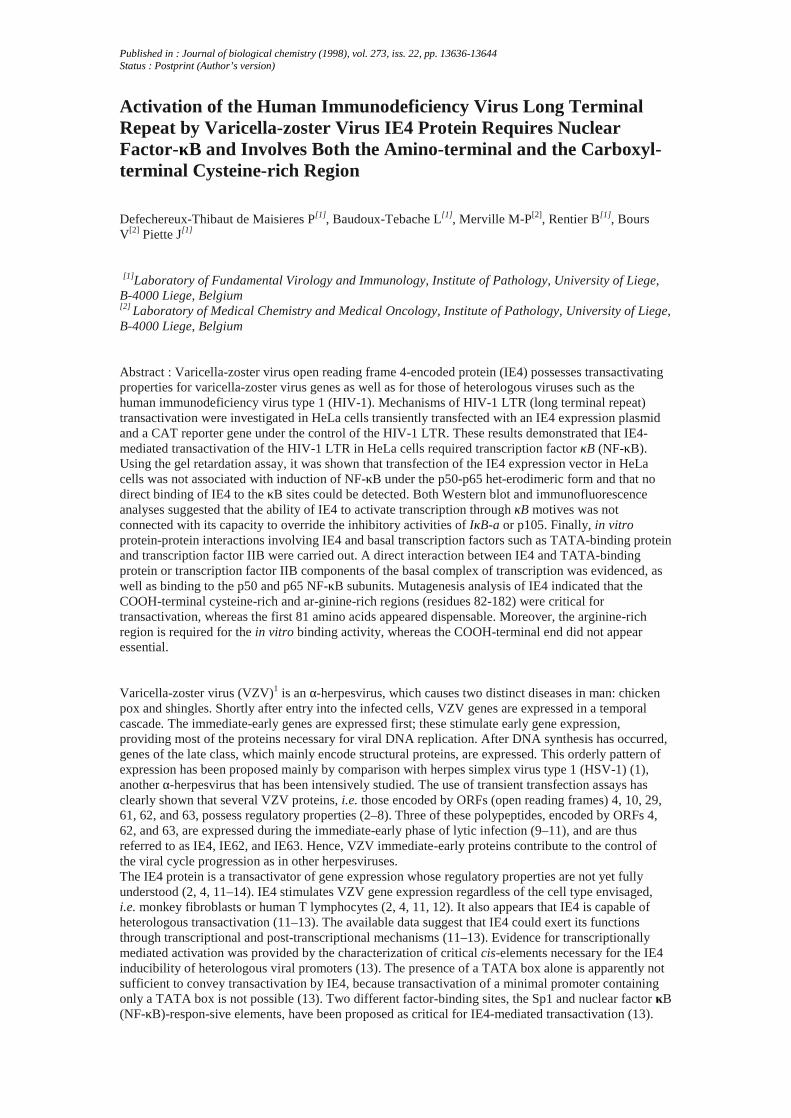
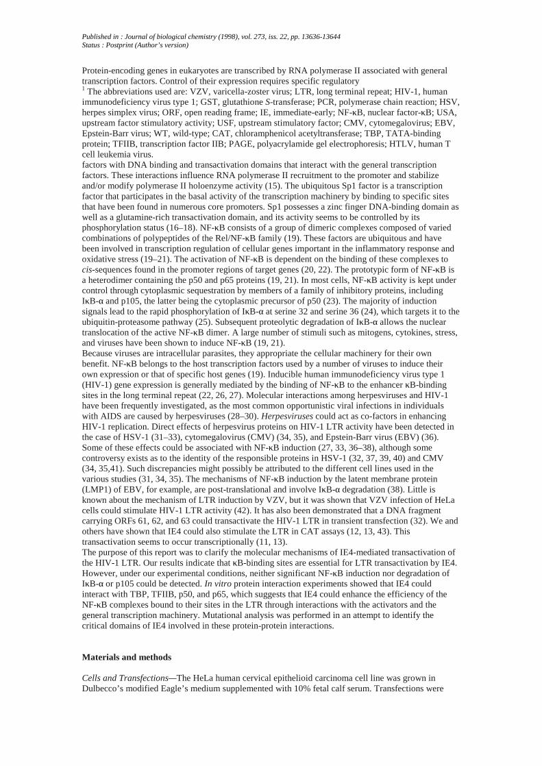
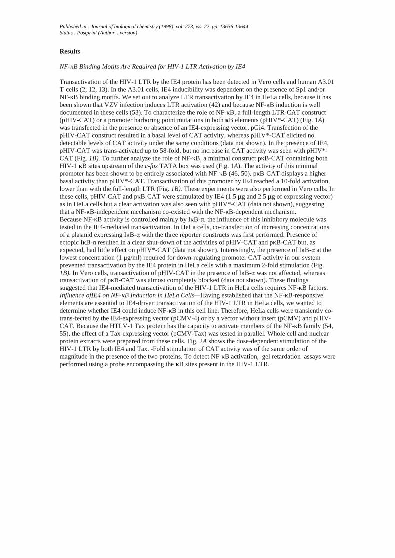
Transactivation of the HIV-1 LTR by the IE4 protein has been detected in Vero cells and human A3.01T-cells (2, 12, 13). In the A3.01 cells, IE4 inducibility was dependent on the presence of Sp1 and/orNF-κB binding motifs. We set out to analyze LTR transactivation by IE4 in HeLa cells, because it hasbeen shown that VZV infection induces LTR activation (42) and because NF-κB induction is welldocumented in these cells (53). To characterize the role of NF-κB, a full-length LTR-CAT construct(pHIV-CAT) or a promoter harboring point mutations in both κB elements (pHIV*-CAT) (Fig. 1A)was transfected in the presence or absence of an IE4-expressing vector, pGi4. Transfection of thepHIV-CAT construct resulted in a basal level of CAT activity, whereas pHIV*-CAT elicited nodetectable levels of CAT activity under the same conditions (data not shown). In the presence of IE4,pHIV-CAT was trans-activated up to 58-fold, but no increase in CAT activity was seen with pHIV*-CAT (Fig. 1B). To further analyze the role of NF-κB, a minimal construct pκB-CAT containing bothHIV-1 κB sites upstream of the c-fos TATA box was used (Fig. 1A). The activity of this minimalpromoter has been shown to be entirely associated with NF-κB (46, 50). pκB-CAT displays a higherbasal activity than pHIV*-CAT. Transactivation of this promoter by IE4 reached a 10-fold activation,lower than with the full-length LTR (Fig. 1B). These experiments were also performed in Vero cells. Inthese cells, pHIV-CAT and pκB-CAT were stimulated by IE4 (1.5 μg and 2.5 μg of expressing vector)as in HeLa cells but a clear activation was also seen with pHIV*-CAT (data not shown), suggestingthat a NF-κB-independent mechanism co-existed with the NF-κB-dependent mechanism.Because NF-κB activity is controlled mainly by IκB-α, the influence of this inhibitory molecule wastested in the IE4-mediated transactivation. In HeLa cells, co-transfection of increasing concentrationsof a plasmid expressing IκB-α with the three reporter constructs was first performed. Presence ofectopic IκB-α resulted in a clear shut-down of the activities of pHIV-CAT and pκB-CAT but, asexpected, had little effect on pHIV*-CAT (data not shown). Interestingly, the presence of IκB-α at thelowest concentration (1 μg/ml) required for down-regulating promoter CAT activity in our systemprevented transactivation by the IE4 protein in HeLa cells with a maximum 2-fold stimulation (Fig.1B). In Vero cells, transactivation of pHIV-CAT in the presence of IκB-α was not affected, whereastransactivation of pκB-CAT was almost completely blocked (data not shown). These findingssuggested that IE4-mediated transactivation of the HIV-1 LTR in HeLa cells requires NF-κB factors.Influence ofIE4 on NF-κB Induction in HeLa Cells—Having established that the NF-κB-responsiveelements are essential to IE4-driven transactivation of the HIV-1 LTR in HeLa cells, we wanted todetermine whether IE4 could induce NF-κB in this cell line. Therefore, HeLa cells were transiently co-trans-fected by the IE4-expressing vector (pCMV-4) or by a vector without insert (pCMV) and pHIV-CAT. Because the HTLV-1 Tax protein has the capacity to activate members of the NF-κB family (54,55), the effect of a Tax-expressing vector (pCMV-Tax) was tested in parallel. Whole cell and nuclearprotein extracts were prepared from these cells. Fig. 2A shows the dose-dependent stimulation of theHIV-1 LTR by both IE4 and Tax. -Fold stimulation of CAT activity was of the same order ofmagnitude in the presence of the two proteins. To detect NF-κB activation, gel retardation assays wereperformed using a probe encompassing the κB sites present in the HIV-1 LTR.
Published in : Journal of biological chemistry (1998), vol. 273, iss. 22, pp. 13636-13644Status : Postprint (Author’s version)
FIG. 1. Stimulation of HIV-1 LTR by IE4 is dependent on NF-κB. A, Schematic representation of theCAT reporter plasmids. LTR sequences extending from -453 to +80 relative to the transcriptioninitiation site were cloned in both HIV-CAT and HIV*-CAT constructs; the point mutations introducedin the κB sites of pHIV*-CAT are indicated. The NF-κB enhancer and Sp1 elements are depicted aswell as the TAR element. The TATA box is the cross-hatched box, and the broken arrow locates theinitiation site. pκB-CAT contains the minimal κB promoter with the c-fos TATA box (stippled box) (57).B, HeLa cells were transfected by lipofection and CAT activity of reporter plasmids was determined 44h after transfection as described under “Materials and Methods.” Each reporter CAT construct (0.5μg) was co-transfected with 1.5 and 2.5 μg of IE4-expressing vector (pGi4). Fold stimulation of CATactivity was calculated relative to the basal level of each reporter plasmid in the presence of carrierDNA alone, arbitrarily set at 1. To test for inhibition of NF-κB activity, co-transfection with 1 μg ofIκB-α effector plasmid was performed. In this case, the stimulations by IE4 were also expressedrelative to the activity of each reporter plasmid in the presence of the pRSV-IκB-α vector alone, whichwas set at 1. Results from four independent experiments are presented with standard errors of themean.
Incubation of nuclear extracts from non-transfected cells (used as an uninduced control) with the κBprobe exhibited a very low level of the specific NF-κB band (Fig. 2B, lane 1). Transfection of pCMVor pCMV-4 had little effect on the amount of the NF-κB complex (Fig. 2B, lanes 2 and 3), whereas astrong induction was seen with pCMV-Tax (Fig. 2B, lane 4). The electrophoretic mobility of theseDNA-protein complexes was identical to that induced by phorbol myristate acetate, a powerful NF-κBinducer (19). These complexes specifically competed with an excess of unlabeled κB oligonucleotides,but an excess of mutant oligonucleotides, which are devoid of NF-κB binding activity, had no effect(data not shown). The immunological characterization of these complexes demonstrated the presenceof p50 and p65 and the absence of p52, c-Rel, and IE4 (data not shown). Therefore, IE4 does not bindto the κB sites. Under these experimental conditions, expression of IE4 is not associated with a nucleartranslocation of the p50-p65 NF-κB complex, whereas expression of Tax induces such a translocation.Having established the effect of IE4 and Tax on the nuclear translocation of NF-κB, we next
Published in : Journal of biological chemistry (1998), vol. 273, iss. 22, pp. 13636-13644Status : Postprint (Author’s version)
investigated the effect of these proteins on the stability of IκB-α, because the classical pathway of NF-κB activation proceeds through degradation of this cytoplasmic inhibitor. Nuclear and cytoplasmicextracts from the cells that were used in the gel retardation assay were used for Western blot analysis.No significant variations in intrinsic IκB-α levels were detected when IE4 was present, whereas a clearcytoplasmic diminution was seen with Tax (data not shown). No modification in the levels of p50, itscytoplasmic precursor p105, or p65 were detected after transfection of theIE4 vector (data not shown). In order to ensure that an effect of IE4 was not masked by IκB-αneosynthesis, we next compared the stabilities of IκB-α in transfected cells exposed to cyclohex-imideto prevent de novo protein synthesis. As shown in Fig. 2C, expression of Tax led to a significantreduction in the cytoplasmic levels of IκB-α relative to that observed in pCMV-transfected cells. Incontrast, expression of IE4 had apparently no effect on the IκB-α cytoplasmic levels. These resultsindicate that the requirement for NF-κB in the IE4-mediated transac-tivation of the HIV-1 LTR is notassociated with a nuclear translocation of NF-κB/Rel family members concomitant with a degradationof cytoplasmic IκB-α.
IE4 Has No Effect on the Cytoplasmic Retention of Ectopic p50 and p65 by the IκB-α and p105Inhibitors
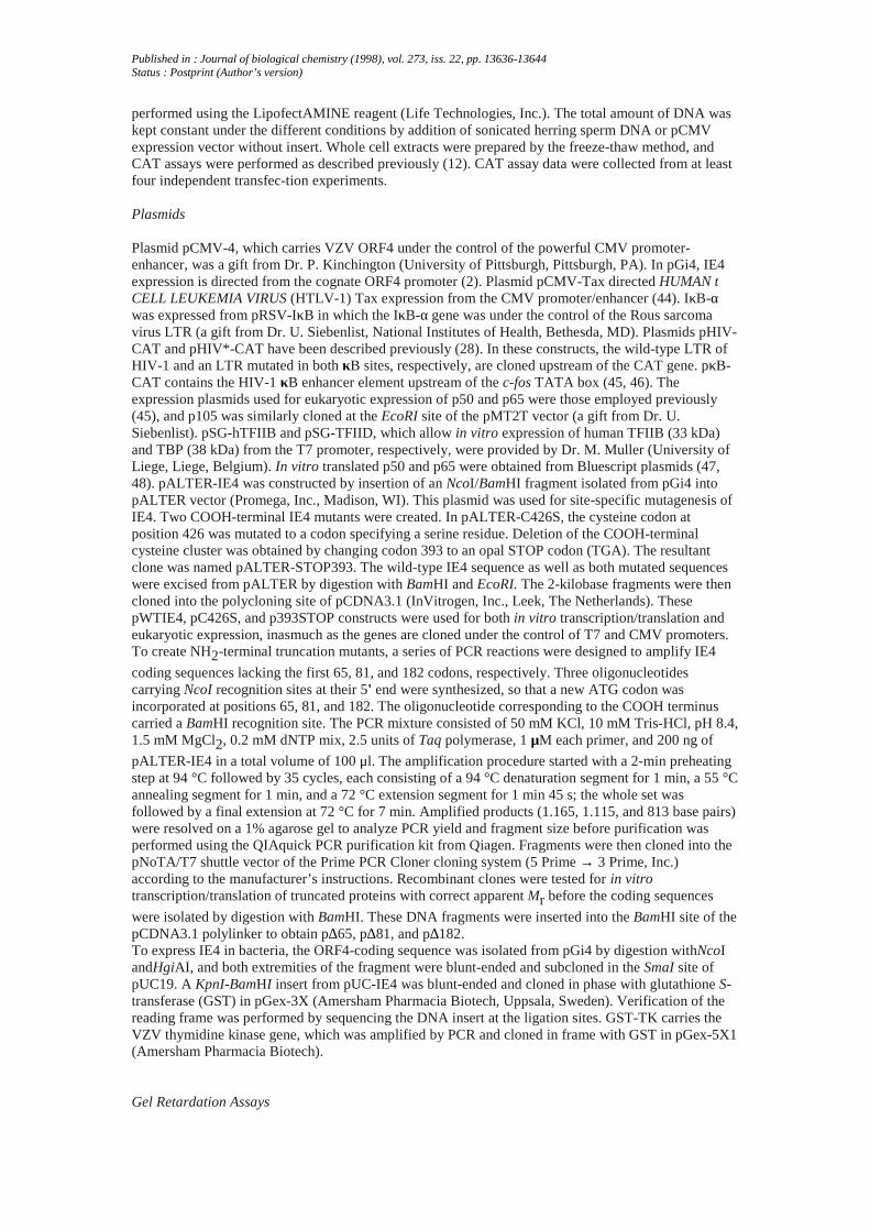
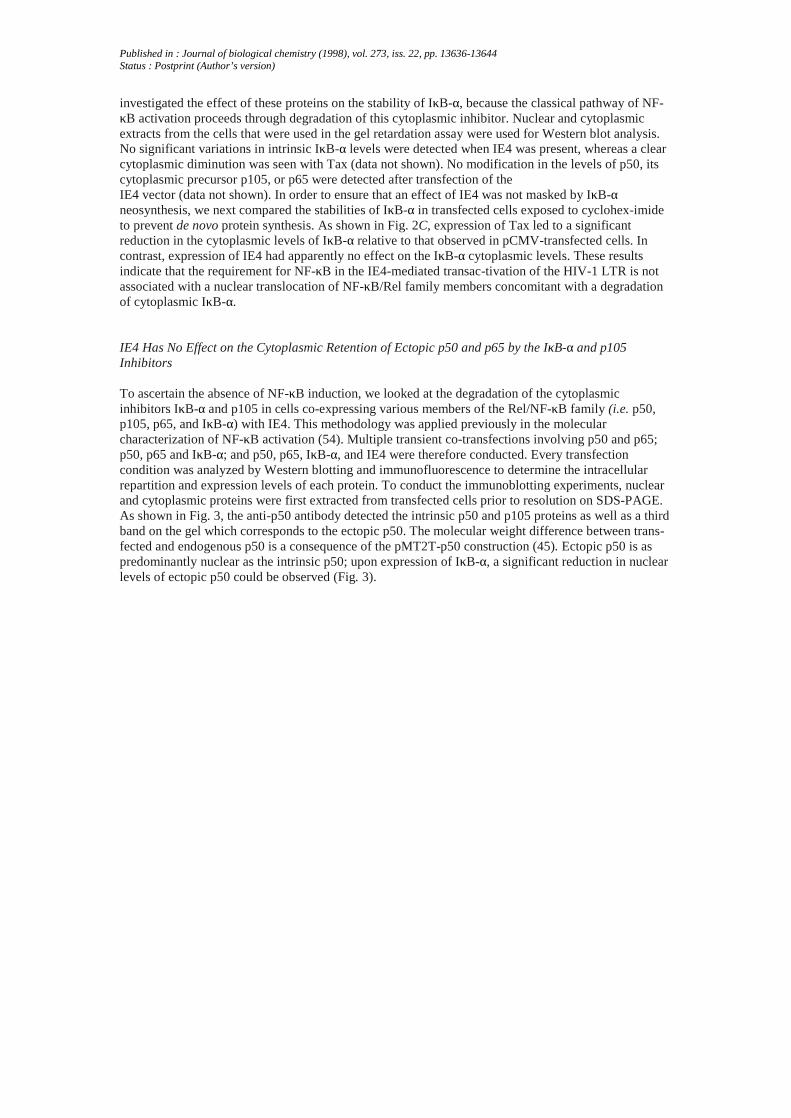
To ascertain the absence of NF-κB induction, we looked at the degradation of the cytoplasmicinhibitors IκB-α and p105 in cells co-expressing various members of the Rel/NF-κB family (i.e. p50,p105, p65, and IκB-α) with IE4. This methodology was applied previously in the molecularcharacterization of NF-κB activation (54). Multiple transient co-transfections involving p50 and p65;p50, p65 and IκB-α; and p50, p65, IκB-α, and IE4 were therefore conducted. Every transfectioncondition was analyzed by Western blotting and immunofluorescence to determine the intracellularrepartition and expression levels of each protein. To conduct the immunoblotting experiments, nuclearand cytoplasmic proteins were first extracted from transfected cells prior to resolution on SDS-PAGE.As shown in Fig. 3, the anti-p50 antibody detected the intrinsic p50 and p105 proteins as well as a thirdband on the gel which corresponds to the ectopic p50. The molecular weight difference between trans-fected and endogenous p50 is a consequence of the pMT2T-p50 construction (45). Ectopic p50 is aspredominantly nuclear as the intrinsic p50; upon expression of IκB-α, a significant reduction in nuclearlevels of ectopic p50 could be observed (Fig. 3).

Published in : Journal of biological chemistry (1998), vol. 273, iss. 22, pp. 13636-13644Status : Postprint (Author’s version)
FIG. 2. NF-κB binding activity in IE4-transfected cells. A, HeLa cells were transfected by lipofectionwith increasing concentrations of IE4 (pCMV-4)- and Tax (pCMV-Tax)-expressing vectors asindicated. Total input DNA was normalized by using the pCMV vector. -Fold stimulation of CATactivity was determined as described in the legend to Fig. 1 and presented as means of threeindependent experiments (± standard error of the mean). B, nuclear extracts were prepared from non-transfected cells and from cells transfected with 2.5 μg of pCMV (vector without insert), 2.5 μg ofpCMV-4 (expressing IE4), and 2.5 μg of pCMV-Tax harvested 24 h after transfection. These extracts
were then incubated with the 32P-labeled probe encompassing the κB elements of the HIV-1 LTR.DNA-protein complexes (specified by the filled arrow) were resolved on a 6% polyacrylamide gel. Theopen arrow refers to a nonspecific band. Seven independent experiments were performed to determineinduction of binding activity. C, IκB-α expression was monitored in cytoplasmic extracts prepared fromcells transfected with the pCMV vector (control), the pCMV-Tax vector, or the pCMV-4 vector. Forty-eight hours after transfection, cytoplasmic extracts were prepared after treatment with cycloheximide(CHX) for the indicated times and immunoblotted with an IκB-α-specific antiserum. The positions ofprestained molecular weight markers are indicated.
No clear effect of IE4 on these levels could be evidenced when the IE4-expressing vector was included.
Published in : Journal of biological chemistry (1998), vol. 273, iss. 22, pp. 13636-13644Status : Postprint (Author’s version)
FIG. 3. Influence of IE4 on ectopic IκB-a degradation in HeLa cells. p50, p65, and IκB-α expressionwas followed in cells co-trans-fected with the p50-, p65-, IκB-α-, and IE4-expressing vectors (1 μgeach). Twenty-four hours after transfection, nuclear and cytoplasmic extracts were fractionated andimmunoblotted with antisera specific for p50, p65, and IκB-α. Two arrows specify the p50 subunits, theupper arrow corresponding to the transfected p50 and the lower arrow indicating the endogenousprotein. Quantification of band intensities was performed by photodensitometry analysis of theautoradiograms.
Similarly, IE4 had no influence on the nuclear p65 levels in the presence of IκB-α (Fig. 3). Theseobservations were connected with the analysis of IκB-α levels. As expected, ectopic IκB-α waspredominantly cytoplasmic (Fig. 3). Presence of IE4 in the transfected cells did not lead to amodification in either cytoplasmic or nuclear levels of IκB-α. The second pathway of investigation,which was to follow intracellular localization of the NF-κB/Rel proteins by immunofluorescence,confirmed that expression of IE4 had no significant effect on the intracellular repartition of p50 andp65 (data not shown).It has been demonstrated that the p105 precursor of the p50 subunit of NF-κB also exhibits IκB-likeactivity (54, 56). Because IE4 appears incapable of overriding cytoplasmic retention of p50 and p65 byIκB-α, we investigated whether it could exert its action on p105. No effect was detected on the intrinsicp105 levels (data not shown). Multiple cotransfections were performed with p105-, p65-, and IE4-expressing vectors followed by immunoblotting and immunofluorescence. Under these conditions, IE4was not capable of influencing p105 or p50 levels, or p65 intracellular repartition (data not shown).These data suggest that the ability of IE4 to activate transcription through κB motives is notcorroborated with a capacity to override the inhibitory activities of p105.
IE4 Interacts with Several Transcription Factors in Vitro—
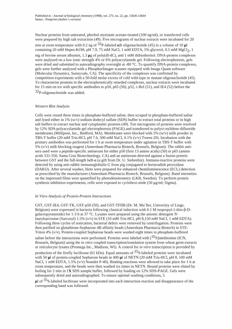
The results obtained from the gel shift assays and the Western blot analysis strongly suggest that theNF-κB-dependent activation of the LTR by IE4 must depend on other mechanisms than thetranslocation of NF-κB to the nucleus. Stimulation of transcription by activators results from multipledirect and indirect interactions between these activators, the co-activators and components of the basaltranscription complex (57). Thus, p65, which possesses an acidic transactivation domain, was shown tointeract with the general transcription factors TBP and TFIIB (58, 59) as well as with the p300 co-activator (60). Due to the lack of information concerning the molecular properties of IE4, its capacityto bind in vitro to TBP and TFIIB was investigated. A fusion protein between GST and IE4 wasexpressed in E. coli and purified on glutathione-Sepharose beads.
Published in : Journal of biological chemistry (1998), vol. 273, iss. 22, pp. 13636-13644Status : Postprint (Author’s version)
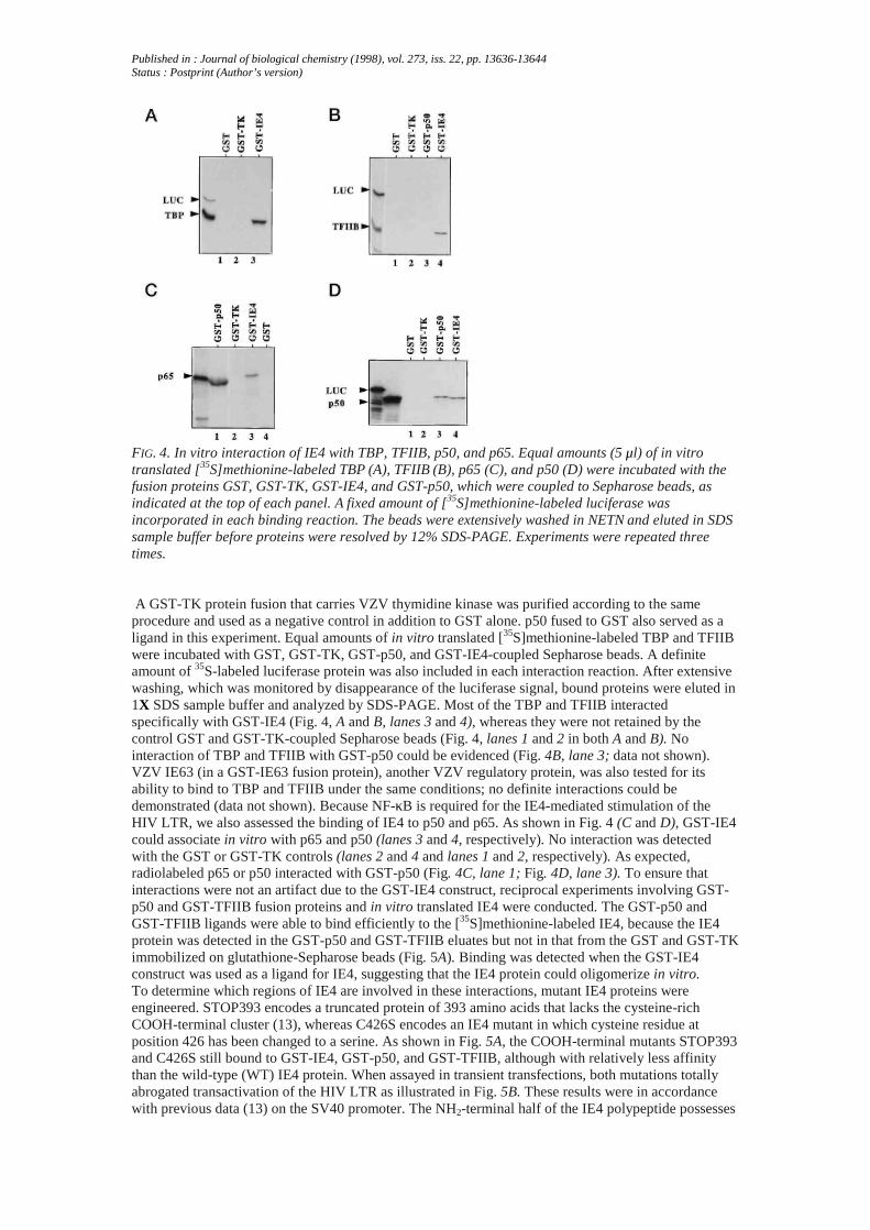
FIG. 4. In vitro interaction of IE4 with TBP, TFIIB, p50, and p65. Equal amounts (5 μl) of in vitrotranslated [35S]methionine-labeled TBP (A), TFIIB (B), p65 (C), and p50 (D) were incubated with thefusion proteins GST, GST-TK, GST-IE4, and GST-p50, which were coupled to Sepharose beads, asindicated at the top of each panel. A fixed amount of [35S]methionine-labeled luciferase wasincorporated in each binding reaction. The beads were extensively washed in NETN and eluted in SDSsample buffer before proteins were resolved by 12% SDS-PAGE. Experiments were repeated threetimes.
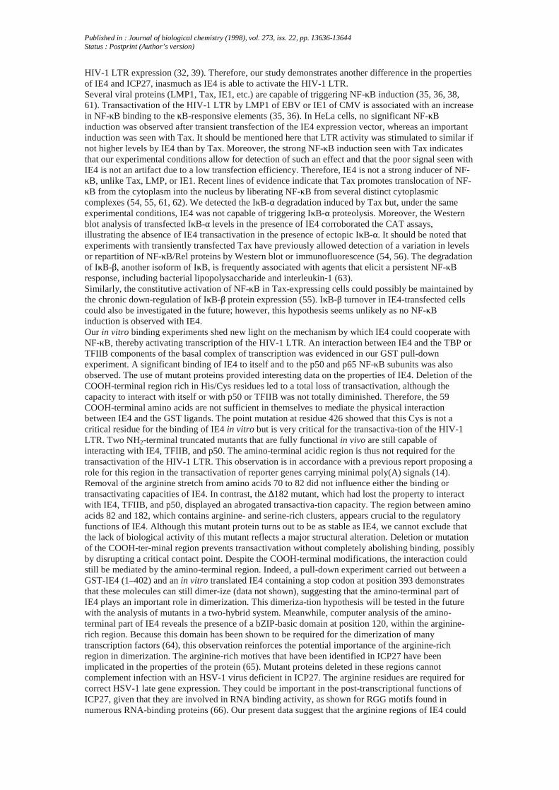
A GST-TK protein fusion that carries VZV thymidine kinase was purified according to the sameprocedure and used as a negative control in addition to GST alone. p50 fused to GST also served as aligand in this experiment. Equal amounts of in vitro translated [35S]methionine-labeled TBP and TFIIBwere incubated with GST, GST-TK, GST-p50, and GST-IE4-coupled Sepharose beads. A definiteamount of 35S-labeled luciferase protein was also included in each interaction reaction. After extensivewashing, which was monitored by disappearance of the luciferase signal, bound proteins were eluted in1X SDS sample buffer and analyzed by SDS-PAGE. Most of the TBP and TFIIB interactedspecifically with GST-IE4 (Fig. 4, A and B, lanes 3 and 4), whereas they were not retained by thecontrol GST and GST-TK-coupled Sepharose beads (Fig. 4, lanes 1 and 2 in both A and B). Nointeraction of TBP and TFIIB with GST-p50 could be evidenced (Fig. 4B, lane 3; data not shown).VZV IE63 (in a GST-IE63 fusion protein), another VZV regulatory protein, was also tested for itsability to bind to TBP and TFIIB under the same conditions; no definite interactions could bedemonstrated (data not shown). Because NF-κB is required for the IE4-mediated stimulation of theHIV LTR, we also assessed the binding of IE4 to p50 and p65. As shown in Fig. 4 (C and D), GST-IE4could associate in vitro with p65 and p50 (lanes 3 and 4, respectively). No interaction was detectedwith the GST or GST-TK controls (lanes 2 and 4 and lanes 1 and 2, respectively). As expected,radiolabeled p65 or p50 interacted with GST-p50 (Fig. 4C, lane 1; Fig. 4D, lane 3). To ensure thatinteractions were not an artifact due to the GST-IE4 construct, reciprocal experiments involving GST-p50 and GST-TFIIB fusion proteins and in vitro translated IE4 were conducted. The GST-p50 andGST-TFIIB ligands were able to bind efficiently to the [35S]methionine-labeled IE4, because the IE4protein was detected in the GST-p50 and GST-TFIIB eluates but not in that from the GST and GST-TKimmobilized on glutathione-Sepharose beads (Fig. 5A). Binding was detected when the GST-IE4construct was used as a ligand for IE4, suggesting that the IE4 protein could oligomerize in vitro.To determine which regions of IE4 are involved in these interactions, mutant IE4 proteins wereengineered. STOP393 encodes a truncated protein of 393 amino acids that lacks the cysteine-richCOOH-terminal cluster (13), whereas C426S encodes an IE4 mutant in which cysteine residue atposition 426 has been changed to a serine. As shown in Fig. 5A, the COOH-terminal mutants STOP393and C426S still bound to GST-IE4, GST-p50, and GST-TFIIB, although with relatively less affinitythan the wild-type (WT) IE4 protein. When assayed in transient transfections, both mutations totallyabrogated transactivation of the HIV LTR as illustrated in Fig. 5B. These results were in accordancewith previous data (13) on the SV40 promoter. The NH2-terminal half of the IE4 polypeptide possesses
Published in : Journal of biological chemistry (1998), vol. 273, iss. 22, pp. 13636-13644Status : Postprint (Author’s version)
an acidic-rich region in the first 65 amino acids as well as arginine-rich stretches interspersed fromamino acids 67 to 182. These regions could potentially be important in the properties of IE4. Therefore,three truncation mutants were tested in the GST-chromatography binding assay. In vitro translated ∆65and ∆81, which lack the acidic region and both the acidic region and an arginine-rich run respectively,bound to GST-IE4, GST-p50, and GST-TFIIB with affinity similar to WT IE4, although signals areweaker. In contrast, an IE4 mutant protein which lacks the entire arginine-rich region, ∆182, failed tointeract with all GST ligands (Fig. 5A). Very interestingly, when their functional properties werestudied in vivo, both ∆65 and ∆81 mutants elicited unmodified transactivating properties on the HIVLTR, whereas ∆182 failed completely to stimulate the HIV-CAT target (Fig. 5B). The arginine-richregion appeared critical for the activation of the HIV-1 LTR, whereas it could not be excluded that thislack of transactivation reflected a major structural change of IE4 induced by the truncation of the first182 amino acids. These data indicate that IE4 was capable of interacting in vitro with itself, TBP,TFIIB, p50, and p65. These interactions were likely dependent on an argi-nine-rich region present inthe NH2-terminal half of the molecule. The COOH-terminal end and the cysteine at position 426 couldalso be important for the binding activity, as the interaction was diminished when the COOH-terminalend was absent or mutated.
Discussion
The molecular mechanisms underlying VZV IE4 regulatory properties are still greatly misunderstood,although it now seems clear that IE4 is a strong transactivator whether expressed alone or in synergywith VZV IE62 (2, 4, 5, 12, 43). In the past, interesting evidence has been presented concerning theidentification of specific upstream elements involved in IE4 inducibility (13). Moreover, the samestudy established that IE4 could act transcriptionally in A3.01 cells using the HIV-1 LTR as a modelsystem for investigation. Our previous work based on mRNA steady-state level analysis had led us toconclude that IE4 could fulfill its functions through transcriptional and post-transcriptional mechanisms(11, 12). Because the transcriptional effect was also studied using the HIV-1 LTR as experimentalsystem, the present work was focused on the pathway of LTR activation.The first main observation from the transfection assays is that the κB-responsive elements are essentialto IE4-mediated activation in HeLa cells, inasmuch as an LTR mutated at both κB sites is refractory tothe action of IE4. Interestingly, this mutant construct was still transactivated in Vero cells, indicatingthat the other cis-acting sequences present in the LTR contribute to IE4 inducibility in these cells. Co-expression of the cytoplasmic inhibitor IκB-α prevented transactivation of the HIV LTR by IE4, whichclearly confirmed that NF-κB is required in HeLa cells.
Published in : Journal of biological chemistry (1998), vol. 273, iss. 22, pp. 13636-13644Status : Postprint (Author’s version)
FIG. 5. Role of the COOH-terminal and NH2-terminal regions of IE4 in its regulatory functions and its
interaction with transcription factor IIB and p50. A, in vitro protein-protein binding by IE4 mutants.
Equal amounts of in vitro translated [35S]methionine-labeled wild-type IE4, STOP393, C426S, and∆65 were incubated with bacterially expressed GST, GST-TK, GST-IE4, GST-TFIIB, and GST-p50,
which were coupled to Sepharose beads, as indicated at the top of each panel. [35S]Methionine-labeled ∆81 and ∆182 were added in larger amounts because the proteins were labeled in vitro with
less efficiency and signals were weaker. A fixed amount of [35S]methionine-labeled luciferase was alsoincorporated in each binding reaction. The beads were extensively washed in NETN and eluted in SDSsample buffer before proteins were resolved by 12% SDS-PAGE. Experiments were repeated threetimes independently. B, stimulation of the HIV-1 LTR by IE4 requires both cysteine-rich COOH-terminal and arginine-rich regions. HeLa cells were co-transfected by lipofection with 0.25 μg ofpHIV-CAT and 0.5 μg of IE4 expression vectors (WTIE4 or mutant proteins) as indicated. CAT activitywas determined 44 h after transfection. -Fold stimulation of CAT activity was calculated relative to thebasal level of reporter plasmid in presence of control plasmid alone, arbitrarily set at 1. The resultsshown are representative of three independent experiments, and error bars represent standard errorvalues. C, schematic representation of wild-type IE4 and mutant proteins. The wild-type IE4 contains ahighly acidic amino-terminal region (dark stippled area), an arginine-rich region (stippled area), anda cysteine-rich carboxyl-terminal cluster (hatched area). The positions are denoted under the bar. Themutation STOP393 is an insertion of a STOP codon at position 393, which leads to the expression of atruncated protein lacking the cysteine-rich cluster. The mutation C426S is the substitution of a serineresidue for a cysteine residue at position 426. The mutation ∆65 is a deletion of amino acids 1-65removing the acidic region. The mutation ∆81 is a deletion of residues 66-81 removing an arginine-rich stretch. The mutation ∆182 is a deletion of residues 82-182 completely removing the arginine-richregion.
Despite some controversy as to the nature of the HSV-1 IE proteins implicated in the transactiva-tionof the LTR by HSV-1, it is currently thought that ICP27, the IE4 homolog in HSV-1, has no effect on
Published in : Journal of biological chemistry (1998), vol. 273, iss. 22, pp. 13636-13644Status : Postprint (Author’s version)
HIV-1 LTR expression (32, 39). Therefore, our study demonstrates another difference in the propertiesof IE4 and ICP27, inasmuch as IE4 is able to activate the HIV-1 LTR.Several viral proteins (LMP1, Tax, IE1, etc.) are capable of triggering NF-κB induction (35, 36, 38,61). Transactivation of the HIV-1 LTR by LMP1 of EBV or IE1 of CMV is associated with an increasein NF-κB binding to the κB-responsive elements (35, 36). In HeLa cells, no significant NF-κBinduction was observed after transient transfection of the IE4 expression vector, whereas an importantinduction was seen with Tax. It should be mentioned here that LTR activity was stimulated to similar ifnot higher levels by IE4 than by Tax. Moreover, the strong NF-κB induction seen with Tax indicatesthat our experimental conditions allow for detection of such an effect and that the poor signal seen withIE4 is not an artifact due to a low transfection efficiency. Therefore, IE4 is not a strong inducer of NF-κB, unlike Tax, LMP, or IE1. Recent lines of evidence indicate that Tax promotes translocation of NF-κB from the cytoplasm into the nucleus by liberating NF-κB from several distinct cytoplasmiccomplexes (54, 55, 61, 62). We detected the IκB-α degradation induced by Tax but, under the sameexperimental conditions, IE4 was not capable of triggering IκB-α proteolysis. Moreover, the Westernblot analysis of transfected IκB-α levels in the presence of IE4 corroborated the CAT assays,illustrating the absence of IE4 transactivation in the presence of ectopic IκB-α. It should be noted thatexperiments with transiently transfected Tax have previously allowed detection of a variation in levelsor repartition of NF-κB/Rel proteins by Western blot or immunofluorescence (54, 56). The degradationof IκB-β, another isoform of IκB, is frequently associated with agents that elicit a persistent NF-κBresponse, including bacterial lipopolysaccharide and interleukin-1 (63).Similarly, the constitutive activation of NF-κB in Tax-expressing cells could possibly be maintained bythe chronic down-regulation of IκB-β protein expression (55). IκB-β turnover in IE4-transfected cellscould also be investigated in the future; however, this hypothesis seems unlikely as no NF-κBinduction is observed with IE4.Our in vitro binding experiments shed new light on the mechanism by which IE4 could cooperate withNF-κB, thereby activating transcription of the HIV-1 LTR. An interaction between IE4 and the TBP orTFIIB components of the basal complex of transcription was evidenced in our GST pull-downexperiment. A significant binding of IE4 to itself and to the p50 and p65 NF-κB subunits was alsoobserved. The use of mutant proteins provided interesting data on the properties of IE4. Deletion of theCOOH-terminal region rich in His/Cys residues led to a total loss of transactivation, although thecapacity to interact with itself or with p50 or TFIIB was not totally diminished. Therefore, the 59COOH-terminal amino acids are not sufficient in themselves to mediate the physical interactionbetween IE4 and the GST ligands. The point mutation at residue 426 showed that this Cys is not acritical residue for the binding of IE4 in vitro but is very critical for the transactiva-tion of the HIV-1LTR. Two NH2-terminal truncated mutants that are fully functional in vivo are still capable ofinteracting with IE4, TFIIB, and p50. The amino-terminal acidic region is thus not required for thetransactivation of the HIV-1 LTR. This observation is in accordance with a previous report proposing arole for this region in the transactivation of reporter genes carrying minimal poly(A) signals (14).Removal of the arginine stretch from amino acids 70 to 82 did not influence either the binding ortransactivating capacities of IE4. In contrast, the ∆182 mutant, which had lost the property to interactwith IE4, TFIIB, and p50, displayed an abrogated transactiva-tion capacity. The region between aminoacids 82 and 182, which contains arginine- and serine-rich clusters, appears crucial to the regulatoryfunctions of IE4. Although this mutant protein turns out to be as stable as IE4, we cannot exclude thatthe lack of biological activity of this mutant reflects a major structural alteration. Deletion or mutationof the COOH-ter-minal region prevents transactivation without completely abolishing binding, possiblyby disrupting a critical contact point. Despite the COOH-terminal modifications, the interaction couldstill be mediated by the amino-terminal region. Indeed, a pull-down experiment carried out between aGST-IE4 (1–402) and an in vitro translated IE4 containing a stop codon at position 393 demonstratesthat these molecules can still dimer-ize (data not shown), suggesting that the amino-terminal part ofIE4 plays an important role in dimerization. This dimeriza-tion hypothesis will be tested in the futurewith the analysis of mutants in a two-hybrid system. Meanwhile, computer analysis of the amino-terminal part of IE4 reveals the presence of a bZIP-basic domain at position 120, within the arginine-rich region. Because this domain has been shown to be required for the dimerization of manytranscription factors (64), this observation reinforces the potential importance of the arginine-richregion in dimerization. The arginine-rich motives that have been identified in ICP27 have beenimplicated in the properties of the protein (65). Mutant proteins deleted in these regions cannotcomplement infection with an HSV-1 virus deficient in ICP27. The arginine residues are required forcorrect HSV-1 late gene expression. They could be important in the post-transcriptional functions ofICP27, given that they are involved in RNA binding activity, as shown for RGG motifs found innumerous RNA-binding proteins (66). Our present data suggest that the arginine regions of IE4 could
Published in : Journal of biological chemistry (1998), vol. 273, iss. 22, pp. 13636-13644Status : Postprint (Author’s version)
be involved in physical protein-protein interactions. Deleting this region either removes a criticaldomain or disrupts the overall structure of the protein in such a way that binding is prevented.Experiments are currently being conducted to investigate the role of these regions in the transcriptionaland post-transcriptional properties of IE4.It has been shown that a GAL4-IE4 chimera transactivates a promoter carrying GAL4-binding sites,suggesting that once IE4 is brought to the vicinity of the promoter it can activate transcription (13). Theinteractions between IE4 and p50-p65 on the one hand and between IE4 and TBP and TFIIB on theother suggest that IE4 could be targeted to the vicinity of the basal transcription complex throughprotein-protein interactions with DNA-binding proteins tethered to their sites and could thereforestabilize or enhance processivity of the multim-eric transcription complex. This activity is reminiscentof a co-activator, which stimulates activator-dependent transcription (67), and of Bcl-3 activity, whichcould tightly associate to p52 homodimers to transactivate through the κB motif (51). p65 also binds toTBP and TFIIB, which contact next to each other the promoter site (58, 59). It could therefore bepossible that the redundant binding of IE4 to both general transcription factors and NF-κB increasesinitiation of transcription. There is accumulating evidence that interactions with multiple transcriptionfactors, activators, and co-activators exist for viral transactivators such as Tax of HTLV-1 or E1A ofadenovirus. E1A has been shown to bind to TBP and p65 to promote LTR transactivation. E1A is alsocapable of binding to p300, a co-activator recruited to a number of unrelated promoters by protein-protein interactions, although this interaction is not involved in LTR transactivation (68). Recently,Tsukada et al. (69) have established that Tax stimulates human pro-interleu-kin-1β promoter by directinteractions with the transcription factors NF-IL6 and Sp-1. It is therefore possible that IE4 represents acritical co-activator used by VZV to divert cellular transcription factors and enhance viral-specific geneexpression. If so, IE4 could act as an adaptator/bridging factor for the VZV IE62 protein. It isinteresting that a co-activating fraction termed USA (upstream factor stimulatory activity) purifiedfrom HeLa cells is required for NF-κB activation of the HIV-1 LTR in cell-free transcription systems(70). A PC1 co-factor originating from this USA fraction was shown to enhance p65-dependentactivation in this cell-free reconstruction of transcription (59). This USA fraction is also required foractivation by USF (upstream stimulatory factor), which is a helix-loop-helix regulatory factor (70).Now, this USF cooperates with IE62 to activate expression of VZV genes 28 and 29 (71). We knowthat IE4 and IE62 act synergistically to stimulate expression of genes unresponsive to IE4 alone (2, 4,12, 43), whereas IE62 induces nuclear translocation of IE4 in transfected cells (52). This suggests verystrongly that the increase in IE62-mediated transactivation by IE4 could potentially involve acollaboration of IE4 with this USA and USF/IE62, which might be involved in the LTR transactivation.Various experiments are currently under way in our laboratory to further define the interplay betweenIE4, IE62, activators, co-activators, and transcription factors both in vitro and in vivo. Direct effects ofIE4 on transcription will be envisaged using in vitro transcription assays. These should help toelucidate the molecular mechanisms implicated in transcriptional activation by the multifunctional IE4.
Acknowledgments
We thank P. Kinchington, J. M. Ostrove, and M. Mu¨ller for providing plasmids. We are very gratefulto U. Siebenlist for the different plasmids and antibodies. We extend our gratitude to E. Janssens fortechnical assistance.
References
[1]. Honess, R. W., and Roizman, B. (1974) J. Virol. 14, 8–19[2]. Inchauspe, G., Nagpal, S., and Ostrove, J. M. (1989) Virology 173, 700–709[3]. Nagpal, S., and Ostrove, J. M. (1991) J. Virol. 65, 5289 –5296[4]. Perera, L. P., Mosca, J. D., Ruyechan, W. T., and Hay, J. (1992) J. Virol. 66, 5298–5304[5]. Moriuchi, H., Moriuchi, M., Straus, S. E., and Cohen, J. I. (1993) J. Virol. 67, 2739–2746[6]. Moriuchi, H., Moriuchi, M., Straus, S. E., and Cohen, J. I. (1993) J. Virol. 67, 4290–4295[7]. Baudoux, L., Defechereux, P., Schoonbroodt, S., Merville, M.-P., Rentier, B., and Piette, J. (1995)Nucleic Acids Res. 23, 1341–1349[8]. Hay, I., and Ruyechan, W. T. (1994) Semin. Virol. 5, 241–248[9]. Forghani, B., Mahalingham, R., Vafai, A., Hurst, J. W., and Dupuis, K. W. (1990) Virus Res. 16,195–210[10]. Debrus, S., Sadzot-Delvaux, C., Nikkels, A. F., Piette, J., and Rentier, B. (1995) J. Virol. 69,
Published in : Journal of biological chemistry (1998), vol. 273, iss. 22, pp. 13636-13644Status : Postprint (Author’s version)
3240–3245[11]. Defechereux, P., Debrus, S., Baudoux, L., Rentier, B., and Piette, J. (1997) J. Virol. 71, 7073–7079[12]. Defechereux, P., Melen, L., Baudoux, L., Merville-Louis, M.-P., Rentier, B., and Piette, J. (1993)J. Virol. 67, 4379–4385[13]. Perera, L. P., Kaushal, S., Kinchington, P. R., Mosca, J. D., Hayward, G. S., and Straus, S. E.(1994) J. Virol. 68, 2468–2477[14]. Moriuchi, H., Moriuchi, M., Debrus, S., Piette, J., and Cohen, J. I. (1995) Virology 207, 376–382[15]. Connaway, R. C., and Connaway, J. W. (1993) Annu. Rev. Biochem. 62, 161–190[16]. Courey, A. J., and Tijan, R. (1988) Cell 55, 887–898[17]. Briggs, M. R., Kadonaga, J. T., Bell, S. P., and Tijan, R. (1986) Science 234,47–52[18]. Pascal, E., and Tijan, R. (1991) Genes Dev. 5, 155–165[19]. Siebenlist, U., Franzoso, G., and Brown, K. (1994) Annu. Rev. Cell. Biol. 10, 405–455[20]. Sen, R., and Baltimore, D. (1986) Cell 46, 705–716[21]. Baeuerle, P. A., and Henkel, T. (1994) Annu. Rev. Immunol. 12, 141–179[22]. Nabel, G., and Baltimore, D. (1987) Nature 326, 711–713[23]. Beg, A. A., Ruben, S. M., Scheinman, R. I., Haskill, S., Rosen, C. A., and Baldwin, A. S., Jr.(1992) Genes Dev. 6, 1899–1913[24]. Brown, K., Gerstberger, S., Carlson, L., Franzoso, G., and Siebenlist, U. (1995) Science 267,1485–1488[25]. Chen, Z., Hagler, J., Palombella, V. J., Melandri, F., Scherer, D., Ballard, D., and Maniatis, T.(1995) Genes Dev. 9, 1586–1597[26]. Gaynor, R. (1992) AIDS 6, 347–363[27]. Gimble, J. M., Duh, E., Ostrove, J. M., Gendelman, H. E., Max, E. E., and Rabson, A. B. (1988)J. Virol. 62, 4104–4112[28]. Gendelman, H. E., Phelps, W., Feigenbaum, L., Ostrove, J. M., Adachi, A., Howley, P. M.,Koury, J., Ginsberg, H. S., and Martin, M. A. (1986) Proc. Natl. Acad. Sci. U. S. A. 83, 9759–9763[29]. Laurence, J. (1990) J. Infect. Dis. 162, 338–346[30]. Moore, P. S., and Chang, Y. (1995) New Engl. J. Med. 332, 1181–1185[31]. Mosca, J. D., Bednarik, D. P., Raj, N. B. K., Rosen, C. A., Sodroski, J. G.,Haseltine, W. A., Hayward, G. S., and Pitha, P. M. (1987) Proc. Natl. Acad. Sci. U. S. A. 84, 7408–7412[32]. Ostrove, J. M., Leonard, J., Weck, K. E., Rabson, A. B., Gendelman, H. E. (1987) J. Virol. 61,3726–3732[33]. Margolis, D. M., Rabson, A. R., Straus, S. E., and Ostrove, J. M. (1992) Virology 186, 788–791[34]. Ghazal, P., Young, J., Giulietti, E., De Mattei, C., Garcia, J., Gaynor, R., Steinerg, R., andNelson, J. A. (1991) J. Virol. 65, 6735–6742[35]. Kim, S., Yu, S. S., and Kim, V. N. (1996) J. Gen. Virol. 77, 83–91[36]. Hammarskjold, M.-L., and Simurda, M. C. (1992) J. Virol. 66, 6496–6501[37]. Vlach, J., and Pitha, P. M. (1992) J. Virol. 66, 3616 –3623[38]. Paine, E., Scheimann, R. I., Baldwin, A. S., Jr., and Raab-Traub, N. (1995) J. Virol. 69, 4572–4576[39]. Albrecht, M. A., DeLuca, N. A., Byrn, R. A., Schaffer, P. A., and Hammer, S. M.(1989) J. Virol. 63, 1861–1868[40]. Schafer, S. L., Vlach, J., and Pitha, P. M. (1996) J. Virol. 70, 6937–6946[41]. Markovitz, D. M., Kenney, S., Kamine, J., Smith, M. S., Davis, M., Huang,E.-S., Rosen, C., and Pagano, J. (1989) Virology 173, 750–754[42]. Rando, R. F., Srinivasan, A., Feingold, J., Gonczol, E., and Plotkin, S. (1987) Oncogene 1, 13–18[43]. Inchaupse, G., and Ostrove, J. M. (1989) Virology 173, 710–714[44]. Rimsky, L., Hauber, J., Dukovich, M., Malim, M. H., Langlois, A., Cullen, B. R., and Greene, W.C. (1988) Nature 335, 738–740[45]. Bours, V., Burd, R. P., Brown, K., Villalobos, J., Park, S., Rysek, R. P., Bravo, R., Kelly, K., andSiebenlist, U. (1992) Mol. Cell. Biol. 12, 685–695[46]. Pierce, J. W., Lenardo, M., and Baltimore, D. (1988) Proc. Natl. Acad. Sci. U. S. A. 85, 1482–1486[47]. Bours, V., Villalobos, J., Burd, P. R., Kelly, K., and Siebenlist, U. (1990) Nature 348, 76–80[48]. Ruben, S. M., Dillon, P. J., Schreck, R., Henkel, T., Chen, C. M., Moher, M., Baeuerle,, P. A.,and Rosen, C. A. (1991) Science 251, 1490–1493[49]. Andrews, N. C., and Fawler, D. V. (1991) Nucleic Acids Res. 19, 2499[50]. Franzoso, G., Bours, V., Park, S., Tomita-Yamagushi, M., Kanno, T., Brown, K., and Siebenlist,
Published in : Journal of biological chemistry (1998), vol. 273, iss. 22, pp. 13636-13644Status : Postprint (Author’s version)
U. (1992) Nature 359, 207–210[51]. Bours, V., Franzoso, G., Azarenko, V., Park, S., Kanno, T., Brown, K., and Siebenlist, U. (1993)Cell 72, 729–739[52]. Defechereux, P., Debrus, S., Baudoux, L., Schoonbroodt, S., Merville-Louis, M.-P., Rentier, B.,and Piette, J. (1996) J. Gen. Virol. 77, 1505–1513[53]. Kretzschmar, M., Meisterernst, M., Scheidereit, C., Li, G., and Roeder, R. G. (1992) Genes Dev.6, 761–774[54]. Munoz, E., Courtois, G., Veschambre, P., Jalinot, P., and Israël, A. (1994) J. Virol. 68, 8035–8044[55]. McKinsey, T. A., Brockman, J. A., Scherer, D. C., Al-Murrani, S. W., Green, P. L., and Ballard,D. W. (1996) Mol. Cell. Biol. 16, 2083–2090[56]. Rice, N. R., Mackichan, M. L., and Israël, A. (1992) Cell 71, 243–253[57]. Janknet, R., and Hunter, T. (1996) Curr. Biol. 6, 951–954[58]. Kerr, L. D., Ransone, L. J., Wamsley, P., Schmitt, M. J., Boyer, T. G., Zhou, Q., Berk, A. J., andVerma, I. M. (1993) Nature 365, 412–419[59]. Schmitz, M. L., Stelzer, G., Altmann, H., Meisterernst, M., and Baeuerle, P. (1995) J. Biol. Chem.270, 7219–7226[60]. Perkins, N. D., Felzien, L. K., Betts, J. C., Leung, K., Beach, D. H., and Nabel, G. J. (1997)Science 275, 523–527[61]. Kanno, T., Brown, K., Franzoso, G., and Siebenlist, U. (1994) Mol. Cell. Biol. 14, 6443- 6451[62]. Kanno, T., Brown, K., and Siebenlist, U. (1995) J. Biol. Chem. 270, 11745–11748[63]. Thomson, J. E., Phillips, R. J., Erdjument-Bromage, H., Tempst, P., and Ghosh, S. (1995) Cell 80,573–582[64]. Ellenberger, T. (1994) Curr. Opin. Struct. Biol. 4, 12–21[65]. Hibbard, M. K., and Sandri-Goldin, R. M. (1995) J. Virol. 69, 4656–4667[66]. Mears, W. E., and Rice, S. A. (1996) J. Virol. 70, 7445–7453[67]. Pugh, B. F., and Tijan, R. (1990) Cell 61, 11187–1197[68]. Parker, S. F., Felzien, L. K., Perkins, N. D., Imperiale, M. J., and Nabel, G. J.(1997) J. Virol. 71, 2004–2012[69]. Tsukada, J., Misago, M., Serino, Y., Ogawa, R., Murakami, S., Nakanishi, M., Tonai, S.,Kominato, Y., Morimoto, I., Auron, P. E., and Eto, S. (1997) Blood 8, 3142–3153[70]. Meisterernst, M., Roy, A. L., Lieu, H. M., and Roeder, R. G. (1991) Cell 66, 981–993[71]. Meier, J. L., Luo, X., Sawadogo, M., and Straus, S. E. (1994) Mol. Cell. Biol. 14, 6896- 6906