ABIN-1 Negatively Regulates μ-Opioid Receptor...
13
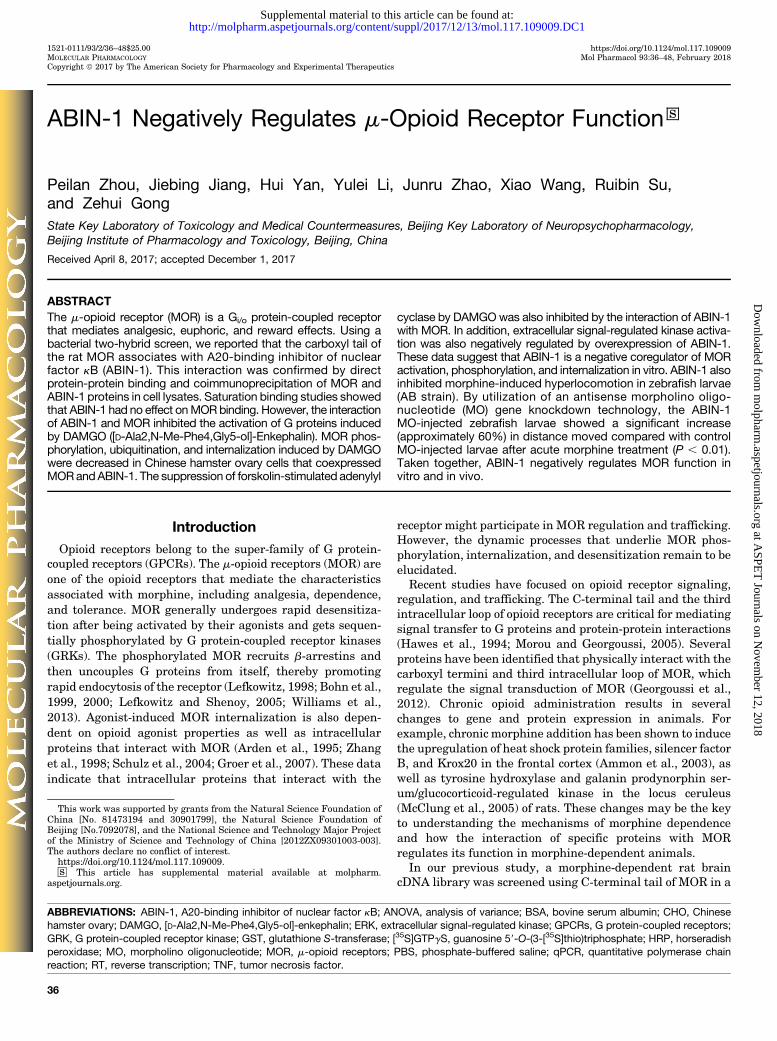
1521-0111/93/2/36–48$25.00 https://doi.org/10.1124/mol.117.109009 MOLECULAR PHARMACOLOGY Mol Pharmacol 93:36–48, February 2018 Copyright ª 2017 by The American Society for Pharmacology and Experimental Therapeutics ABIN-1 Negatively Regulates m-Opioid Receptor Function s Peilan Zhou, Jiebing Jiang, Hui Yan, Yulei Li, Junru Zhao, Xiao Wang, Ruibin Su, and Zehui Gong State Key Laboratory of Toxicology and Medical Countermeasures, Beijing Key Laboratory of Neuropsychopharmacology, Beijing Institute of Pharmacology and Toxicology, Beijing, China Received April 8, 2017; accepted December 1, 2017 ABSTRACT The m-opioid receptor (MOR) is a G i/o protein-coupled receptor that mediates analgesic, euphoric, and reward effects. Using a bacterial two-hybrid screen, we reported that the carboxyl tail of the rat MOR associates with A20-binding inhibitor of nuclear factor kB (ABIN-1). This interaction was confirmed by direct protein-protein binding and coimmunoprecipitation of MOR and ABIN-1 proteins in cell lysates. Saturation binding studies showed that ABIN-1 had no effect on MOR binding. However, the interaction of ABIN-1 and MOR inhibited the activation of G proteins induced by DAMGO ([D-Ala2,N-Me-Phe4,Gly5-ol]-Enkephalin). MOR phos- phorylation, ubiquitination, and internalization induced by DAMGO were decreased in Chinese hamster ovary cells that coexpressed MOR and ABIN-1. The suppression of forskolin-stimulated adenylyl cyclase by DAMGO was also inhibited by the interaction of ABIN-1 with MOR. In addition, extracellular signal-regulated kinase activa- tion was also negatively regulated by overexpression of ABIN-1. These data suggest that ABIN-1 is a negative coregulator of MOR activation, phosphorylation, and internalization in vitro. ABIN-1 also inhibited morphine-induced hyperlocomotion in zebrafish larvae (AB strain). By utilization of an antisense morpholino oligo- nucleotide (MO) gene knockdown technology, the ABIN-1 MO-injected zebrafish larvae showed a significant increase (approximately 60%) in distance moved compared with control MO-injected larvae after acute morphine treatment (P , 0.01). Taken together, ABIN-1 negatively regulates MOR function in vitro and in vivo. Introduction Opioid receptors belong to the super-family of G protein- coupled receptors (GPCRs). The m-opioid receptors (MOR) are one of the opioid receptors that mediate the characteristics associated with morphine, including analgesia, dependence, and tolerance. MOR generally undergoes rapid desensitiza- tion after being activated by their agonists and gets sequen- tially phosphorylated by G protein-coupled receptor kinases (GRKs). The phosphorylated MOR recruits b-arrestins and then uncouples G proteins from itself, thereby promoting rapid endocytosis of the receptor (Lefkowitz, 1998; Bohn et al., 1999, 2000; Lefkowitz and Shenoy, 2005; Williams et al., 2013). Agonist-induced MOR internalization is also depen- dent on opioid agonist properties as well as intracellular proteins that interact with MOR (Arden et al., 1995; Zhang et al., 1998; Schulz et al., 2004; Groer et al., 2007). These data indicate that intracellular proteins that interact with the receptor might participate in MOR regulation and trafficking. However, the dynamic processes that underlie MOR phos- phorylation, internalization, and desensitization remain to be elucidated. Recent studies have focused on opioid receptor signaling, regulation, and trafficking. The C-terminal tail and the third intracellular loop of opioid receptors are critical for mediating signal transfer to G proteins and protein-protein interactions (Hawes et al., 1994; Morou and Georgoussi, 2005). Several proteins have been identified that physically interact with the carboxyl termini and third intracellular loop of MOR, which regulate the signal transduction of MOR (Georgoussi et al., 2012). Chronic opioid administration results in several changes to gene and protein expression in animals. For example, chronic morphine addition has been shown to induce the upregulation of heat shock protein families, silencer factor B, and Krox20 in the frontal cortex (Ammon et al., 2003), as well as tyrosine hydroxylase and galanin prodynorphin ser- um/glucocorticoid-regulated kinase in the locus ceruleus (McClung et al., 2005) of rats. These changes may be the key to understanding the mechanisms of morphine dependence and how the interaction of specific proteins with MOR regulates its function in morphine-dependent animals. In our previous study, a morphine-dependent rat brain cDNA library was screened using C-terminal tail of MOR in a This work was supported by grants from the Natural Science Foundation of China [No. 81473194 and 30901799], the Natural Science Foundation of Beijing [No.7092078], and the National Science and Technology Major Project of the Ministry of Science and Technology of China [2012ZX09301003-003]. The authors declare no conflict of interest. https://doi.org/10.1124/mol.117.109009. s This article has supplemental material available at molpharm. aspetjournals.org. ABBREVIATIONS: ABIN-1, A20-binding inhibitor of nuclear factor kB; ANOVA, analysis of variance; BSA, bovine serum albumin; CHO, Chinese hamster ovary; DAMGO, [D-Ala2,N-Me-Phe4,Gly5-ol]-enkephalin; ERK, extracellular signal-regulated kinase; GPCRs, G protein-coupled receptors; GRK, G protein-coupled receptor kinase; GST, glutathione S-transferase; [ 35 S]GTPgS, guanosine 59-O-(3-[ 35 S]thio)triphosphate; HRP, horseradish peroxidase; MO, morpholino oligonucleotide; MOR, m-opioid receptors; PBS, phosphate-buffered saline; qPCR, quantitative polymerase chain reaction; RT, reverse transcription; TNF, tumor necrosis factor. 36 http://molpharm.aspetjournals.org/content/suppl/2017/12/13/mol.117.109009.DC1 Supplemental material to this article can be found at: at ASPET Journals on November 12, 2018 molpharm.aspetjournals.org Downloaded from
-
Upload
vuongkhanh -
Category
Documents
-
view
214 -
download
0
Transcript of ABIN-1 Negatively Regulates μ-Opioid Receptor...
1521-0111/93/2/36–48$25.00 https://doi.org/10.1124/mol.117.109009MOLECULAR PHARMACOLOGY Mol Pharmacol 93:36–48, February 2018Copyright ª 2017 by The American Society for Pharmacology and Experimental Therapeutics
ABIN-1 Negatively Regulates m-Opioid Receptor Function s
Peilan Zhou, Jiebing Jiang, Hui Yan, Yulei Li, Junru Zhao, Xiao Wang, Ruibin Su,and Zehui GongState Key Laboratory of Toxicology and Medical Countermeasures, Beijing Key Laboratory of Neuropsychopharmacology,Beijing Institute of Pharmacology and Toxicology, Beijing, China
Received April 8, 2017; accepted December 1, 2017
ABSTRACTThe m-opioid receptor (MOR) is a Gi/o protein-coupled receptorthat mediates analgesic, euphoric, and reward effects. Using abacterial two-hybrid screen, we reported that the carboxyl tail ofthe rat MOR associates with A20-binding inhibitor of nuclearfactor kB (ABIN-1). This interaction was confirmed by directprotein-protein binding and coimmunoprecipitation of MOR andABIN-1 proteins in cell lysates. Saturation binding studies showedthat ABIN-1 had no effect onMORbinding. However, the interactionof ABIN-1 and MOR inhibited the activation of G proteins inducedby DAMGO ([D-Ala2,N-Me-Phe4,Gly5-ol]-Enkephalin). MOR phos-phorylation, ubiquitination, and internalization induced by DAMGOwere decreased in Chinese hamster ovary cells that coexpressedMORandABIN-1. The suppression of forskolin-stimulated adenylyl
cyclase by DAMGOwas also inhibited by the interaction of ABIN-1with MOR. In addition, extracellular signal-regulated kinase activa-tion was also negatively regulated by overexpression of ABIN-1.These data suggest that ABIN-1 is a negative coregulator of MORactivation, phosphorylation, and internalization in vitro. ABIN-1 alsoinhibited morphine-induced hyperlocomotion in zebrafish larvae(AB strain). By utilization of an antisense morpholino oligo-nucleotide (MO) gene knockdown technology, the ABIN-1MO-injected zebrafish larvae showed a significant increase(approximately 60%) in distance moved compared with controlMO-injected larvae after acute morphine treatment (P , 0.01).Taken together, ABIN-1 negatively regulates MOR function invitro and in vivo.
IntroductionOpioid receptors belong to the super-family of G protein-
coupled receptors (GPCRs). The m-opioid receptors (MOR) areone of the opioid receptors that mediate the characteristicsassociated with morphine, including analgesia, dependence,and tolerance. MOR generally undergoes rapid desensitiza-tion after being activated by their agonists and gets sequen-tially phosphorylated by G protein-coupled receptor kinases(GRKs). The phosphorylated MOR recruits b-arrestins andthen uncouples G proteins from itself, thereby promotingrapid endocytosis of the receptor (Lefkowitz, 1998; Bohn et al.,1999, 2000; Lefkowitz and Shenoy, 2005; Williams et al.,2013). Agonist-induced MOR internalization is also depen-dent on opioid agonist properties as well as intracellularproteins that interact with MOR (Arden et al., 1995; Zhanget al., 1998; Schulz et al., 2004; Groer et al., 2007). These dataindicate that intracellular proteins that interact with the
receptor might participate in MOR regulation and trafficking.However, the dynamic processes that underlie MOR phos-phorylation, internalization, and desensitization remain to beelucidated.Recent studies have focused on opioid receptor signaling,
regulation, and trafficking. The C-terminal tail and the thirdintracellular loop of opioid receptors are critical for mediatingsignal transfer to G proteins and protein-protein interactions(Hawes et al., 1994; Morou and Georgoussi, 2005). Severalproteins have been identified that physically interact with thecarboxyl termini and third intracellular loop of MOR, whichregulate the signal transduction of MOR (Georgoussi et al.,2012). Chronic opioid administration results in severalchanges to gene and protein expression in animals. Forexample, chronic morphine addition has been shown to inducethe upregulation of heat shock protein families, silencer factorB, and Krox20 in the frontal cortex (Ammon et al., 2003), aswell as tyrosine hydroxylase and galanin prodynorphin ser-um/glucocorticoid-regulated kinase in the locus ceruleus(McClung et al., 2005) of rats. These changes may be the keyto understanding the mechanisms of morphine dependenceand how the interaction of specific proteins with MORregulates its function in morphine-dependent animals.In our previous study, a morphine-dependent rat brain
cDNA library was screened using C-terminal tail of MOR in a
This work was supported by grants from the Natural Science Foundation ofChina [No. 81473194 and 30901799], the Natural Science Foundation ofBeijing [No.7092078], and the National Science and Technology Major Projectof the Ministry of Science and Technology of China [2012ZX09301003-003].The authors declare no conflict of interest.
https://doi.org/10.1124/mol.117.109009.s This article has supplemental material available at molpharm.
aspetjournals.org.
ABBREVIATIONS: ABIN-1, A20-binding inhibitor of nuclear factor kB; ANOVA, analysis of variance; BSA, bovine serum albumin; CHO, Chinesehamster ovary; DAMGO, [D-Ala2,N-Me-Phe4,Gly5-ol]-enkephalin; ERK, extracellular signal-regulated kinase; GPCRs, G protein-coupled receptors;GRK, G protein-coupled receptor kinase; GST, glutathione S-transferase; [35S]GTPgS, guanosine 59-O-(3-[35S]thio)triphosphate; HRP, horseradishperoxidase; MO, morpholino oligonucleotide; MOR, m-opioid receptors; PBS, phosphate-buffered saline; qPCR, quantitative polymerase chainreaction; RT, reverse transcription; TNF, tumor necrosis factor.
36
http://molpharm.aspetjournals.org/content/suppl/2017/12/13/mol.117.109009.DC1Supplemental material to this article can be found at:
at ASPE
T Journals on N
ovember 12, 2018
molpharm
.aspetjournals.orgD
ownloaded from
bacterial two-hybrid system. A20-binding inhibitor of nuclearfactor (NF)-kB 1 (ABIN-1, also known as TNIP1, Naf1, andVAN) was one of the 19 known proteins that were identified(Zhou et al., 2015). ABIN-1 is predominantly expressed in thehuman lymphatic system and blood (Verstrepen et al., 2009).Little is known about the function of ABIN-1 in the centralnervous system and the ABIN-1-mediated regulation of MORsignaling. In our present study, we are the first to provideevidence that ABIN-1 interacts withMORand this interactionnegatively regulates MOR function in Chinese hamster ovary(CHO) cells and zebrafish (Danio rerio) larvae.
Materials and MethodsColumn Overlay Assays. The C-terminus of rat MOR (aa 402–
460) was subcloned into vector pGEX-4T-3 for expression in Escher-ichia coli strain DH5a as a glutathione S-transferase (GST) fusion.Full-lengthABIN-1 (BC008186, BCCM/LMBP)was subcloned into thepET-28a vector (Novagen, Madison, WI) to generate a fusion His-ABIN-1 protein. His-ABIN-1 was expressed in E. coli strain BL21 andaffinity-purified using a Pro-Bond Nickel-chelating resin. Bindingassay betweenGST-C andHis-ABIN-1 fusion proteins was carried outas previously described (Zhou et al., 2015). GST-C and GST wereimmobilized on glutathione agarose beads and incubatedwith purifiedHis-ABIN-1 at 4°C for 16 hours. Bound proteins were eluted with10 mM glutathione in 50 mM Tris buffer (pH 8.0). Fractions of elutedproteins were separated by SDS/PAGE, and immunoblotting wasvisualized using the enhanced chemiluminescence method followingovernight incubation at 4°C withmonoclonal anti-polyhistidine (1:500dilution; Santa Cruz Biotechnology, Dallas, TX) or polyclonal anti-GST antibodies (1:1000 dilution; Sigma-Aldrich, St. Louis,MO). Equalamounts of protein were added to each lane, based on the amounts ofGST and His-ABIN-1estimated by immunoblotting.
Immunofluorescence Staining. The colocalization of ABIN-1with MOR using immunocytochemistry was carried out as pre-viously described (Zhou et al., 2015). Chinese hamster ovary cellsthat were stably transfected with DDK-tagged MOR (MOR-CHO),ABIN-1 (ABIN-1-CHO), or cotransfected with DDK-taggedMOR andABIN-1 (MOR-ABIN-1-CHO) were maintained in F12 media con-taining 10% fetal bovine serum (FBS) (plus 200 mg/ml geneticin withor without 40 mg/ml hygromycin). The C-terminal single flag-taggedMOR was subcloned into pCMV6 Entry (DDK-tagged MOR)(RC210383; OriGene, Rockville, MD). Cells were seeded on glasscoverslips in 24-well plates at 70–80% confluence and then treatedwith or without 5 mM DAMGO for 30 minutes. After washing inphosphate-buffered saline (PBS) and fixing in 3% freshly preparedparaformaldehyde for 20 minutes, the cells were once more washedin PBS and blocked in PBS containing 1% bovine serum albumin(BSA) and 3% normal donkey serum for 1 hour. Cells were thenpermeabilized with PBS/0.2% Triton X-100 for 5 minutes. Afterwashing again in PBS, the cells were incubated with 20 mg/ml M2anti-FLAG antibody (Sigma-Aldrich) with or without 20 mg/mlpolyclonal anti-ABIN-1 antibody (Cell Signaling, Danvers, MA) atroom temperature for 90 minutes. After washing in PBS, the cellswere incubated in the dark with rhodamine-conjugated AffiniPuregoat anti-mouse IgG (1:100; Santa Cruz Biotechnology), Alexa Fluor488 goat anti-mouse or rabbit IgG (1:200 dilution; Invitrogen, Karlsruhe,Germany) to determine colocalization, or with Alexa Fluor 488 goat anti-mouse IgG (1:200 dilution; Invitrogen) for 1 hour at room temperatureto determine MOR internalization. After washing in PBS with orwithout Hoechst (10 mg/ml), the cells were mounted onto glass slidesand examined using a laser-scanning Zeiss LSM510 confocal microscopeat the following settings: 488 nm excitation and 527 nm emission forfluorescein isothiocyanate (FITC), 543 nm excitation and 560 nmemission for tetramethyl rhodamine (TRITC), 460 nm excitation and535 nm emission for Hoechst staining.
Human neuroblastoma SH-SY5Y cells were also seeded on cover-slips and cultured for 24 hours. The immunocytochemistry processwas similar to MOR-CHO cells. To detect the expression of MOR andABIN-1, 0.3 mg/ml monoclonal rabbit anti-MOR (Abcam, Cambridge,UK), 20 mg/ml polyclonal rabbit anti-ABIN-1 and Alexa Fluor488(green)-coupled goat anti-rabbit IgG were used.
MOR-CHO and MOR-ABIN-1-CHO cells were seeded in 96-wellplates at a density of 8� 103 cells/well. After incubation for 24 hours at37°C with 5% CO2, the culture mediumwas replaced with F12 culturemedium without FBS for 2 hours. Media was then replaced with F12containing 0 or 10 mM DAMGO for 30 minutes. Cell fixation andantibody incubation were carried out as described above. The cellswere imaged using IN Cell Analyzer 2000 (20� objective; GE Health-care Life Sciences, Pittsburgh, PA) with the following settings:exposure time of 500 milliseconds/field, 475 nm excitation, and535 nm emission for FITC. The images were analyzed using theGranularity Analysis Module.
Coimmunoprecipitation. MOR-CHO, ABIN-1-CHO, and MOR-ABIN-1-CHO cells in 60-mm plates at 90% confluence were washedtwice with ice-cold PBS and lysed in 200 ml RIPA1 buffer (1% NonidetP-40, 0.05% deoxycholic acid, 0.1% SDS, 50 mM Tris-HCl (pH 8.0),150 mM NaCl) supplemented with a standard protease inhibitorcocktail (Roche Applied Science, Mannheim, Germany) for 30 minuteson ice. The lysate was centrifuged at 15,000g for 15 minutes at 4°C.Supernatant (50 ml) was removed to determine protein concentrationand permit equal protein loading and immunoblotting. An additional350 ml of extraction buffer was then added to give a final volume of500 ml, and the lysate was incubated overnight at 4°C with 2 mg of M2anti-Flag antibody (Sigma-Aldrich). Subsequently, 40 ml of proteinA/G agarose (Santa Cruz Biotechnology) was added for 2 hours at 4°C.Immunoprecipitates were collected by centrifugation at 3000g for5 minutes at 4°C, and then washed three times with 1 ml of extractionbuffer before being eluted with 50 ml of 1� SDS sample buffer at 95°Cfor 5 minutes. Eluted samples (20 ml) were separated by SDS/PAGE,and immunoblot analysis was performed using a polyclonal anti-ABIN-1 antibody (1:800; Cell Signaling) or anti-FLAG M2- horserad-ish peroxidase (HRP) antibody (1:1000; Sigma-Aldrich) to determineprotein levels.
SH-SY5Y cells were also used for coimmunoprecipitation experi-ments to verify endogenous ABIN-1 andMOR interaction. The processwas similar to that used for CHO cells. SH-SY5Y cell lysates wereincubated overnight at 4°C with 1 mg of polyclonal anti-MOR antibody(Millipore Sigma, Burlington, MA). Eluted samples were separated bySDS/PAGE, and immunoblotting analysis was performed using poly-clonal anti-ABIN-1 antibody (1:800; Cell Signaling) to detect ABIN-1and monoclonal anti-MOR antibody (1:500; Abcam) to detect MOR.
Receptor Radioligand Binding. Cell membranes from MOR-CHO and MOR-ABIN-1-CHO cells were prepared as described pre-viously (Li et al., 2002a). Membrane proteins (20 mg/tube) wereincubated with 0.06–1.2 nM [3H]-diprenorphine (50 Ci/mmol; Perki-nElmer, Waltham, MA). Nonspecific binding was evaluated in thepresence of 10 mMnaloxone. The binding reactions were carried out induplicate in 50mMTris-HCl buffer (pH7.4) at 37°C for 30minutes in atotal volume of 0.5ml and terminated by inserting the assay tubes intoice-cold water. The membrane-bound [3H]-diprenorphine was rapidlyseparated by filtration through GF/C filters. The filters were washedthree times with 5 ml of ice-cold Tris-HCl buffer, and filter-boundradioactivity was counted using a liquid scintillation counter (LS6500;Beckman Inc., Brea, CA).
[35S]-GTPgS Binding Assay. MOR-CHO and MOR-ABIN-1-CHOcells were harvested in Tris/EGTA/MgCl2 (50 mM Tris-HCl, 10 mMEGTA, 5 mM MgCl2, pH 7.4). The cells were subsequently dispersedby agitation. After centrifugation at 500g, the cell pellet was resuspendedin ice-cold Tris/EGTA/MgCl2 and passed through a 29G3/8 syringeneedle at least 10 times. The pellet was then centrifuged at 20,000gfor 30 minutes, resuspended in Tris/EGTA/MgCl2, and passed againthrough the syringe needle and centrifuged in the samemanner. Finally,the pellet was resuspended in membrane buffer (50 mM Tris-HCl,
ABIN-1 Inhibits m-Opioid Receptor Function 37
at ASPE
T Journals on N
ovember 12, 2018
molpharm
.aspetjournals.orgD
ownloaded from
100 mM NaCl, 10 mM MgCl2, 1 mM EDTA, 1 mM dithiothreitol, 0.1%BSA, pH 7.4). Membrane protein (20 mg) was incubated with DAMGO(10211 to 1025 M) in buffer A (100mMTris, 100mMNaCl, 5 mMMgCl2,pH 7.4) containing [35S]-GTPgS (200 pM) (1250 Ci/mmol; PerkinElmer)and 30 mM guanosine 59-diphosphate (GDP; Sigma-Aldrich) in a totalvolume of 0.5 ml. Nonspecific binding was determined following in-cubation in the presence of 40 mM unlabeled GTPgS with 0.2 nM [35S]-GTPgS. Basal nonspecific binding was determined following incubationin the presence of [35S]-GTPgS (0.2 nM).Basal bindingwas defined as thelevel of [35S]-GTPgS binding in the absence of agonist. After 1 hour ofincubation at 25°C, binding was terminated in ice-cold water, and themixture was then passed through GF/C filters under reduced pressureand rinsed three times with ice-cold buffer B (50 mM Tris-HCl, 50 mMNaCl, 5 mM MgCl2, pH 7.4). The level of radioactivity on the filters wasdetermined by liquid scintillation counting using a LS 6500 scintillationcounter (Beckman Coulter, Fullerton, CA). Net-stimulated [35S]-GTPgSbinding was defined as stimulated binding minus nonspecific binding.Percentage stimulation was defined as (net stimulated binding/basalbindingminus nonspecific binding)� 100%. EC50 values were calculatedusing nonlinear regression.
Phosphorylation and Ubiquitination of MOR. MOR-CHOand MOR-ABIN-1-CHO cells were plated onto 60-mm dishes andgrown to 80–90% confluence. Cells were exposed to 0 or 5 mMDAMGOfor 30 minutes then cooled to 4°C by washing with ice-cold PBS.Proteins were lysed for 30 minutes at 4°C with 0.2 ml of RIPA1 buffersupplemented with protease inhibitors. The lysate was centrifuged at15,000g for 15minutes at 4°C. The supernatantwas then removed andsolubilized proteins were incubated with 1 mg of anti-FLAG antibody(Sigma-Aldrich) for 2 hours. A total of 40 ml of protein A/G (Santa CruzBiotechnology) was then added for an overnight incubation at 4°C toimmunoprecipitate DDK-tagged MOR. The immunoprecipitated pro-teins were washed three times with RIPA1 buffer before being elutedwith 50 ml of 1� SDS sample buffer at 95°C for 5 minutes or 56°C for20 minutes. Eluted samples (20 ml) were separated using SDS/PAGEand immunoblotting was performed using an anti-phospho-MOR(Ser375) antibody (1:1000; Cell Signaling) or anti-FLAG M2-HRPantibody to verify equal receptor levels. Anti-MOR antibody (1:1000;Millipore Sigma) was also used to verify receptor expression levels.
MOR-CHO and MOR-ABIN-1-CHO cells plated in 100-mm disheswere grown to 80–90% confluence and treated with 0 or 5 mMDAMGOfor 30 minutes. The cells were then lysed with solubilization buffer(50mMTrisHCl, 150mMNaCl, 1mMEDTA, 2%TritonX-100, 20mMN-ethylmaleimide, pH 7.4) supplemented with protease inhibitors for30 minutes at 4°C, and subsequently centrifuged at 15,000g for30 minutes. Solubilized proteins were incubated overnight at 4°Cwith anti-FLAG affinity gel (Sigma-Aldrich) or anti-ubiquitin anti-body to immunoprecipitate DDK tagged-MOR or ubiquitinated MOR.The immunoprecipitated proteins were washed three times with Tris-buffered saline before being eluted with 20 ml of Laemmli samplebuffer without 2-mercaptoethanol or dithiothreitol at 95°C for 5 min-utes or 56°C for 20 minutes. Eluted samples were separated usingSDS/PAGE, and immunoblotting was performed using an anti-ubiquitin antibody (1:500; Santa Cruz Biotechnology), anti-Flagantibody (1:1000; Sigma-Aldrich) or anti-MOR antibody (1:1000;Millipore Sigma). Blots were then stripped (Restore Western BlotStripping Buffer; Pierce, Thermo Fisher Scientific,Waltham,MA) andreprobed with an anti-FLAG M2-HRP antibody (Sigma-Aldrich) toverify equal receptor levels.
cAMP Assays by ELISA. Stably transfected MOR-CHO andMOR-ABIN-1-CHO cells were harvested using Versene dissociationsolution (Gibco/Invitrogen, Thermo Fisher Scientific) and washedwith Hanks’ balanced salt solution buffer. The cells were thenresuspended at a density of 2 � 106/ml in stimulation buffer (Hanks’balanced salt solution containing 5 mM HEPES, 0.1% BSA, and0.05 mM 3-isobutyl-1-methylxanthine). Alexa Fluor647-labeled anti-bodies (PerkinElmer) were added to the final cell suspension prior tothe addition of forskolin, in the presence of DAMGO. Followingincubation at 37°C for either, 1, 5, 15, or 20 minutes, the detection
mix was added and the signal was read using the Victor 2DInstrument (PerkinElmer). The LANCE signal was subsequentlymeasured after a 1-hour incubation step. The LANCE signal obtainedat 665 nM was used to directly analyze cAMP standard curves andcellular cAMP levels. The signal at 615 nM was used to identifydispensing or quenching issues. cAMP inhibition was defined asLANCE signal (DAMGO minus forskolin)/(basal minus forskolin) �100%.
Extracellular Signal-Regulated Protein Kinase Activation.MOR-CHO and MOR-ABIN-1-CHO cells were plated onto 60-mmdishes and grown to 80–90% confluence. Cells were exposed to 0 or5 mM DAMGO for 10 minutes then cooled to 4°C by washing with ice-cold PBS. Proteins were extracted for 30 minutes at 4°C with 0.2 ml ofRIPA1 buffer supplemented with protease inhibitors. The lysate wascentrifuged at 15,000g for 15 minutes at 4°C. The whole-cell proteinwas analyzed by Western blotting using a phospho-p44/42MAPK[extracellular signal-regulated protein kinase (ERK) 1/2] (Thr202/Tyr204) antibody (Cell Signaling). Blots were then stripped (RestoreWestern Blot Stripping Buffer; Pierce) and reprobed with p44/42MAPK (ERK1/2) antibody (Cell Signaling).
Microinjection of Zebrafish Embryos and Morphine Chal-lenge. Morpholino antisense oligos (MO) were designed and manu-factured by Gene Tools (Philomath, OR). The ABIN-1 MO sequencewas 59-GGCCTTTTCCCTCCATCTGTCACCC-39. The control MO, 59-CCTCTTACCTCAGTTACAATTTATA-39, was used as a morpholinoinjection control. A second MO and control, e1i1 59-GTCAGTTTCA-GAGCCCTCACCTAAA-39 and e1i1-mispair 59-GTAAGTTTAAGAC-CCCTAACGTAAA-39, were also used to validate the locomotion ofABIN-1 knockdown larvae following morphine administration. Tostudy the effects of ABIN-1 overexpression on larvae, mABIN-1(LMBP5392) was subcloned into pEGFPn1 vector (Clontech, Moun-tain View, CA) to generate an ABIN-1-GFP-tagged fusion protein, andpEGFPn1 empty vector was used as the control. Morpholinos andcDNA were diluted to 250 mM injection stocks in RNase-free water.
Micropipettes were fabricated by heating and pulling borosilicateglass capillary tubes (Zheng tian yi Inc., Beijing, China) using amicropipette puller device (Flaming/Brown P-97; Sutter InstrumentsInc., Novato, CA). Zebrafish randomly mate during the first few hoursof each morning. The resultant embryos were subsequently collectedat 1-cell stage and placed in 1.5% agarose in E3mediumplatesmoldedwithwedge-shaped troughs that were used to hold the embryos duringinjection (Hogan et al., 2008; Yuan and Sun, 2009).
Morpholino injection volumes ranged from 1 to 2 nl. Injectionpressure was varied to compensate for variable needle bore sizes,thereby ensuring appropriate injection volumes. After injection,embryos were removed from the holding plates and transferred toPetri dishes with E3 media. Media was replaced on a daily basis inaddition to removal of nonviable embryos. Only normally developingembryos were included in subsequent dosing experiments. Embryoshatched around 3 days postfertilization (dpf) and the larvae werenurtured to 7 dpf in a 28°C incubator. On the seventh day, media waschanged to E3mediumwith orwithoutmorphine (at a concentration of10 mg/l) for 1 hour. To determine the specificity of morphine on thelarvae, naloxone (at a concentration of 10 mg/l) was added into E3medium for 30 minutes prior to morphine addition. The larvae werethen transferred to individual wells (of 48-well plates) containing100 ml of fresh E3 medium. The plate was placed on the bench of theZebrabox (ViewPoint Behavior Technology, France) and a 10-minutetrial for each larvae was recorded using a digital camera (up to60 frames/second at 640� 480; ViewPoint Behavior Technology). Eachgroup (control MO, control MO 1 morphine, ABIN-1 knockdown,ABIN-1 knockdown1morphine, ABIN-1 knockin, ABIN-1 knockin1morphine) had 24 or 36 zebrafish larvae. Trials were analyzed usingZebraLab 3.3 software (ViewPoint Behavior Technology). Each exper-iment was performed at least twice. After the track recorded trial, thelarvae were assessed for expression of ABIN-1 using quantitativepolymerase chain reaction (qPCR). RNA was extracted from the larvaeusing the RNeasyMini kit according to the manufacturer’s instructions
38 Zhou et al.
at ASPE
T Journals on N
ovember 12, 2018
molpharm
.aspetjournals.orgD
ownloaded from
(Qiagen, Hilden, Germany). First-strand cDNA was synthesized in afinal reaction volume of 20 ml. Each reaction mixture contained 1 mg ofRNA, 5� first strand buffer (Invitrogen), 1 ml of Oligo(dT)18 primer(10 mM), 1 ml of deoxyribonucleoside 59triphosphates (dNTPs 10 mM),1 ml of moloney murine leukemia virus (MML-V 200 IU/ml), 1 ml ofribonuclease inhibitor (40 IU/ml), and nuclease-free water. Reverse tran-scription reactions were performed for 1 hour at 40°C, then 10 minutes at70°C. SYBR green-based qPCR was performed next using an ABI PRISM7300 Sequence Detection System (Applied Biosystems, Foster City, CA)with the followingprimers:mABIN-1: forwardprimer59-GACCGTGAACG-CATGAATGA-39, reverse primer 59- GACCCTGGGCCTGCAGCTT-39;DrABIN-1: forwardprimer59-GGAGGACGTCTGACCCTACGT-39, reverseprimer 59-AACGGCCCGCAGTATGG-39; b-Actin: forward primer 59-TC-TGTGTGGATTGGTGGCTCTA-39, reverse primer 59- CTGCTTGCTGAT-CCACATCTG-39. All experiments were performed in duplicate andrepeated at least three times. Three controls aimed at detecting genomicDNA contamination in the RNA samples during the reverse transcrip-tion (RT) or qPCR reactions were included in all experiments. An RTmixture that lacked reverse transcriptase and an RT mixture thatcontained reverse transcriptase but no RNA were used as negativecontrols (reaction mixture without cDNA template). The data werecollected andanalyzedusingOneStepSoftwareAppliedBiosystems (ABI7300; Waltham, MA). Relative quantification was performed using thecomparative threshold (Ct) method (DDCt) after determining referenceCt values for b-Actin and the target gene (ABIN-1).
Statistical Analysis. Statistical and curve-fitting analyses wereperformed using PRISM 5.0 (GraphPad Software Inc., La Jolla, CA).The datawere expressed asmean6S.D. Two-way analysis of variance(ANOVA) followed by Bonferroni post-tests or one-way ANOVA wereused for the analysis of multiple treatments. A value of P , 0.05 wasconsidered statistically significant.
ResultsABIN-1 Interacts with the C-Terminus of MOR
Column overlay experiments were performed to confirm thedirect interaction between MOR and ABIN-1. GST fusionproteins of the C-terminus of MOR (GST-C) and the hexahis-tidine fusion protein ABIN-1 (aa 1–647) were generated.Glutathione-agarose beads, bound to GST fusion proteins orGST, were incubated with purified His-ABIN-1. The resin-bound proteins were eluted with glutathione elution bufferand analyzed by immunoblotting with anti-polyhistidineantibodies. Positive bands corresponding to His-ABIN-1 weredetected in the lanes with eluate containing GST-C, whereasno bands were detected in the lanes with GST. To ensure thatapproximately equal amounts of elutedGST andGST-C fusionproteins were loaded on the gel, nitrocellulose membraneswere probed with anti-GST antibodies. His-ABIN-1 was alsodetected byWestern blotting to ensure equal loading (Fig. 1A).
ABIN-1 Interaction with MOR by Coimmunoprecipitationand Immunofluorescence Colocalization
Coimmunoprecipitation experiments were performed todetermine the direct interaction between MOR and overex-pressed ABIN-1. DDK-tagged MOR was immunoprecipitatedfrom lysates from MOR-CHO, ABIN-1-CHO, or MOR-ABIN-1-CHO cells, using a monoclonal anti-Flag antibody. The immu-noprecipitates were immunoblotted with antibodies againstABIN-1. As shown in Fig. 1B, ABIN-1 was detected in immuno-precipitates from MOR-ABIN-1-CHO cells, whereas no bandswere detected in MOR-CHO or ABIN-1-CHO cells, suggestingthat MOR physically associates with ABIN-1 in vitro. Endoge-nous ABIN-1 interaction with MOR was detected in SH-SY5Y
cells by coimmunoprecipitation. ABIN-1 was also coimmunopre-cipitated with MOR from SH-SY5Y cells (Fig. 1C). ABIN-1 didnot coimmunoprecipitated with normal rat IgG.Confocal microscopy revealed that DDK-tagged MOR was
predominantly distributed in the plasma membrane in MOR-ABIN-1-CHO cells (Fig. 1D, a). ABIN-1 was mainly distrib-uted in the cytoplasm and a distinct corona of staining wasobserved close to the plasmamembrane (Fig. 1D, b). Followingthe coexpression of MOR and ABIN-1, there was clear over-lapping of signals corresponding to the presence of the twoproteins at the cell surface. The orange areas represent thecolocalization of MOR and ABIN-1 (Fig. 1D, c). In MOR-CHOcells, MOR was predominantly distributed in the plasmamembrane (Fig. 1D, d). In ABIN-1-CHO cells, ABIN-1 wasmainly distributed in the cytoplasm (Fig. 1D, e). In SH-SY5Ycells, endogenous MOR was predominantly distributed in theplasma membrane (Fig. 1D, f), and ABIN-1 was mainlydistributed in the cytoplasm (Fig. 1D, h).
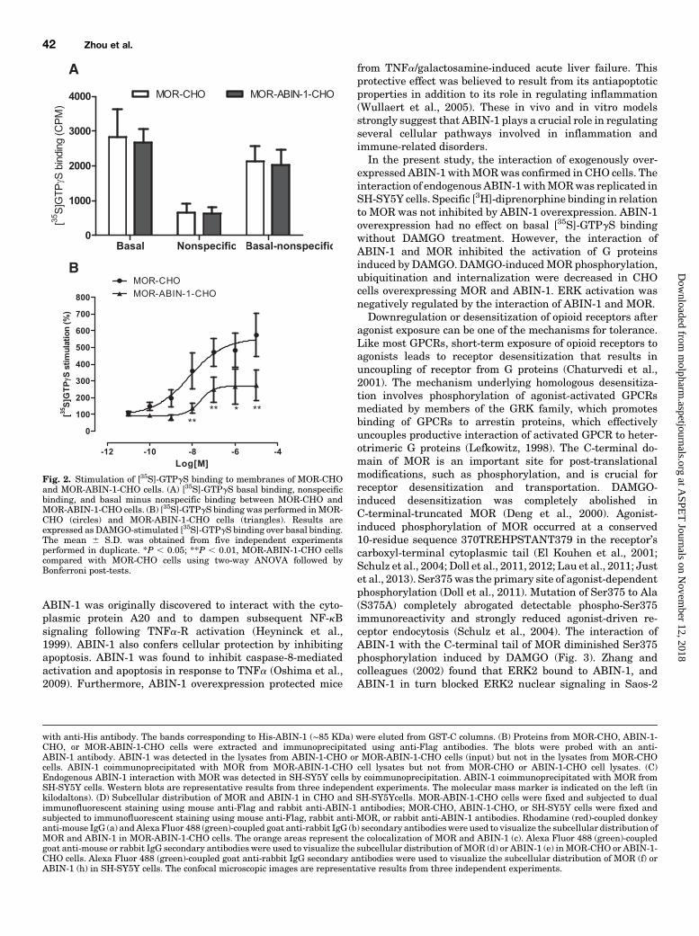
ABIN-1 Has No Effect on Ligand Binding but Inhibited [35S]-GTPgS Binding with MOR
Saturation binding studies (n 5 6) revealed no substantialdifference between MOR-CHO and MOR-ABIN-1-CHO cells.The binding properties of MOR with [3H]-diprenorphine inMOR-CHOcells (Bmax, 2.5360.84 pmol/mg;Kd, 0.4460.14 nM)were similar to those inMOR-ABIN-1-CHO cells (Bmax, 2.0560.64 pmol/mg; Kd, 0.33 6 0.12 nM). Thus, ABIN-1 doesnot appear to affect the binding affinity of diprenorphinefor MOR.To investigate whether ABIN-1 interferes with G protein
activation byMOR, the stimulation of [35S]-GTPgS binding byDAMGO was examined. No difference was observed in [35S]-GTPgS basal binding, nonspecific binding, or basal minusnonspecific binding between MOR-CHO and MOR-ABIN-1-CHO cells (Fig. 2A). DAMGO potently stimulated [35S]-GTPgSbinding in a concentration-dependent manner (EC50 5 18.0 65.4 nM) in MOR-CHO cells, with a maximal value of 574.8 699.1% compared with basal levels. In contrast, DAMGO-mediated stimulation of [35S]-GTPgS binding in MOR-ABIN-1-CHO cells was attenuated (EC50 5 45.9 6 10.3 nM), with amaximal value of 274.4 6 93.1% compared with basal levels(Fig. 2B).
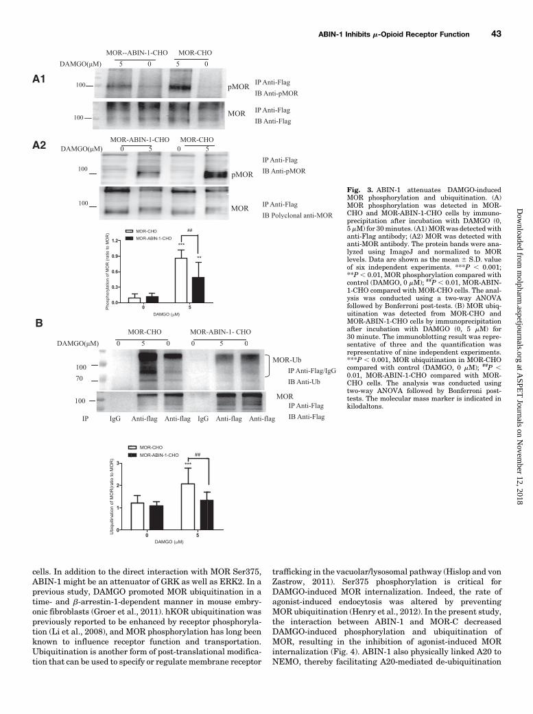
ABIN-1 Inhibited MOR Phosphorylation, Ubiquitination, andInternalization
The basal level of MOR phosphorylation at Ser375 wassimilar betweenMOR-CHO andMOR-ABIN-1-CHO cells. AfterDAMGO (5mM) treatment of 30minutes, MOR phosphorylationat Ser375 increased approximately 9-fold inMOR-CHO cells and5-fold in MOR-ABIN-1-CHO cells compared with the phosphor-ylation of MOR without DAMGO exposure. MOR phosphoryla-tion at Ser375 induced by DAMGO in MOR-ABIN-1-CHO cellswas significantly lower (by 30–40%) comparedwith that ofMOR-CHO cells (Fig. 3A, P , 0.01).Since receptor ubiquitination could be influenced by re-
ceptor phosphorylation, we examined the effect of ABIN-1on agonist-induced MOR ubiquitination in MOR-CHO andMOR-ABIN-1-CHO cells. MOR ubiquitination induced byDAMGO (5 mM) was significantly increased in MOR-CHOcells (P, 0.001) but was impaired in MOR-ABIN-1-CHO cells(P , 0.01, Fig. 3B). Ubiquitinated MOR were also detected
ABIN-1 Inhibits m-Opioid Receptor Function 39
at ASPE
T Journals on N
ovember 12, 2018
molpharm
.aspetjournals.orgD
ownloaded from
using anti-Flag antibody or anti-MOR antibody in MOR-CHOand MOR-ABIN-1-CHO cells (Supplemental Fig. 1). It isprobable that ABIN-1 associates with MOR and then inhibitsthe phosphorylation and ubiquitination of MOR induced byDAMGO.To determine whether this postulated inhibition of MOR
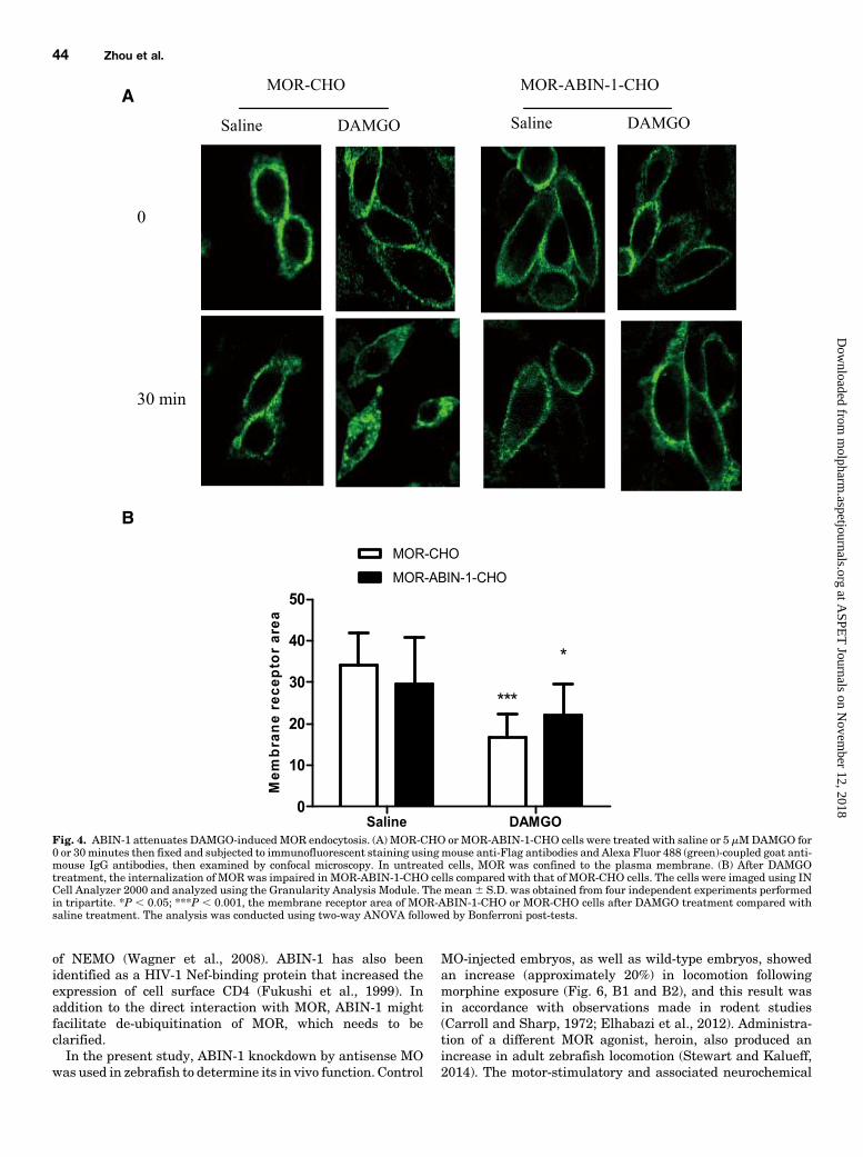
phosphorylation and ubiquitination affected agonist-inducedMOR endocytosis, we examined MOR internalization. MORwas expressed primarily on the plasma membrane, asassessed by immunolabeling both in MOR-CHO and MOR-ABIN-1-CHO cells (Fig. 4A). MOR were not internalizedfollowing saline treatment in either MOR-CHO or MOR-ABIN-1-CHO cells (Fig. 4A). Exposure to DAMGO (5 mM)resulted in rapid internalization of MOR into the cytoplasm ofMOR-CHO cells, but this internalization was impaired inMOR-ABIN-1-CHO cells. It should be noted that the mem-brane receptor area of MOR-ABIN-1-CHO cells was similar tothat of MOR-CHO cells after saline treatment. After DAMGOtreatment, the membrane receptor area of MOR-CHO cellsdecreased significantly (P , 0.001). The membrane receptorarea of MOR-ABIN-1-CHO also decreased (P , 0.05) com-pared with saline treatment (Fig. 4B). These results suggestthat internalization of MOR is partially inhibited in thepresence of ABIN-1.
The Effect of ABIN-1 on cAMP Accumulation and ERKActivity Following Acute Agonist Treatment
MOR is thought to inhibit adenylyl cyclase via Gi/Go
proteins. Hence, we aimed to determine whether the interac-tion of ABIN-1 and MOR interfered with G protein activationafter agonist treatment. The effect of ABIN-1 onMOR signalingwas measured by an assay that measured forskolin-stimulatedcAMPaccumulation byDAMGO inMOR-CHOorMOR-ABIN-1-CHO cells. In MOR-CHO cells, DAMGO (3.2 � 1029–1025 M)inhibited in a concentration-dependent manner forskolin-stimulated cAMP production after incubation for 15 minutes,with a maximum cAMP inhibition of 66.0 6 21.34%. DAMGO(3.2 � 1029–1025 M) also in a concentration-dependentmanner inhibited forskolin-stimulated cAMP production inMOR-ABIN-1-CHO cells, but the maximum inhibition wassignificantly lower than that of MOR-CHO cells (P, 0.05, Fig.5A1). The cAMP inhibition following DAMGO (5.0 � 1026 M)incubation for 5 and 15 minutes were 39.95% and 62.95%,respectively, in MOR-CHO cells, which was higher than thecAMP inhibition of 18.35% and 33.23%, respectively, in MOR-ABIN-1-CHO cells (P , 0.05, P , 0.01, Fig. 5A2).The agonist-induced activation of MOR in CHO cells has
previously been shown to cause a rapid and transient increasein mitogen-activated protein kinase (MAPK) activity (Fukudaet al., 1996). ERK is the best-studied member of the MAPKfamily and plays a critical role in intracellular signal trans-duction (Ji and Woolf, 2001). To assess ERK activity in thecurrent study, MOR-CHO and MOR-ABIN-1-CHO cells wereexposed to DAMGO (0 or 5 mM) for 10 minutes, and levels ofphospho-ERK1/2/ERK1/2 were analyzed by Western blotting.ERK1/2 levels did not vary after DAMGO treatment either inMOR-CHO cells or MOR-ABIN-1-CHO cells. Moreover, basallevels of phospho-ERK1/2 were similar in the two cell types.However, following 5 mMDAMGO treatment, phospho-ERK1/2 levels increased to eight times to that of untreated MOR-CHO cells (P , 0.001), whereas phospho-ERK1/2 levels were
upregulated by about three times after DAMGO treatmentin MOR-ABIN-1-CHO cells. The levels of phospho-ERK1/2 inMOR-ABIN-1-CHO cells were significantly lower than that ofMOR-CHO cells following 5 mMDAMGO treatment (P, 0.05)(Fig. 5B).Analysis of Zebrafish Larvae Mobility. The mobility
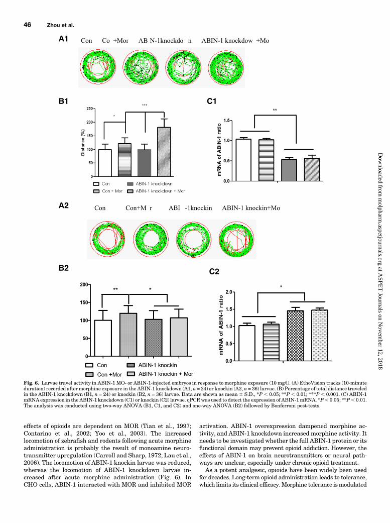
parameter of the ZebraLab 3.3 software is able to detectdistance traveled following stationary state to movement ofspecimens. In the control MO-injected larvae, morphine-exposed larvae showed a 20% increase in distance traveledcompared with saline-treated larvae (P, 0.05) (Fig. 6, A1 andB1). Following morphine treatment in ABIN-1 MO-injectedzebrafish larvae, there was a significant increase in distancetraveled (about 60% compared with the control MO-injectedzebrafish larvae) (P, 0.001) (Fig. 6, A1 and B1). There was nosignificant differences in mobility following comparison withnoninjected, control MO-injected or ABIN-1 MO-injectedlarvae following saline treatment. The ABIN-1 mRNAlevels of ABIN-1 MO-injected larvae were about 50% of thosein control MO-injected or noninjected larvae (Fig. 6C1) asdetermined by qPCR using forward primer 5-GGAGGACG-TCTGACCCTACGT-3 and reverse primer 5-AACGGCCCG-CAGTATGG-3. In the control pEGFPn1-injected cohorts,morphine-exposed larvae showed a 20% increase in distancetraveled compared with saline-treated larvae (P , 0.01).Following morphine treatment, the ABIN-1 knockin larvaeshowed a 15% decrease in distance traveled compared withthe control pEGFPn1-injected larvae (P, 0.05). In the ABIN-1 knockin larvae, morphine-exposed larvae showed no obviousvariation in distance traveled compared with saline-treatedlarvae (Fig. 6, A2 andB2). TheABIN-1mRNA levels for ABIN-1 knockin larvae were about 40–50% higher than those for thecontrol larvae (Fig. 6C2) determined by qPCR using forwardprimer 5-GACCGTGAACGCATGAATGA-3 and reverse primer 5-GACCCTGGGCCTGCAGCTT-3. The locomotion of a secondABIN-1 MO-injected larvae also increased substantially followingmorphine administration. Furthermore, the locomotion of thelarvae following acute morphine administration was antagonizedby pretreatment with naloxone (Supplemental Fig. 2).
DiscussionThe bacterial two-hybrid system that we used exhibited
high screening efficiency. When employed in our previousstudy, this system facilitated the identification of a total of19 proteins that interactedwith ratMOR-C (Zhou et al., 2015).Among the MOR-C-interacting proteins we found, calmodulin1 and heat shock protein 70 were also reported to interact withopioid receptors (Wang et al., 1999, 2000;Wannemacher et al.,2008). One of the 19MOR-C interacting proteins, ABIN-1, wasreported to be elevated in different brain regions followingchronic morphine treatment (Zhou et al., 2015). In the presentstudy, the activity of MOR was negatively regulated by ABIN-1 both in vitro and in vivo.ABIN-1 is a protein that inhibits both transduction of
transmembrane receptors such as tumor necrosis factor(TNF)a-receptor (TNFa-R), epidermal growth factor receptor(EGFR), and Toll-like receptor, and the activity of nuclearreceptors peroxisome proliferator-activated receptor and ret-inoic acid receptor (Mauro et al., 2006; Gurevich and Anes-kievich, 2009; Flores et al., 2011; Ramirez et al., 2012). Thesereceptors play key roles in regulating inflammatory diseases.
40 Zhou et al.
at ASPE
T Journals on N
ovember 12, 2018
molpharm
.aspetjournals.orgD
ownloaded from
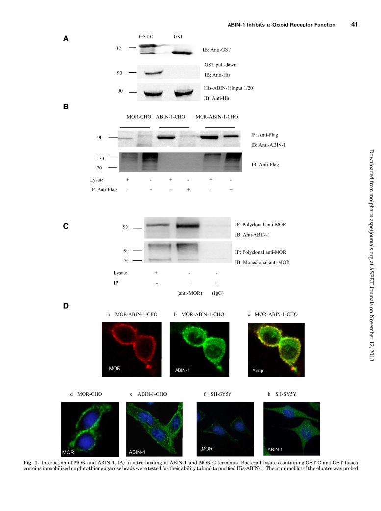
Fig. 1. Interaction of MOR and ABIN-1. (A) In vitro binding of ABIN-1 and MOR C-terminus. Bacterial lysates containing GST-C and GST fusionproteins immobilized on glutathione agarose beads were tested for their ability to bind to purified His-ABIN-1. The immunoblot of the eluates was probed
ABIN-1 Inhibits m-Opioid Receptor Function 41
at ASPE
T Journals on N
ovember 12, 2018
molpharm
.aspetjournals.orgD
ownloaded from
ABIN-1 was originally discovered to interact with the cyto-plasmic protein A20 and to dampen subsequent NF-kBsignaling following TNFa-R activation (Heyninck et al.,1999). ABIN-1 also confers cellular protection by inhibitingapoptosis. ABIN-1 was found to inhibit caspase-8-mediatedactivation and apoptosis in response to TNFa (Oshima et al.,2009). Furthermore, ABIN-1 overexpression protected mice
from TNFa/galactosamine-induced acute liver failure. Thisprotective effect was believed to result from its antiapoptoticproperties in addition to its role in regulating inflammation(Wullaert et al., 2005). These in vivo and in vitro modelsstrongly suggest that ABIN-1 plays a crucial role in regulatingseveral cellular pathways involved in inflammation andimmune-related disorders.In the present study, the interaction of exogenously over-
expressed ABIN-1 withMORwas confirmed in CHO cells. Theinteraction of endogenous ABIN-1withMORwas replicated inSH-SY5Y cells. Specific [3H]-diprenorphine binding in relationto MOR was not inhibited by ABIN-1 overexpression. ABIN-1overexpression had no effect on basal [35S]-GTPgS bindingwithout DAMGO treatment. However, the interaction ofABIN-1 and MOR inhibited the activation of G proteinsinduced by DAMGO. DAMGO-inducedMOR phosphorylation,ubiquitination and internalization were decreased in CHOcells overexpressing MOR and ABIN-1. ERK activation wasnegatively regulated by the interaction of ABIN-1 and MOR.Downregulation or desensitization of opioid receptors after
agonist exposure can be one of the mechanisms for tolerance.Like most GPCRs, short-term exposure of opioid receptors toagonists leads to receptor desensitization that results inuncoupling of receptor from G proteins (Chaturvedi et al.,2001). The mechanism underlying homologous desensitiza-tion involves phosphorylation of agonist-activated GPCRsmediated by members of the GRK family, which promotesbinding of GPCRs to arrestin proteins, which effectivelyuncouples productive interaction of activated GPCR to heter-otrimeric G proteins (Lefkowitz, 1998). The C-terminal do-main of MOR is an important site for post-translationalmodifications, such as phosphorylation, and is crucial forreceptor desensitization and transportation. DAMGO-induced desensitization was completely abolished inC-terminal-truncated MOR (Deng et al., 2000). Agonist-induced phosphorylation of MOR occurred at a conserved10-residue sequence 370TREHPSTANT379 in the receptor’scarboxyl-terminal cytoplasmic tail (El Kouhen et al., 2001;Schulz et al., 2004; Doll et al., 2011, 2012; Lau et al., 2011; Justet al., 2013). Ser375was the primary site of agonist-dependentphosphorylation (Doll et al., 2011). Mutation of Ser375 to Ala(S375A) completely abrogated detectable phospho-Ser375immunoreactivity and strongly reduced agonist-driven re-ceptor endocytosis (Schulz et al., 2004). The interaction ofABIN-1 with the C-terminal tail of MOR diminished Ser375phosphorylation induced by DAMGO (Fig. 3). Zhang andcolleagues (2002) found that ERK2 bound to ABIN-1, andABIN-1 in turn blocked ERK2 nuclear signaling in Saos-2
with anti-His antibody. The bands corresponding to His-ABIN-1 (∼85 KDa) were eluted from GST-C columns. (B) Proteins from MOR-CHO, ABIN-1-CHO, or MOR-ABIN-1-CHO cells were extracted and immunoprecipitated using anti-Flag antibodies. The blots were probed with an anti-ABIN-1 antibody. ABIN-1 was detected in the lysates from ABIN-1-CHO or MOR-ABIN-1-CHO cells (input) but not in the lysates from MOR-CHOcells. ABIN-1 coimmunoprecipitated with MOR from MOR-ABIN-1-CHO cell lysates but not from MOR-CHO or ABIN-1-CHO cell lysates. (C)Endogenous ABIN-1 interaction with MOR was detected in SH-SY5Y cells by coimmunoprecipitation. ABIN-1 coimmunoprecipitated with MOR fromSH-SY5Y cells. Western blots are representative results from three independent experiments. The molecular mass marker is indicated on the left (inkilodaltons). (D) Subcellular distribution of MOR and ABIN-1 in CHO and SH-SY5Ycells. MOR-ABIN-1-CHO cells were fixed and subjected to dualimmunofluorescent staining using mouse anti-Flag and rabbit anti-ABIN-1 antibodies; MOR-CHO, ABIN-1-CHO, or SH-SY5Y cells were fixed andsubjected to immunofluorescent staining using mouse anti-Flag, rabbit anti-MOR, or rabbit anti-ABIN-1 antibodies. Rhodamine (red)-coupled donkeyanti-mouse IgG (a) andAlexaFluor 488 (green)-coupled goat anti-rabbit IgG (b) secondary antibodieswere used to visualize the subcellular distribution ofMOR and ABIN-1 in MOR-ABIN-1-CHO cells. The orange areas represent the colocalization of MOR and ABIN-1 (c). Alexa Fluor 488 (green)-coupledgoat anti-mouse or rabbit IgG secondary antibodies were used to visualize the subcellular distribution of MOR (d) or ABIN-1 (e) inMOR-CHO or ABIN-1-CHO cells. Alexa Fluor 488 (green)-coupled goat anti-rabbit IgG secondary antibodies were used to visualize the subcellular distribution of MOR (f) orABIN-1 (h) in SH-SY5Y cells. The confocal microscopic images are representative results from three independent experiments.
Fig. 2. Stimulation of [35S]-GTPgS binding to membranes of MOR-CHOand MOR-ABIN-1-CHO cells. (A) [35S]-GTPgS basal binding, nonspecificbinding, and basal minus nonspecific binding between MOR-CHO andMOR-ABIN-1-CHO cells. (B) [35S]-GTPgS binding was performed inMOR-CHO (circles) and MOR-ABIN-1-CHO cells (triangles). Results areexpressed as DAMGO-stimulated [35S]-GTPgS binding over basal binding.The mean 6 S.D. was obtained from five independent experimentsperformed in duplicate. *P , 0.05; **P , 0.01, MOR-ABIN-1-CHO cellscompared with MOR-CHO cells using two-way ANOVA followed byBonferroni post-tests.
42 Zhou et al.
at ASPE
T Journals on N
ovember 12, 2018
molpharm
.aspetjournals.orgD
ownloaded from
cells. In addition to the direct interaction with MOR Ser375,ABIN-1 might be an attenuator of GRK as well as ERK2. In aprevious study, DAMGO promoted MOR ubiquitination in atime- and b-arrestin-1-dependent manner in mouse embry-onic fibroblasts (Groer et al., 2011). hKOR ubiquitination waspreviously reported to be enhanced by receptor phosphoryla-tion (Li et al., 2008), and MOR phosphorylation has long beenknown to influence receptor function and transportation.Ubiquitination is another form of post-translational modifica-tion that can be used to specify or regulatemembrane receptor
trafficking in the vacuolar/lysosomal pathway (Hislop and vonZastrow, 2011). Ser375 phosphorylation is critical forDAMGO-induced MOR internalization. Indeed, the rate ofagonist-induced endocytosis was altered by preventingMOR ubiquitination (Henry et al., 2012). In the present study,the interaction between ABIN-1 and MOR-C decreasedDAMGO-induced phosphorylation and ubiquitination ofMOR, resulting in the inhibition of agonist-induced MORinternalization (Fig. 4). ABIN-1 also physically linked A20 toNEMO, thereby facilitating A20-mediated de-ubiquitination
Fig. 3. ABIN-1 attenuates DAMGO-inducedMOR phosphorylation and ubiquitination. (A)MOR phosphorylation was detected in MOR-CHO and MOR-ABIN-1-CHO cells by immuno-precipitation after incubation with DAMGO (0,5mM) for 30minutes. (A1)MORwas detected withanti-Flag antibody; (A2) MOR was detected withanti-MOR antibody. The protein bands were ana-lyzed using ImageJ and normalized to MORlevels. Data are shown as the mean 6 S.D. valueof six independent experiments. ***P , 0.001;**P , 0.01, MOR phosphorylation compared withcontrol (DAMGO, 0 mM); ##P , 0.01, MOR-ABIN-1-CHO compared with MOR-CHO cells. The anal-ysis was conducted using a two-way ANOVAfollowed by Bonferroni post-tests. (B) MOR ubiq-uitination was detected from MOR-CHO andMOR-ABIN-1-CHO cells by immunoprecipitationafter incubation with DAMGO (0, 5 mM) for30 minute. The immunoblotting result was repre-sentative of three and the quantification wasrepresentative of nine independent experiments.***P , 0.001, MOR ubiquitination in MOR-CHOcompared with control (DAMGO, 0 mM); ##P ,0.01, MOR-ABIN-1-CHO compared with MOR-CHO cells. The analysis was conducted usingtwo-way ANOVA followed by Bonferroni post-tests. The molecular mass marker is indicated inkilodaltons.
ABIN-1 Inhibits m-Opioid Receptor Function 43
at ASPE
T Journals on N
ovember 12, 2018
molpharm
.aspetjournals.orgD
ownloaded from
of NEMO (Wagner et al., 2008). ABIN-1 has also beenidentified as a HIV-1 Nef-binding protein that increased theexpression of cell surface CD4 (Fukushi et al., 1999). Inaddition to the direct interaction with MOR, ABIN-1 mightfacilitate de-ubiquitination of MOR, which needs to beclarified.In the present study, ABIN-1 knockdown by antisense MO
was used in zebrafish to determine its in vivo function. Control
MO-injected embryos, as well as wild-type embryos, showedan increase (approximately 20%) in locomotion followingmorphine exposure (Fig. 6, B1 and B2), and this result wasin accordance with observations made in rodent studies(Carroll and Sharp, 1972; Elhabazi et al., 2012). Administra-tion of a different MOR agonist, heroin, also produced anincrease in adult zebrafish locomotion (Stewart and Kalueff,2014). The motor-stimulatory and associated neurochemical
Fig. 4. ABIN-1 attenuates DAMGO-inducedMOR endocytosis. (A) MOR-CHO orMOR-ABIN-1-CHO cells were treated with saline or 5 mMDAMGO for0 or 30minutes then fixed and subjected to immunofluorescent staining usingmouse anti-Flag antibodies and Alexa Fluor 488 (green)-coupled goat anti-mouse IgG antibodies, then examined by confocal microscopy. In untreated cells, MOR was confined to the plasma membrane. (B) After DAMGOtreatment, the internalization of MOR was impaired in MOR-ABIN-1-CHO cells compared with that of MOR-CHO cells. The cells were imaged using INCell Analyzer 2000 and analyzed using the Granularity Analysis Module. The mean6 S.D. was obtained from four independent experiments performedin tripartite. *P , 0.05; ***P , 0.001, the membrane receptor area of MOR-ABIN-1-CHO or MOR-CHO cells after DAMGO treatment compared withsaline treatment. The analysis was conducted using two-way ANOVA followed by Bonferroni post-tests.
44 Zhou et al.
at ASPE
T Journals on N
ovember 12, 2018
molpharm
.aspetjournals.orgD
ownloaded from
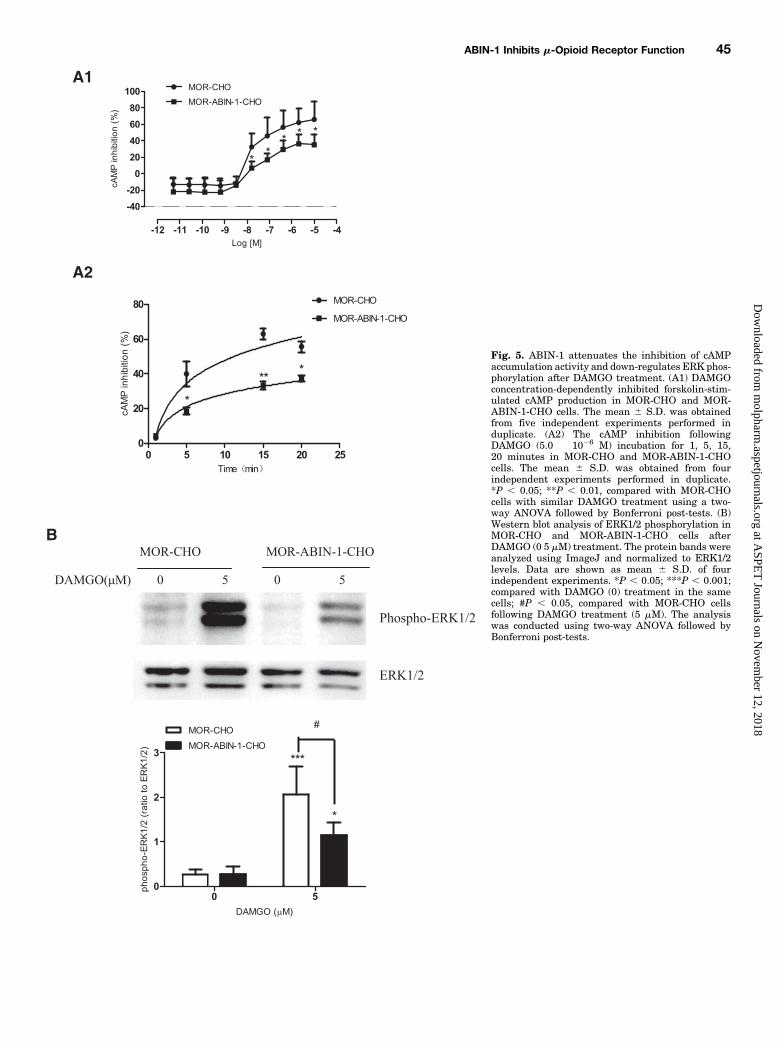
Fig. 5. ABIN-1 attenuates the inhibition of cAMPaccumulation activity and down-regulates ERK phos-phorylation after DAMGO treatment. (A1) DAMGOconcentration-dependently inhibited forskolin-stim-ulated cAMP production in MOR-CHO and MOR-ABIN-1-CHO cells. The mean 6 S.D. was obtainedfrom five independent experiments performed induplicate. (A2) The cAMP inhibition followingDAMGO (5.0 � 1026 M) incubation for 1, 5, 15,20 minutes in MOR-CHO and MOR-ABIN-1-CHOcells. The mean 6 S.D. was obtained from fourindependent experiments performed in duplicate.*P , 0.05; **P , 0.01, compared with MOR-CHOcells with similar DAMGO treatment using a two-way ANOVA followed by Bonferroni post-tests. (B)Western blot analysis of ERK1/2 phosphorylation inMOR-CHO and MOR-ABIN-1-CHO cells afterDAMGO (0 5 mM) treatment. The protein bands wereanalyzed using ImageJ and normalized to ERK1/2levels. Data are shown as mean 6 S.D. of fourindependent experiments. *P , 0.05; ***P , 0.001;compared with DAMGO (0) treatment in the samecells; #P , 0.05, compared with MOR-CHO cellsfollowing DAMGO treatment (5 mM). The analysiswas conducted using two-way ANOVA followed byBonferroni post-tests.
ABIN-1 Inhibits m-Opioid Receptor Function 45
at ASPE
T Journals on N
ovember 12, 2018
molpharm
.aspetjournals.orgD
ownloaded from
effects of opioids are dependent on MOR (Tian et al., 1997;Contarino et al., 2002; Yoo et al., 2003). The increasedlocomotion of zebrafish and rodents following acute morphineadministration is probably the result of monoamine neuro-transmitter upregulation (Carroll and Sharp, 1972; Lau et al.,2006). The locomotion of ABIN-1 knockin larvae was reduced,whereas the locomotion of ABIN-1 knockdown larvae in-creased after acute morphine administration (Fig. 6). InCHO cells, ABIN-1 interacted with MOR and inhibited MOR
activation. ABIN-1 overexpression dampened morphine ac-tivity, and ABIN-1 knockdown increased morphine activity. Itneeds to be investigated whether the full ABIN-1 protein or itsfunctional domain may prevent opioid addiction. However, theeffects of ABIN-1 on brain neurotransmitters or neural path-ways are unclear, especially under chronic opioid treatment.As a potent analgesic, opioids have been widely been used
for decades. Long-term opioid administration leads to tolerance,which limits its clinical efficacy.Morphine tolerance ismodulated
Fig. 6. Larvae travel activity in ABIN-1 MO- or ABIN-1-injected embryos in response to morphine exposure (10 mg/l). (A) EthoVision tracks (10-minuteduration) recorded after morphine exposure in the ABIN-1 knockdown (A1, n = 24) or knockin (A2, n = 36) larvae. (B) Percentage of total distance traveledin the ABIN-1 knockdown (B1, n = 24) or knockin (B2, n = 36) larvae. Data are shown as mean 6 S.D., *P , 0.05; **P , 0.01; ***P , 0.001. (C) ABIN-1mRNA expression in the ABIN-1 knockdown (C1) or knockin (C2) larvae. qPCRwas used to detect the expression of ABIN-1mRNA. *P, 0.05; **P, 0.01.The analysis was conducted using two-way ANOVA (B1, C1, and C2) and one-way ANOVA (B2) followed by Bonferroni post-tests.
46 Zhou et al.
at ASPE
T Journals on N
ovember 12, 2018
molpharm
.aspetjournals.orgD
ownloaded from
by proinflammatory cytokines such as TNF-a (Mao et al., 1995;Shen et al., 2012). Chronic morphine infusion has been demon-strated to induce a significant increase in TNF-a mRNA expres-sions of tolerant rats. Administration of the TNF-a inhibitoretanercept reduced proinflammatory cytokine production andmicroglial activation, thus preserving the antinociceptive effectsof morphine (Li et al., 2002b). ABIN-1 increased significantly inthe frontal cortex, striatum, and ventral tegmental are of ratsafter chronic morphine exposure (Zhou et al., 2015). The increaseinNF-kBsignalingbyTNF-awas inhibited by theupregulation ofABIN-1 in the cytoplasm after chronic morphine exposure, whichmay prevent excessive tolerance or addiction to morphine. Inaddition to its anti-inflammatory effects, ABIN-1 also protectscells by inhibiting apoptosis. ABIN-1 overexpression may protectmorphine-induced apoptosis and preserve the antinociceptiveeffects ofmorphine.Morphine has been shown to induce apoptoticcell death in vitro (Goswami et al., 1998; Singhal et al., 2000, 2002;Yin et al., 2000; Tegeder et al., 2003).Morphine has been shown toinduce apoptosis of human microglia and neurons (Hu et al.,2002), which is consistent with studies in rodent peritonealmacrophages and a macrophage cell line (Singhal et al., 2000).Caspase-3 is involved in the execution phase ofmorphine-inducedapoptosis (Hu et al., 2002). In vivo,morphine also increases cortexand amygdala apoptosis with cleaved caspase-3-positive cellssignificantly increased after repeated administration to neonatalrats (Bajic et al., 2013). Apoptotic cell death was also increasedfollowing morphine administration associated with developmentof antinociceptive tolerance.In summary, this study is the first to provide evidence that
ABIN-1 protein interacts with MOR, leading to the suppres-sion of MOR activation, phosphorylation, and internalization.ABIN-1 also inhibits morphine-induced hyperlocomotion inzebrafish larvae. Therefore, ABIN-1 appears to be an impor-tant inhibitor for agonist-bound MOR. Immunoinflammatoryfactors are involved in the formation of opioid tolerance.ABIN-1 has been reported to inhibit the transcription ofinflammatory factors through theNF-kBpathway. In additionto functioning as a inhibitor for agonist-bound MOR, ABIN-1may be an important regulator for opioid tolerance. The exactmechanism by which ABIN-1 regulates chronic morphinetolerance needs to be investigated.
Acknowledgments
The authors thankDr. Antonio Leonardi of theUniversity of Naples(Naples, Italy) for kindly providing the pcDNA3.1-FLAG-hABIN-1plasmid, and Dr. Lili W (Beijing Institute of Pharmacology andToxicology, Beijing, China) for technical assistance.
Authorship Contributions
Participated in research design: Zhou, Su, Gong.Conducted experiments: Zhou, Jiang, Yan, Li, Zhao, Wang.Contributed new reagents or analytic tools: Yan, Li.Performed data analysis: Zhou, Yan.Wrote or contributed to the writing of the manuscript: Zhou, Su.
References
Ammon S, Mayer P, Riechert U, Tischmeyer H, and Höllt V (2003) Microarrayanalysis of genes expressed in the frontal cortex of rats chronically treated withmorphine and after naloxone precipitated withdrawal. Brain Res Mol Brain Res112:113–125.
Arden JR, Segredo V, Wang Z, Lameh J, and Sadée W (1995) Phosphorylation andagonist-specific intracellular trafficking of an epitope-tagged mu-opioid receptorexpressed in HEK 293 cells. J Neurochem 65:1636–1645.
Bajic D, Commons KG, and Soriano SG (2013) Morphine-enhanced apoptosis in se-lective brain regions of neonatal rats. Int J Dev Neurosci 31:258–266.
Bohn LM, Gainetdinov RR, Lin FT, Lefkowitz RJ, and Caron MG (2000) Mu-opioidreceptor desensitization by beta-arrestin-2 determines morphine tolerance but notdependence. Nature 408:720–723.
Bohn LM, Lefkowitz RJ, Gainetdinov RR, Peppel K, Caron MG, and Lin FT (1999)Enhanced morphine analgesia inmice lacking beta-arrestin 2. Science 286:2495–2498.
Carroll BJ and Sharp PT (1972) Monoamine mediation of the morphine-inducedactivation of mice. Br J Pharmacol 46:124–139.
Chaturvedi K, Bandari P, Chinen N, and Howells RD (2001) Proteasome involvementin agonist-induced down-regulation of mu and delta opioid receptors. J Biol Chem276:12345–12355.
Contarino A, Picetti R, Matthes HW, Koob GF, Kieffer BL, and Gold LH (2002) Lackof reward and locomotor stimulation induced by heroin in mu-opioid receptor-deficient mice. Eur J Pharmacol 446:103–109.
Deng HB, Yu Y, Pak Y, O’Dowd BF, George SR, Surratt CK, Uhl GR, and Wang JB(2000) Role for the C-terminus in agonist-induced mu opioid receptor phosphory-lation and desensitization. Biochemistry 39:5492–5499.
Doll C, Konietzko J, Pöll F, Koch T, Höllt V, and Schulz S (2011) Agonist-selectivepatterns of m-opioid receptor phosphorylation revealed by phosphosite-specificantibodies. Br J Pharmacol 164:298–307.
Doll C, Pöll F, Peuker K, Loktev A, Glück L, and Schulz S (2012) Decipheringm-opioid receptor phosphorylation and dephosphorylation in HEK293 cells. Br JPharmacol 167:1259–1270.
Elhabazi K, Trigo JM, Mollereau C, Moulédous L, Zajac JM, Bihel F, Schmitt M,Bourguignon JJ, Meziane H, Petit-demoulière B, et al. (2012) Involvement ofneuropeptide FF receptors in neuroadaptive responses to acute and chronic opiatetreatments. Br J Pharmacol 165:424–435.
El Kouhen R, Burd AL, Erickson-Herbrandson LJ, Chang CY, Law PY, and Loh HH(2001) Phosphorylation of Ser363, Thr370, and Ser375 residues within the carboxyltail differentially regulates mu-opioid receptor internalization. J Biol Chem 276:12774–12780.
Flores AM, Gurevich I, Zhang C, Ramirez VP, Devens TR, and Aneskievich BJ (2011)TNIP1 is a corepressor of agonist-bound PPARs. Arch Biochem Biophys 516:58–66.
Fukuda K, Kato S, Morikawa H, Shoda T, and Mori K (1996) Functional coupling ofthe delta-, mu-, and kappa-opioid receptors to mitogen-activated protein kinase andarachidonate release in Chinese hamster ovary cells. J Neurochem 67:1309–1316.
Fukushi M, Dixon J, Kimura T, Tsurutani N, Dixon MJ, and Yamamoto N (1999)Identification and cloning of a novel cellular protein Naf1, Nef-associated factor 1,that increases cell surface CD4 expression. FEBS Lett 442:83–88.
Georgoussi Z, Georganta EM, and Milligan G (2012) The other side of opioid receptorsignalling: regulation by protein-protein interaction. Curr Drug Targets 13:80–102.
Goswami R, Dawson SA, and Dawson G (1998) Cyclic AMP protects against staur-osporine and wortmannin-induced apoptosis and opioid-enhanced apoptosis inboth embryonic and immortalized (F-11kappa7) neurons. J Neurochem 70:1376–1382.
Groer CE, Schmid CL, Jaeger AM, and Bohn LM (2011) Agonist-directed interactionswith specific beta-arrestins determine mu-opioid receptor trafficking, ubiquitina-tion, and dephosphorylation. J Biol Chem 286:31731–31741.
Groer CE, Tidgewell K, Moyer RA, Harding WW, Rothman RB, Prisinzano TE,and Bohn LM (2007) An opioid agonist that does not induce mu-opioid receptor–arrestin interactions or receptor internalization. Mol Pharmacol 71:549–557.
Gurevich I and Aneskievich BJ (2009) Liganded RARalpha and RARgamma interactwith but are repressed by TNIP1. Biochem Biophys Res Commun 389:409–414.
Hawes BE, Luttrell LM, Exum ST, and Lefkowitz RJ (1994) Inhibition of G protein-coupled receptor signaling by expression of cytoplasmic domains of the receptor. JBiol Chem 269:15776–15785.
Henry AG, Hislop JN, Grove J, Thorn K, Marsh M, and von Zastrow M (2012) Regu-lation of endocytic clathrin dynamics by cargo ubiquitination. Dev Cell 23:519–532.
Heyninck K, De Valck D, Vanden Berghe W, Van Criekinge W, Contreras R, Fiers W,Haegeman G, and Beyaert R (1999) The zinc finger protein A20 inhibits TNF-induced NF-kappaB-dependent gene expression by interfering with an RIP- orTRAF2-mediated transactivation signal and directly binds to a novel NF-kappaB-inhibiting protein ABIN. J Cell Biol 145:1471–1482.
Hislop JN and von Zastrow M (2011) Role of ubiquitination in endocytic trafficking ofG-protein-coupled receptors. Traffic 12:137–148.
Hogan BM, Verkade H, Lieschke GJ, and Heath JK (2008) Manipulation of geneexpression during zebrafish embryonic development using transient approaches.Methods Mol Biol 469:273–300.
Hu S, Sheng WS, Lokensgard JR, and Peterson PK (2002) Morphine induces apo-ptosis of human microglia and neurons. Neuropharmacology 42:829–836.
Ji RR and Woolf CJ (2001) Neuronal plasticity and signal transduction in nociceptiveneurons: implications for the initiation and maintenance of pathological pain.Neurobiol Dis 8:1–10.
Just S, Illing S, Trester-Zedlitz M, Lau EK, Kotowski SJ, Miess E, Mann A, Doll C,Trinidad JC, Burlingame AL, et al. (2013) Differentiation of opioid drug effects byhierarchical multi-site phosphorylation. Mol Pharmacol 83:633–639.
Lau B, Bretaud S, Huang Y, Lin E, and Guo S (2006) Dissociation of food and opiatepreference by a genetic mutation in zebrafish. Genes Brain Behav 5:497–505.
Lau EK, Trester-Zedlitz M, Trinidad JC, Kotowski SJ, Krutchinsky AN, BurlingameAL, and von Zastrow M (2011) Quantitative encoding of the effect of a partialagonist on individual opioid receptors by multisite phosphorylation and thresholddetection. Sci Signal 4:ra52.
Lefkowitz RJ (1998) G protein-coupled receptors. III. New roles for receptor kinasesand beta-arrestins in receptor signaling and desensitization. J Biol Chem 273:18677–18680.
Lefkowitz RJ and Shenoy SK (2005) Transduction of receptor signals by beta-arrestins. Science 308:512–517.
Li J, Li JG, Chen C, Zhang F, and Liu-Chen LY (2002a) Molecular basis of differencesin (-)(trans)-3,4-dichloro-N-methyl-N-[2-(1-pyrrolidiny)-cyclohexyl]benzeneacetamide-induced desensitization and phosphorylation between human and rat kappa-opioidreceptors expressed in Chinese hamster ovary cells. Mol Pharmacol 61:73–84.
ABIN-1 Inhibits m-Opioid Receptor Function 47
at ASPE
T Journals on N
ovember 12, 2018
molpharm
.aspetjournals.orgD
ownloaded from
Li JG, Haines DS, and Liu-Chen LY (2008) Agonist-promoted Lys63-linked poly-ubiquitination of the human kappa-opioid receptor is involved in receptor down-regulation. Mol Pharmacol 73:1319–1330.
Li Y, Wang X, Tian S, Guo CJ, Douglas SD, and Ho WZ (2002b) Methadone enhanceshuman immunodeficiency virus infection of human immune cells. J Infect Dis 185:118–122.
Mao J, Price DD, and Mayer DJ (1995) Mechanisms of hyperalgesia and morphinetolerance: a current view of their possible interactions. Pain 62:259–274.
Mauro C, Pacifico F, Lavorgna A, Mellone S, Iannetti A, Acquaviva R, Formisano S,Vito P, and Leonardi A (2006) ABIN-1 binds to NEMO/IKKgamma and co-operateswith A20 in inhibiting NF-kappaB. J Biol Chem 281:18482–18488.
McClung CA, Nestler EJ, and Zachariou V (2005) Regulation of gene expression bychronic morphine and morphine withdrawal in the locus ceruleus and ventraltegmental area. J Neurosci 25:6005–6015.
Morou E and Georgoussi Z (2005) Expression of the third intracellular loop of thedelta-opioid receptor inhibits signaling by opioid receptors and other G protein-coupled receptors. J Pharmacol Exp Ther 315:1368–1379.
Oshima S, Turer EE, Callahan JA, Chai S, Advincula R, Barrera J, Shifrin N, Lee B,Benedict Yen TS, Woo T, et al. (2009) ABIN-1 is a ubiquitin sensor that restrictscell death and sustains embryonic development. Nature 457:906–909.
Ramirez VP, Gurevich I, and Aneskievich BJ (2012) Emerging roles for TNIP1 inregulating post-receptor signaling. Cytokine Growth Factor Rev 23:109–118.
Schulz S, Mayer D, Pfeiffer M, Stumm R, Koch T, and Höllt V (2004) Morphineinduces terminal micro-opioid receptor desensitization by sustained phosphoryla-tion of serine-375. EMBO J 23:3282–3289.
Shen CH, Tsai RY, and Wong CS (2012) Role of neuroinflammation in morphinetolerance: effect of tumor necrosis factor-a. Acta Anaesthesiol Taiwan 50:178–182.
Singhal PC, Bhaskaran M, Patel J, Patel K, Kasinath BS, Duraisamy S, Franki N,Reddy K, and Kapasi AA (2002) Role of p38 mitogen-activated protein kinasephosphorylation and Fas-Fas ligand interaction in morphine-induced macrophageapoptosis. J Immunol 168:4025–4033.
Singhal PC, Kapasi AA, Franki N, and Reddy K (2000) Morphine-induced macrophageapoptosis: the role of transforming growth factor-beta. Immunology 100:57–62.
Stewart AM and Kalueff AV (2014) The behavioral effects of acute D9-tetrahydro-cannabinol and heroin (diacetylmorphine) exposure in adult zebrafish. Brain Res1543:109–119.
Tegeder I, Grösch S, Schmidtko A, Häussler A, Schmidt H, Niederberger E, Scholich K,and Geisslinger G (2003) G protein-independent G1 cell cycle block and apoptosiswith morphine in adenocarcinoma cells: involvement of p53 phosphorylation. CancerRes 63:1846–1852.
Tian M, Broxmeyer HE, Fan Y, Lai Z, Zhang S, Aronica S, Cooper S, Bigsby RM,Steinmetz R, Engle SJ, et al. (1997) Altered hematopoiesis, behavior, and sexualfunction in mu opioid receptor-deficient mice. J Exp Med 185:1517–1522.
Verstrepen L, Carpentier I, Verhelst K, and Beyaert R (2009) ABINs: A20 bindinginhibitors of NF-kappa B and apoptosis signaling. Biochem Pharmacol 78:105–114.
Wagner S, Carpentier I, Rogov V, Kreike M, Ikeda F, Löhr F, Wu CJ, Ashwell JD,Dötsch V, Dikic I, et al. (2008) Ubiquitin binding mediates the NF-kappaB in-hibitory potential of ABIN proteins. Oncogene 27:3739–3745.
Wang D, Sadée W, and Quillan JM (1999) Calmodulin binding to G protein-couplingdomain of opioid receptors. J Biol Chem 274:22081–22088.
Wang D, Surratt CK, and Sadée W (2000) Calmodulin regulation of basal andagonist-stimulated G protein coupling by the mu-opioid receptor (OP(3)) inmorphine-pretreated cell. J Neurochem 75:763–771.
Wannemacher KM, Terskiy A, Bian S, Yadav PN, Li H, and Howells RD (2008)Purification and mass spectrometric analysis of the kappa opioid receptor. BrainRes 1230:13–26.
Williams JT, Ingram SL, Henderson G, Chavkin C, von Zastrow M, Schulz S, Koch T,Evans CJ, and Christie MJ (2013) Regulation of m-opioid receptors: de-sensitization, phosphorylation, internalization, and tolerance. Pharmacol Rev 65:223–254.
Wullaert A, Wielockx B, Van Huffel S, Bogaert V, De Geest B, Papeleu P, Schotte P,El Bakkouri K, Heyninck K, Libert C, and Beyaert R (2005) Adenoviral genetransfer of ABIN-1 protects mice from TNF/galactosamine-induced acute liverfailure and lethality. Hepatology 42:381–389.
Yin D, Tuthill D, Mufson RA, and Shi Y (2000) Chronic restraint stress promoteslymphocyte apoptosis by modulating CD95 expression. J Exp Med 191:1423–1428.
Yoo JH, Yang EM, Lee SY, Loh HH, Ho IK, and Jang CG (2003) Differential effects ofmorphine and cocaine on locomotor activity and sensitization in mu-opioid receptorknockout mice. Neurosci Lett 344:37–40.
Yuan S and Sun Z (2009) Microinjection of mRNA and morpholino antisense oligo-nucleotides inzebrafish embryos. J Vis Exp 27: pii: 1113. DOI:10.3791/1113.
Zhang J, Ferguson SS, Barak LS, Bodduluri SR, Laporte SA, Law PY, and Caron MG(1998) Role for G protein-coupled receptor kinase in agonist-specific regulation ofmu-opioid receptor responsiveness. Proc Natl Acad Sci USA 95:7157–7162.
Zhou P, Jiang J, Dong Z, Yan H, You Z, Su R, and Gong Z (2015) The proteinsinteracting with C-terminal of m receptor are identified by bacterial two-hybridsystem from brain cDNA library in morphine-dependent rats. Life Sci 143:156–167.
Address correspondence to: Dr. Ruibin Su, Beijing Institute of Pharmacol-ogy and Toxicology, 27th Taiping Rd., Beijing 100850, China. E-mail:[email protected] or Dr. Peilan Zhou, Beijing Institute of Pharmacology andToxicology, 27th Taiping Rd., Beijing 100850, China. E-mail: [email protected]
48 Zhou et al.
at ASPE
T Journals on N
ovember 12, 2018
molpharm
.aspetjournals.orgD
ownloaded from