a of nt β catenin signaling pathway in hepatocellulaR ... of Wnt b-catenin.pdf9 DOI:...
13
9 DOI: 10.5114/PJP.2015.51148 POL J PATHOL 2015; 66 (1): 9-21 Wnt/Fzd/β-catenin signaling pathway in liver The Wnt/Fzd/β-catenin signaling pathway plays a significant role in liver physiology and pathology. Its involvement was demonstrated in development of liver and in morphogenesis of biliary ducts. It main- tains correct homeostasis of liver cells in postnatal life [1], and it influences development of structure and metabolic activity of the hepatic acinus, allowing for growth and regeneration of the liver. It protects the liver from the effects of toxic agents and oxidative stress [2]. Also mechanisms of carcinogenesis, related both to hepatoblastoma and to primary hepatocel- lular carcinoma (HCC) involve the pathway [1, 3]. Interactions of the pathway have been described with other pathways accelerating hepatocyte proliferation. Detailed mechanisms of HCC development remain unknown and determination of a simple model of hepatocarcinogenesis with involvement of the Wnt signaling pathway has proven to be difficult [1]. Due to the fact that incidence of chronic hepatitis C REVIEW PAPER A LTERATIONS OF WNT / β - CATENIN SIGNALING PATHWAY IN HEPATOCELLULAR CARCINOMAS ASSOCIATED WITH HEPATITIS C VIRUS KAROL ROGACKI 1 , ALDONA KASPRZAK 1 , ADRIAN S TęPIńSKI 2 1 Chair and Department of Histology and Embryology, Poznan University of Medical Sciences, Poznan, Poland 2 Heliodor Święcicki Clinical Hospital, Poznan, Poland The Wnt/Fzd/β-catenin signaling pathway plays a significant role in physiology and pathology of the liver. The role of β-catenin is linked mainly to the canonical pathway of the system. Phosphorylation of β-catenin and abnormalities in function of the E-cadherin-catenin unit lead to loss of intercellular junctions, progression in liver fibrosis, and development of cirrhosis and hepatocellular carcinoma (HCC). Progression of liver diseases is noted to be accompanied by disturbances in β-cat- enin expression (mainly with its overexpression), with its cytoplasmic or nuclear translocation and with lowered expression of E-cadherin. Increase in transcriptional activity of β-catenin is associated mainly with mutations of CTNNB1. Detailed mechanisms of HCC development are not known. More β-catenin mutations are manifested in hepatitis C virus (HCV)-associated than in HBV-related HCC. In recent years the role of nonstructural proteins and of the core protein of HCV has been accentuated in induction of the Wnt path- way. HCV proteins affect in a double manner expression of E-cadherin, including modulation of the Wnt pathway and reduction of E-cadherin expression at the transcriptional level. This review presents current data on mechanisms of hepatocarcinogenesis involv- ing participation of the Wnt canonical pathway and, in particular, interaction of Wnt pathway components with HCV genome products in the process. Key words: Wnt canonical pathway, β-catenin/E-cadherin complex, hepatitis C virus, hepatocarcinogenesis.
-
Upload
nguyennhan -
Category
Documents
-
view
217 -
download
0
Transcript of a of nt β catenin signaling pathway in hepatocellulaR ... of Wnt b-catenin.pdf9 DOI:...
9
DOI: 10.5114/pjp.2015.51148 pOl j pathOl 2015; 66 (1): 9-21
Wnt/Fzd/β-catenin signaling pathway in liver
The Wnt/Fzd/β-catenin signaling pathway plays a significant role in liver physiology and pathology. Its involvement was demonstrated in development of liver and in morphogenesis of biliary ducts. It main-tains correct homeostasis of liver cells in postnatal life [1], and it influences development of structure and metabolic activity of the hepatic acinus, allowing for growth and regeneration of the liver. It protects the
liver from the effects of toxic agents and oxidative stress [2]. Also mechanisms of carcinogenesis, related both to hepatoblastoma and to primary hepatocel-lular carcinoma (HCC) involve the pathway [1, 3]. Interactions of the pathway have been described with other pathways accelerating hepatocyte proliferation. Detailed mechanisms of HCC development remain unknown and determination of a simple model of hepatocarcinogenesis with involvement of the Wnt signaling pathway has proven to be difficult [1]. Due to the fact that incidence of chronic hepatitis C
Review papeR
alteRations of wnt/β-catenin signaling pathway in hepatocellulaR caRcinomas associated with hepatitis c viRus
KarOl rOgacKI1, alDOna KasprzaK1, aDrIan stępIńsKI2
1Chair and Department of Histology and Embryology, Poznan University of Medical Sciences, Poznan, Poland2Heliodor Święcicki Clinical Hospital, Poznan, Poland
The Wnt/Fzd/β-catenin signaling pathway plays a significant role in physiology and pathology of the liver. The role of β-catenin is linked mainly to the canonical pathway of the system. Phosphorylation of β-catenin and abnormalities in function of the E-cadherin-catenin unit lead to loss of intercellular junctions, progression in liver fibrosis, and development of cirrhosis and hepatocellular carcinoma (HCC). Progression of liver diseases is noted to be accompanied by disturbances in β-cat-enin expression (mainly with its overexpression), with its cytoplasmic or nuclear translocation and with lowered expression of E-cadherin. Increase in transcriptional activity of β-catenin is associated mainly with mutations of CTNNB1. Detailed mechanisms of HCC development are not known. More β-catenin mutations are manifested in hepatitis C virus (HCV)-associated than in HBV-related HCC. In recent years the role of nonstructural proteins and of the core protein of HCV has been accentuated in induction of the Wnt path-way. HCV proteins affect in a double manner expression of E-cadherin, including modulation of the Wnt pathway and reduction of E-cadherin expression at the transcriptional level. This review presents current data on mechanisms of hepatocarcinogenesis involv-ing participation of the Wnt canonical pathway and, in particular, interaction of Wnt pathway components with HCV genome products in the process.
Key words: Wnt canonical pathway, β-catenin/E-cadherin complex, hepatitis C virus, hepatocarcinogenesis.
10
Karol rogacKi, aldona KasprzaK, adrian stępińsKi
is increasing, leading to development of end-stage liver disease including cirrhosis and HCC [4], it is im- portant to define mechanisms of hepatitis C virus (HCV)-induced liver carcinogenesis with involve-ment of the Wnt/β-catenin signaling pathway. Al-ready at the beginning of this century more numer-ous mutations of β-catenin were found to develop in HCV-associated HCC than with HBV-related HCC [5]. Recent years have brought the results of studies on direct interactions between oncogenic HCV pro-teins and components of the Wnt/β-catenin signal-ing pathway [6-10].
Double role of β-catenin
In its inactivated state, β-catenin is phosphorylated at its serine (SER)/threonine (THR) residues. It rep-resents a component of a large cytoplasmic protein complex with glycogen synthase kinase 3β (GSK-3β), casein kinase 1 (CK1), the product of the APC (ad-enomatous polyposis coli) suppressor gene and axin/conductin [11, 12]. The complex controls intra-
cellular levels of β-catenin mainly through protein phosphorylation. Phosphorylation of the β-catenin N-terminus represents a pre-requirement for recog-nition by β-TrCP of an ubiquitin ligase E component, with its subsequent degradation in proteasomes. At the first stage phosphorylation of serine takes place in position 45 (SER45) by CK1α/ε, and then SER33, SER37 and THR41 by GSK3β [13]. Control of β-cat-enin phosphorylation also involves the Diversin pro-tein: while CK1α binds directly to axin, CK1ε links the ankyrin fragment of the Diversin protein, form-ing a degradation complex [14]. Phosphorylation of β-catenin by GSK3β is much more effective in the presence of axin, and overexpression of conductin additionally augments degradation of β-catenin. In neoplastic tumors (including those in the liver) ex-pression of conductin (but not axin) is frequently el-evated and may represent an early diagnostic marker of certain tumors [15]. APC protein represents an-other protein involved in formation of the β-catenin destructive complex. The sites for β-catenin binding are located in its central portion [16]. The critical
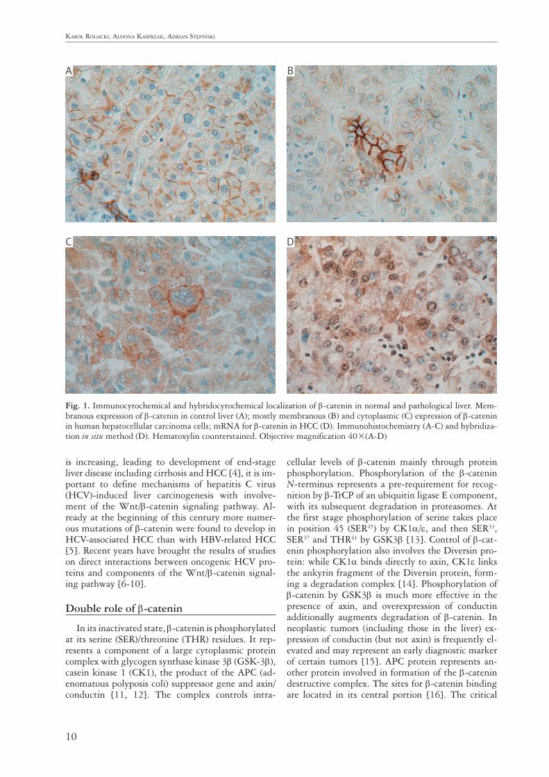
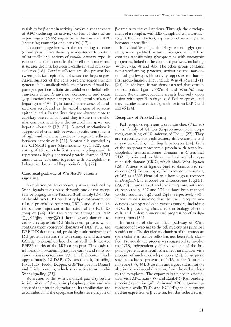
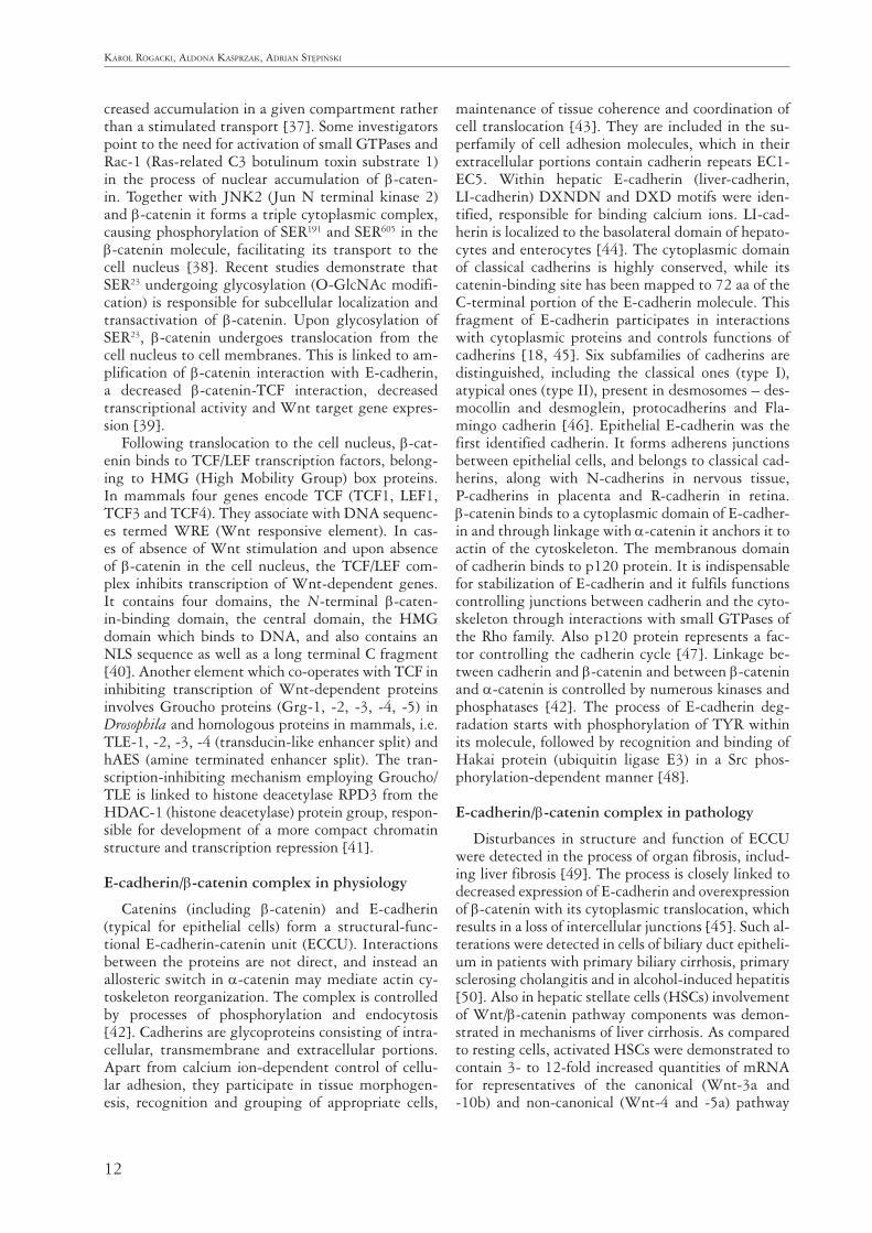
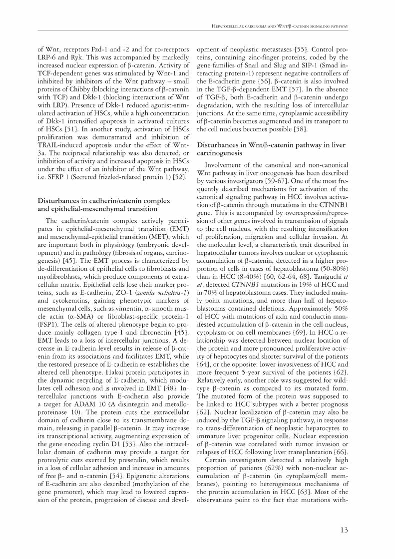
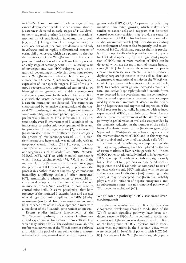
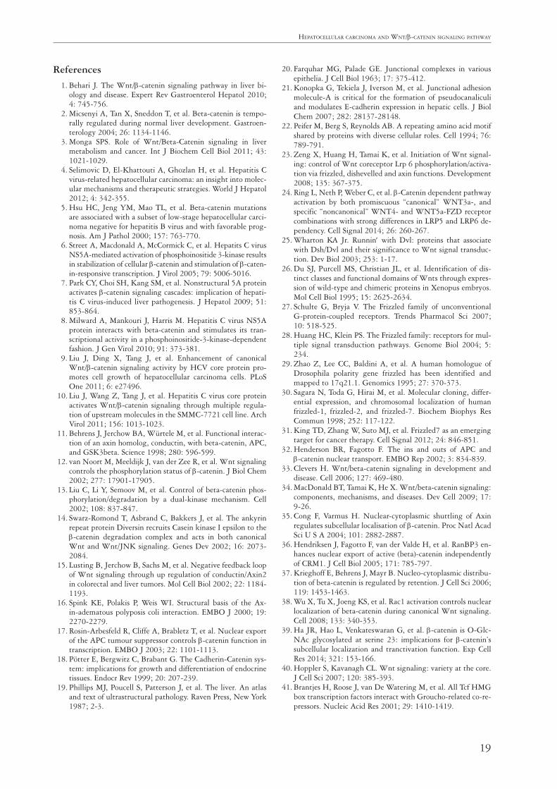
Fig. 1. Immunocytochemical and hybridocytochemical localization of β-catenin in normal and pathological liver. Mem-branous expression of β-catenin in control liver (A); mostly membranous (B) and cytoplasmic (C) expression of β-catenin in human hepatocellular carcinoma cells; mRNA for β-catenin in HCC (D). Immunohistochemistry (A-C) and hybridiza-tion in situ method (D). Hematoxylin counterstained. Objective magnification 40×(A-D)
A
C
B
D
11
Hepatocellular carcinoma and Wnt/β-catenin signaling patHWay
variables for β-catenin activity involve nuclear export of APC (reducing its activity) or loss of the nuclear export signal (NES) sequence in the mutated APC (increasing transcriptional activity) [17].
β-catenin, together with the remaining catenins (α and γ) and E-cadherin, participates in formation of intercellular junctions of zonulae adherens type. It is located at the inner side of the cell membrane, and it secures the link between E-cadherin and cell cyto-skeleton [18]. Zonulae adherens are also present be-tween polarized epithelial cells, such as hepatocytes. Apical surfaces of the cells represent regions which generate bile canaliculi while membranes of basal he-patocyte portions adjoin sinusoidal endothelial cells. Junctions of zonula adherens, desmosome and nexus (gap junction) types are present on lateral surfaces of hepatocytes [19]. Tight junctions are areas of local-ized contact, found in the apical region of adjacent epithelial cells. In the liver they are situated close to capillary bile canaliculi, and they isolate the canalic-ular compartment from the intercellular space and hepatic sinusoids [19, 20]. A novel mechanism is suggested of cross-talk between specific components of tight and adherens junctions to regulate adhesion between hepatic cells [21]. β-catenin is encoded by the CTNNB1 gene (chromosome 3p21-p22), con-sisting of 16 exons (the first is a non-coding exon). It represents a highly conserved protein, formed of 781 amino acids (aa), and, together with plakoglobin, it belongs to the armadillo protein family [22].
Canonical pathway of Wnt/Fzd/β-catenin signaling
Stimulation of the canonical pathway induced by Wnt ligands takes place through one of the recep-tors belonging to the Frizzled (Fzd) family [23]. Out of the old two LRP (low density lipoprotein-receptor related protein) co-receptors, LRP-5 and -6, the lat-ter is more important in formation of the Fzd-LRP complex [24]. The Fzd receptor, through its PDZ (PSD-95/dics large/ZO-1 homologous) domain, re-cruits a cytoplasmic Dvl (disheveled) protein, which contains three conserved domains of DIX, PDZ and DEP. DIX domains and, probably, multimerization of Dvl protein, recruits the axin complex and activates GSK3β to phosphorylate the intracellularly located PPPSP motifs of the LRP co-receptor. This leads to inhibition of β-catenin phosphorylation and to its ac-cumulation in cytoplasm [23]. The Dvl protein binds approximately 18 DAPs (Dvl-associated), including Nkd, Idax, Frodo, Dapper, GBP/Frat, Stbm, Daam1 and Pricle proteins, which may activate or inhibit Wnt signaling [25].
Activation of the Wnt canonical pathway results in inhibition of β-catenin phosphorylation and ab-sence of the protein degradation. Its stabilization and accumulation in the cytoplasm facilitates transport of
β-catenin to the cell nucleus. Through the develop-ment of a complex with LEF (lymphoid enhancer fac-tor)/TCF (T cell factor), expression of various genes becomes intensified.
Individual Wnt ligands (19 cystein-rich glycopro-teins) were qualified to form two groups. The first contains transforming glycoproteins with oncogenic properties, linked to the canonical pathway, including Wnt-1, -3a, -8 and -8b. The other group contains non-transforming proteins, activating the non-ca-nonical pathway with activity opposite to that of first group ligands. They include Wnt-4, -5a and -11 [26]. In addition, it was demonstrated that certain non-canonical ligands (Wnt-4 and Wnt-5a) may induce β-catenin-dependent signals but only upon fusion with specific subtypes of Fzd receptors, and they manifest a selective dependence from LRP-5 and LRP-6 [24].
Receptors of Frizzled family
Fzd receptors represent a separate class (Frizzled) in the family of GPCRs (G-protein-coupled recep-tors), consisting of 10 isoforms of Fzd1-10 [27]. They are responsible for proliferation, differentiation and migration of cells, including hepatocytes [24]. Each of the receptors represents a protein with seven hy-drophobic transmembrane domains, a C-terminal PDZ domain and an N-terminal extracellular cys-teine-rich domain (CRD), which binds Wnt ligands [28]. Various Wnt ligands bind to distinct Fzd re-ceptors [27]. For example, Fzd2 receptor, consisting of 565 aa (56% identical to a homologous receptor in Drosophila), is encoded on chromosome 17q21.1. [29, 30]. Human Fzd1 and Fzd7 receptors, with size of, respectively, 647 and 574 aa, have been mapped to chromosomes 7q21 and 2q33, respectively [30]. Recent reports indicate that the Fzd7 receptor un-dergoes overexpression in various tumors, including HCC. It plays a significant role in biology of stem cells, and in development and progression of malig-nant tumors [31].
In function of the canonical pathway of Wnt, transport of β-catenin to the cell nucleus has principal significance. The detailed mechanism of the transport (particularly in tumor cells) has not been fully clari-fied. Previously the process was suggested to involve the NLS, independently of involvement of the im-portin protein, as a result of a direct interaction with proteins of nuclear envelope pores [32]. Subsequent studies excluded presence of NLS in the β-catenin molecule [33, 34]. β-catenin undergoes translocation also in the reciprocal direction, from the cell nucleus to the cytoplasm. The export takes place in associa-tion with APC, axin [35] and RanBP3 (Ran binding protein 3) proteins [36]. Axin and APC augment cy-toplasmic while TCF4 and BCL9/Pygopus augment nuclear expression of β-catenin, but this reflects its in-
12
Karol rogacKi, aldona KasprzaK, adrian stępińsKi
creased accumulation in a given compartment rather than a stimulated transport [37]. Some investigators point to the need for activation of small GTPases and Rac-1 (Ras-related C3 botulinum toxin substrate 1) in the process of nuclear accumulation of β-caten-in. Together with JNK2 (Jun N terminal kinase 2) and β-catenin it forms a triple cytoplasmic complex, causing phosphorylation of SER191 and SER605 in the β-catenin molecule, facilitating its transport to the cell nucleus [38]. Recent studies demonstrate that SER23 undergoing glycosylation (O-GlcNAc modifi-cation) is responsible for subcellular localization and transactivation of β-catenin. Upon glycosylation of SER23, β-catenin undergoes translocation from the cell nucleus to cell membranes. This is linked to am-plification of β-catenin interaction with E-cadherin, a decreased β-catenin-TCF interaction, decreased transcriptional activity and Wnt target gene expres-sion [39].
Following translocation to the cell nucleus, β-cat-enin binds to TCF/LEF transcription factors, belong-ing to HMG (High Mobility Group) box proteins. In mammals four genes encode TCF (TCF1, LEF1, TCF3 and TCF4). They associate with DNA sequenc-es termed WRE (Wnt responsive element). In cas-es of absence of Wnt stimulation and upon absence of β-catenin in the cell nucleus, the TCF/LEF com-plex inhibits transcription of Wnt-dependent genes. It contains four domains, the N-terminal β-caten-in-binding domain, the central domain, the HMG domain which binds to DNA, and also contains an NLS sequence as well as a long terminal C fragment [40]. Another element which co-operates with TCF in inhibiting transcription of Wnt-dependent proteins involves Groucho proteins (Grg-1, -2, -3, -4, -5) in Drosophila and homologous proteins in mammals, i.e. TLE-1, -2, -3, -4 (transducin-like enhancer split) and hAES (amine terminated enhancer split). The tran-scription-inhibiting mechanism employing Groucho/TLE is linked to histone deacetylase RPD3 from the HDAC-1 (histone deacetylase) protein group, respon-sible for development of a more compact chromatin structure and transcription repression [41].
E-cadherin/β-catenin complex in physiology
Catenins (including β-catenin) and E-cadherin (typical for epithelial cells) form a structural-func-tional E-cadherin-catenin unit (ECCU). Interactions between the proteins are not direct, and instead an allosteric switch in α-catenin may mediate actin cy-toskeleton reorganization. The complex is controlled by processes of phosphorylation and endocytosis [42]. Cadherins are glycoproteins consisting of intra-cellular, transmembrane and extracellular portions. Apart from calcium ion-dependent control of cellu-lar adhesion, they participate in tissue morphogen-esis, recognition and grouping of appropriate cells,
maintenance of tissue coherence and coordination of cell translocation [43]. They are included in the su-perfamily of cell adhesion molecules, which in their extracellular portions contain cadherin repeats EC1-EC5. Within hepatic E-cadherin (liver-cadherin, LI-cadherin) DXNDN and DXD motifs were iden-tified, responsible for binding calcium ions. LI-cad-herin is localized to the basolateral domain of hepato-cytes and enterocytes [44]. The cytoplasmic domain of classical cadherins is highly conserved, while its catenin-binding site has been mapped to 72 aa of the C-terminal portion of the E-cadherin molecule. This fragment of E-cadherin participates in interactions with cytoplasmic proteins and controls functions of cadherins [18, 45]. Six subfamilies of cadherins are distinguished, including the classical ones (type I), atypical ones (type II), present in desmosomes – des-mocollin and desmoglein, protocadherins and Fla-mingo cadherin [46]. Epithelial E-cadherin was the first identified cadherin. It forms adherens junctions between epithelial cells, and belongs to classical cad-herins, along with N-cadherins in nervous tissue, P-cadherins in placenta and R-cadherin in retina. β-catenin binds to a cytoplasmic domain of E-cadher-in and through linkage with α-catenin it anchors it to actin of the cytoskeleton. The membranous domain of cadherin binds to p120 protein. It is indispensable for stabilization of E-cadherin and it fulfils functions controlling junctions between cadherin and the cyto-skeleton through interactions with small GTPases of the Rho family. Also p120 protein represents a fac-tor controlling the cadherin cycle [47]. Linkage be-tween cadherin and β-catenin and between β-catenin and α-catenin is controlled by numerous kinases and phosphatases [42]. The process of E-cadherin deg-radation starts with phosphorylation of TYR within its molecule, followed by recognition and binding of Hakai protein (ubiquitin ligase E3) in a Src phos-phorylation-dependent manner [48].
E-cadherin/β-catenin complex in pathology
Disturbances in structure and function of ECCU were detected in the process of organ fibrosis, includ-ing liver fibrosis [49]. The process is closely linked to decreased expression of E-cadherin and overexpression of β-catenin with its cytoplasmic translocation, which results in a loss of intercellular junctions [45]. Such al-terations were detected in cells of biliary duct epitheli-um in patients with primary biliary cirrhosis, primary sclerosing cholangitis and in alcohol-induced hepatitis [50]. Also in hepatic stellate cells (HSCs) involvement of Wnt/β-catenin pathway components was demon-strated in mechanisms of liver cirrhosis. As compared to resting cells, activated HSCs were demonstrated to contain 3- to 12-fold increased quantities of mRNA for representatives of the canonical (Wnt-3a and -10b) and non-canonical (Wnt-4 and -5a) pathway
13
Hepatocellular carcinoma and Wnt/β-catenin signaling patHWay
of Wnt, receptors Fzd-1 and -2 and for co-receptors LRP-6 and Ryk. This was accompanied by markedly increased nuclear expression of β-catenin. Activity of TCF-dependent genes was stimulated by Wnt-1 and inhibited by inhibitors of the Wnt pathway – small proteins of Chibby (blocking interactions of β-catenin with TCF) and Dkk-1 (blocking interactions of Wnt with LRP). Presence of Dkk-1 reduced agonist-stim-ulated activation of HSCs, while a high concentration of Dkk-1 intensified apoptosis in activated cultures of HSCs [51]. In another study, activation of HSCs proliferation was demonstrated and inhibition of TRAIL-induced apoptosis under the effect of Wnt-3a. The reciprocal relationship was also detected, or inhibition of activity and increased apoptosis in HSCs under the effect of an inhibitor of the Wnt pathway, i.e. SFRP 1 (Secreted frizzled-related protein 1) [52].
Disturbances in cadherin/catenin complex and epithelial-mesenchymal transition
The cadherin/catenin complex actively partici-pates in epithelial-mesenchymal transition (EMT) and mesenchymal-epithelial transition (MET), which are important both in physiology (embryonic devel-opment) and in pathology (fibrosis of organs, carcino-genesis) [45]. The EMT process is characterized by de-differentiation of epithelial cells to fibroblasts and myofibroblasts, which produce components of extra-cellular matrix. Epithelial cells lose their marker pro-teins, such as E-cadherin, ZO-1 (zonula occludens-1) and cytokeratins, gaining phenotypic markers of mesenchymal cells, such as vimentin, α-smooth mus-cle actin (α-SMA) or fibroblast-specific protein-1 (FSP1). The cells of altered phenotype begin to pro-duce mainly collagen type I and fibronectin [45]. EMT leads to a loss of intercellular junctions. A de-crease in E-cadherin level results in release of β-cat-enin from its associations and facilitates EMT, while the restored presence of E-cadherin re-establishes the altered cell phenotype. Hakai protein participates in the dynamic recycling of E-cadherin, which modu-lates cell adhesion and is involved in EMT [48]. In-tercellular junctions with E-cadherin also provide a target for ADAM 10 (A disintegrin and metallo-proteinase 10). The protein cuts the extracellular domain of cadherin close to its transmembrane do-main, releasing in parallel β-catenin. It may increase its transcriptional activity, augmenting expression of the gene encoding cyclin D1 [53]. Also the intracel-lular domain of cadherin may provide a target for proteolytic cuts exerted by presenilin, which results in a loss of cellular adhesion and increase in amounts of free β- and α-catenin [54]. Epigenetic alterations of E-cadherin are also described (methylation of the gene promoter), which may lead to lowered expres-sion of the protein, progression of disease and devel-
opment of neoplastic metastases [55]. Control pro-teins, containing zinc-finger proteins, coded by the gene families of Snail and Slug and SIP-1 (Smad in-teracting protein-1) represent negative controllers of the E-cadherin gene [56]. β-catenin is also involved in the TGF-β-dependent EMT [57]. In the absence of TGF-β, both E-cadherin and β-catenin undergo degradation, with the resulting loss of intercellular junctions. At the same time, cytoplasmic accessibility of β-catenin becomes augmented and its transport to the cell nucleus becomes possible [58].
Disturbances in Wnt/β-catenin pathway in liver carcinogenesis
Involvement of the canonical and non-canonical Wnt pathway in liver oncogenesis has been described by various investigators [59-67]. One of the most fre-quently described mechanisms for activation of the canonical signaling pathway in HCC involves activa-tion of β-catenin through mutations in the CTNNB1 gene. This is accompanied by overexpression/repres-sion of other genes involved in transmission of signals to the cell nucleus, with the resulting intensification of proliferation, migration and cellular invasion. At the molecular level, a characteristic trait described in hepatocellular tumors involves nuclear or cytoplasmic accumulation of β-catenin, detected in a higher pro-portion of cells in cases of hepatoblastoma (50-80%) than in HCC (8-40%) [60, 62-64, 68]. Taniguchi et al. detected CTNNB1 mutations in 19% of HCC and in 70% of hepatoblastoma cases. They included main-ly point mutations, and more than half of hepato-blastomas contained deletions. Approximately 50% of HCC with mutations of axin and conductin man-ifested accumulation of β-catenin in the cell nucleus, cytoplasm or on cell membranes [69]. In HCC a re-lationship was detected between nuclear location of the protein and more pronounced proliferative activ-ity of hepatocytes and shorter survival of the patients [64], or the opposite: lower invasiveness of HCC and more frequent 5-year survival of the patients [62]. Relatively early, another role was suggested for wild-type β-catenin as compared to its mutated form. The mutated form of the protein was supposed to be linked to HCC subtypes with a better prognosis [62]. Nuclear localization of β-catenin may also be induced by the TGF-β signaling pathway, in response to trans-differentiation of neoplastic hepatocytes to immature liver progenitor cells. Nuclear expression of β-catenin was correlated with tumor invasion or relapses of HCC following liver transplantation [66].
Certain investigators detected a relatively high proportion of patients (62%) with non-nuclear ac-cumulation of β-catenin (in cytoplasm/cell mem-branes), pointing to heterogeneous mechanisms of the protein accumulation in HCC [63]. Most of the observations point to the fact that mutations with-
14
Karol rogacKi, aldona KasprzaK, adrian stępińsKi
in CTNNB1 are manifested in a later stage of liver cancer development while nuclear accumulation of β-catenin is detected in early stages of HCC devel-opment, suggesting other (distinct from mutations) mechanisms of stabilization involving the protein [63, 70, 71]. Using a model of transgenic mice, nu-clear localization of β-catenin was demonstrated only in adenoma and in highly differentiated cancers of eosinophil phenotype, which also pointed to the fact that activation of the Wnt/β-catenin pathway with protein translocation of the cell nucleus represents an early stage of carcinogenesis [72]. Following years of investigations, two HCC subtypes were distin-guished, depending on molecular alterations related to the Wnt/β-catenin pathway. The first one, with a mutation in CTNNB1, is characterized by increased expression of liver-specific targets. HCC of this sub-group represents well-differentiated tumors of a low histological malignancy, with stable chromosomes and a good prognosis. In the other subtype of HCC, also with the Wnt/β-catenin pathway activated, no β-catenin mutations are detected. The tumors are characterized by extensive dysregulation of the clas-sical Wnt pathway, a significant degree of chromo-some instability, aggressive phenotype, and they are preferentially linked to HBV infection [71, 73]. In-terestingly, even if involvement of β-catenin is of key importance to embryonic development of liver and for processes of liver regeneration [2], activation of β-catenin itself remains insufficient to initiate per se the process of liver carcinogenesis [67, 74]: a tran-sient hepatocyte hyperplasia was noted only, with no neoplastic transformation [74]. However, the acti-vated β-catenin may cooperate with other pathways of oncogenesis, such as insulin/IGF-1/IRS-1/MAPK, H-RAS, MET, AKT or with chemical compounds which initiate carcinogenesis [74, 75]. Even if the mutated form of β-catenin is insufficient to trigger the process of HCC development, it promotes the process in another manner (increasing chromosome instability, amplifying action of other oncogenes) [67]. Amazingly, a phenomenon of sevenfold in-crease in development of liver tumors was detected in mice with CTNNB1 knockout, as compared to control mice [76]. It seems paradoxical that both presence of the mutated β-catenin form and absence of wild type β-catenin amplifies the DEN (diethyl nitrosamine)-induced liver carcinogenesis in mice [67]. Mechanisms of HCC development in mice with a knockout of the β-catenin gene remain unknown.
Recent studies indicate involvement of the Wnt/β-catenin pathway in processes of self-renew-al and expansion of liver cancer stem cells (CSCs), which may initiate HCC. The evidence is available for preferential activation of the Wnt/β-catenin pathway also within the pool of stem cells within a mature, regenerating liver, termed oval cells or hepatic pro-
genitor cells (HPCs) [77]. As progenitor cells, they manifest uninhibited growth, which makes them similar to cancer cells and suggests that disturbed control over their division may provide a cause for development of HCC. This has been corroborated in studies on animal models [78]. Liver diseases leading to development of cancer also frequently lead to acti-vation of HPCs, which may suggest that it is precise-ly this group of cells which provides a starting point for HCC development [79]. In a significant propor-tion of HCC, one or more markers of HPCs can be detected, which are absent in normal mature hepato-cytes [80, 81]. In the oval cells, stimulated to prolif-eration, an increase was detected in Wnt-3-induced dephosphorylated β-catenin in the cell nucleus and augmented transcriptional activity in the Wnt/β-cat-enin/TCF pathway, with activation of the cell cycle [82]. In another investigation, increased amounts of total and active (dephosphorylated) β-catenin forms were detected in the cytoplasm and the cell nucleus. The increased expression of β-catenin was accompa-nied by increased amounts of Wnt-1 in the neigh-boring hepatocytes and augmented expression of the Fzd-2 receptor in oval cells, in parallel with reduced expression of WIF-1, an inhibitor of Wnt. An ad-ditional proof for involvement of the Wnt/β-catenin pathway in proliferation of oval cells was provided by the dramatic reduction in the number of the cells in livers of rodents devoid of the β-catenin gene [83]. Signals of the Wnt/β-catenin pathway may also affect the microenvironment of HCC and in this way may affect survival and growth of neoplastic cells [67].
β-catenin and E-cadherin, as components of the Wnt signaling pathway, have been placed on the list of serum markers of liver carcinogenesis [84]. In sera of HCC patients (etiologically linked to infection with HCV genotype 4) with liver cirrhosis, significantly higher levels of four proteins were detected, includ-ing β-catenin and E-cadherin, as compared to sera of patients with chronic HCV infection with no cancer and sera of control individuals [84]. Summing up the above, it may be accepted that β-catenin probably plays a role in initiation of hepatic oncogenesis and, at subsequent stages, the non-canonical pathway of Wnt becomes mobilized [67].
Wnt/β-catenin pathway in HCV-associated liver carcinogenesis
Studies on involvement of HCV in liver car-cinogenesis developing through modulation of the Wnt/β-catenin signaling pathway have been con-ducted since the 1990s. At the beginning, nuclear ac-cumulation of β-catenin was demonstrated in HCC, on the background of HCV infection and in associ-ation with mutations in the β-catenin gene, which were detected in 26-41% of patients with HCC [61, 85]. Activation of the Wnt/β-catenin signaling path-
15
Hepatocellular carcinoma and Wnt/β-catenin signaling patHWay
way and its involvement in liver carcinogenesis were also linked to axin mutations [86], inactivation of GSK-3β [87], dephosphorylation of β-catenin [59] and up-regulation of Fzd-7 [88]. Zhang et al. demon-strated that also the up-regulated microRNA-155 (miR-155), markedly increased in HCV-infected pa-tients, activates the Wnt signaling pathway with nu-clear accumulation of β-catenin and the accompany-ing increase in cyclin D1, c-Myc, and survivin. It was also determined that a direct and functional target of miR-155 involved APC [89]. However, it was not until in vitro studies were conducted that interactions between HCV proteins and the Wnt/β-catenin sig-naling pathway were clarified. In HepG2 cell lines both NS5A protein and the entire HCV polyprotein were demonstrated to be responsible for the increase in β-catenin level (protein accumulation and stabili-zation, decreased degradation in proteasomes) in cells with expression of the HCV genome products. This was developing in the mechanism of a reduced activ-ity manifested by the FKHR (forkhead transcription factor) and increased phosphorylation of GSK-3β [6]. Thus, the elevated cellular level of β-catenin resulted from activation of the PI3K/Akt signaling pathway. This caused augmented transcription of β-caten-in-dependent genes and was supposed to facilitate neoplastic transformation of HCV-infected hepato-cytes. Involvement of NS5A protein in activation of the Wnt/β-catenin signaling pathway was confirmed in subsequent studies [7], documenting direct acti-vation of endogenous, unphosphorylated wild-type β-catenin by NS5A protein and co-localization of the two proteins in cytoplasm of HepG2 cells. The mech-anism of β-catenin accumulation at the protein level, also through inactivation of GSK-3β, was confirmed. In addition, the investigators proved that NS5A pro-tein may directly interact with β-catenin through its N-terminus and the ARM 1-6 region of β-catenin [7]. The authors also succeeded in demonstrating that the N terminus of NS5A affects TCF-4-depen-dent transcriptional activity. In other studies, evi-dence was provided for a role of NS5A in binding of the p85 regulatory subunit of phosphoinositide-3 kinase (PIK3) and, in consequence, in stabilization of β-catenin, independently of effector kinases for PIK3, i.e. Akt and GSK-3β. Both ends of the NS5A protein (N and C) were found indispensable for the direct binding of β-catenin and for full activation of the pro-tein within the Wnt pathway [8]. Recent studies of Higgs et al. demonstrated a direct role for NS5A pro-tein in β-catenin-dependent c-Myc expression [90].
Direct activation of the Wnt/β-catenin pathway was demonstrated in an in vitro model also separately for the core (C) protein of HCV [9, 10, 91]. HCV-core transfected Huh7 cells up-regulated Wnt-1 and WISP-2 transcription [91]. The cells demonstrated intensified proliferation, DNA synthesis and pro-
gression of the cell cycle [91]. In both studies by Liu et al., core protein of HCV amplified the TCF-depen-dent transcriptional activity, intensified expression and stabilized β-catenin at the protein level in Huh7 cells through inactivation of GSK-3β. It proved to be responsible for amplification of cell proliferation and promotion of tumor growth following action of one of the Wnt pathway ligands, the Wnt-3a protein [9, 10]. Core protein of HCV increases active β-cat-enin and nuclear accumulation in SMMC-7721 cells. Up-regulation of gene expression involving many Wnt ligands (Wnt-2, -3, -3a, -10a, -10b, Fzd-1, -2, -3, -6, -7, -9, and LRP5/6 co-receptors) was demonstrated [10]. HCV also affects in a twofold way expression of E-cadherin, indirectly by modula-tion of the Wnt/β-catenin pathway and directly with mediation of HCV core protein. C protein diminishes expression of E-cadherin at the transcriptional level, through methylation of CpG islands in the promoter of the CDH1 gene [92, 93].
Recent studies brought proof for HCV involve-ment also in EMT [94-96]. In cultures of HCC cells infected with genotype 1b or 2a of HCV, increased expression of numerous EMT markers (including vi-mentin, snail, slug and twist proteins) was demon-strated and a decrease in E-cadherin expression, as well as an altered phenotype of hepatocytes, with higher expression of fibroblast-specific protein 1 (FSP-1) and elevated levels of β-catenin phosphorylated at Ser552 [94]. Grégoire et al. suggested that neither Hedgehog nor β-catenin is required for NS5A-me-diated EMT [96]. The study of Quan et al. strong-ly suggests that the HCV core-induced epigenetic silencing of SFRP (secreted frizzled-related protein) family may lead to activation of the Wnt signaling pathway and increase HCC aggressiveness through induction of EMT [97].
Clinicopathological role of β-catenin and E-cadherin expression in hepatocellular carcinomas
β-catenin represents a recognized oncogene, and both qualitative (pattern of expression) and quanti-tative evaluation of tissue expression of the protein permitted genetically distinct subsets of HCC to be distinguished [5, 62, 71, 73]. In most HCCs, a vari-able percentage of cells is noted with abnormal local-ization of β-catenin (i.e. cytoplasmic, nuclear, or C/N) [59, 61-66, 68, 69, 101]. Nuclear localization of the protein most frequently correlated with somatic mu-tations of β-catenin [5, 59, 62, 102], although de-scriptions of nuclear accumulation of the protein are available in cases free of the gene mutation [63]. The percentage of cells with β-catenin mutation in HCC is quantitatively quite variable (from a few to a few dozen percent) [59, 63, 64, 68, 103]. Mutations
16
Karol rogacKi, aldona KasprzaK, adrian stępińsKi
in the β-catenin gene seem to be more frequent in HCC with the background of HCV than HBV infec-tion [5].
In HCC most frequently tissue overexpression of the protein is noted [63, 101, 103], but studies are also available which manifest lower expression of the protein in cancer than in the control [62, 104, our own unpublished data]. Recently, a subgroup of patients with HCC has been distinguished (~15%) with complete absence of tissue β-catenin expression [105].
Most positive correlations between invasive char-acter of HCC, high metastatic potential of HCC, poorer cellular differentiation, and shorter survival of patients involve manifestation of nuclear expres-sion or overexpression of β-catenin, independently of localization of the protein [63, 64, 103]. On the other hand, individual studies describing reduced expression of β-catenin [62, our own unpublished data], or even its absence in HCC in a proportion of the patients [105], document absence of significant correlations between the expression on one hand and invasiveness and prognosis of HCC on the other [62], and in the case with complete absence of the protein significantly lower fibrosis and inflammation, but unremarkable differences in proliferation [105]. At present, attempts are being undertaken to evaluate numerous immunohistochemical markers (in parallel with β-catenin) of a high negative predictive value in HCC, such as glutamine synthase (one of the tran-scriptional targets of β-catenin) [105].
Changes in expression of the other ECCU com-ponent, i.e. E-cadherin, in HCC are more frequently linked to epigenetic alterations in the CDH1 pro-moter than to gene mutations [55, 102]. In HCC mainly a decrease in tissue expression of E-cadher-in used to be described, as compared to the control [104, our own unpublished observations]. However, also variable (both decreased and augmented) ex-pression of the protein was described in the studied group of HCC [102]. Individual studies documented increased accumulation of the protein in HCC cells [106]. No nuclear localization of E-cadherin was de-scribed. In cases with parallel examination of both ECCU proteins the decreased expression of E-cadher-in and overexpression of β-catenin was found to be correlated with lymph node invasion, poor patholog-ical stage, TNM stage, and worse prognosis [101]. Correlations were demonstrated between lowered expression of E-cadherin (or its loss) on one hand
and advanced stage, poorly differentiated histology and relapse of HCC following operation on the other [107].
Until now, the variability of tissue expression manifested by β-catenin and E-cadherin in the en-tire HCC group has not permitted the proteins to be recognized as independent prognostic indices in HCC [104, our own unpublished observations]. Examina-tion of the proteins’ expression is not recommended in the routine histopathological diagnosis of HCC. Nevertheless, the quoted results of studies point to complex relationships between tissue expression of the principal representative of the Wnt canonical pathway (β-catenin) and E-cadherin on one hand and histopathological indices of HCC invasion or clinical data of the patients on the other. In our opinion, fur-ther studies should be devoted to developing a more uniform scale for quantitative evaluation of the pro-teins in tissue material which would allow one to draw more reliable conclusions from meta-analysis of the data. In cases of HCV-associated HCC in parallel to expression of β-catenin and E-cadherin, it would be important to examine tissue expression of HCV viral proteins (core, non-structural proteins) [our own unpublished data].
In HCC treatment using therapy targeted at the Wnt/β-catenin pathway, inhibitors of the pathway remain in preclinical evaluation, and only a few com-pounds have started to reach the phase I clinical trials [review of the topic: 67]. In the opinion of the au-thors, an ideal antagonist of the Wnt pathway would involve a drug which would exert its action in the cell nucleus. In Poland the only registered systemic drug for HCC targeted therapy involves the multiki-nase inhibitor sorafenib [108]. Targeted therapy in HCC requires analysis of multiple serum and tissue biomarkers. Uniform quantitative analysis in cases of tissue expression manifested by Wnt/β-catenin pathway proteins may prove to be an invaluable tool in classification for treatment. The individualized targeted therapeutic strategies in HCC should also take into account molecular interactions between the Wnt pathway and fragments of the HCV genome.
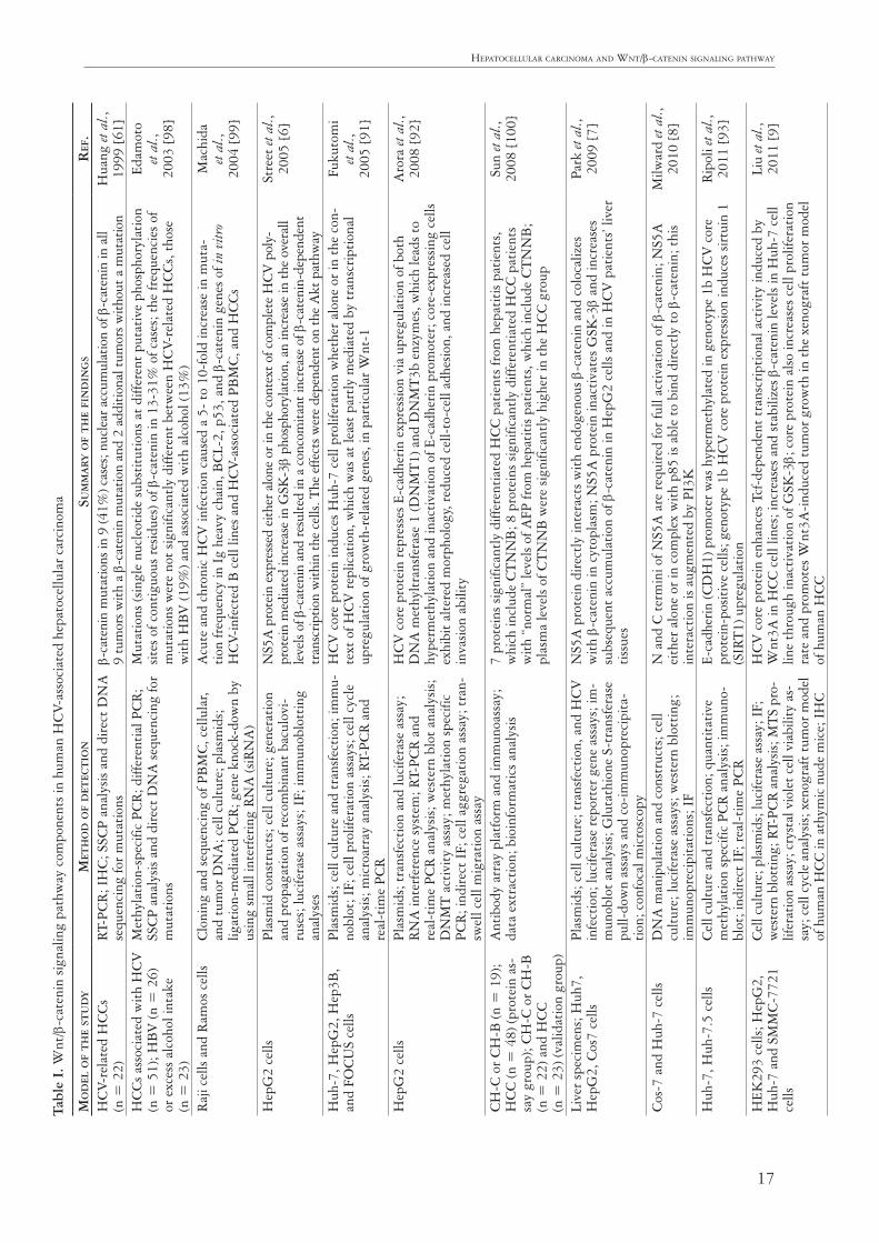
The most important in vitro and in vivo studies on Wnt/β-catenin signaling pathway components in HCV-related hepatocellular carcinomas are summa-rized in Table I.
The authors declare no conflict of interest.
17
Hepatocellular carcinoma and Wnt/β-catenin signaling patHWayTa
ble
I. W
nt/β
-cat
enin
sig
nalin
g pa
thw
ay c
ompo
nent
s in
hum
an H
CV-
asso
ciat
ed h
epat
ocel
lula
r ca
rcin
oma
mo
del
of
th
e st
ud
ym
eth
od
of
det
ect
ion
sum
ma
Ry o
f t
he
fin
din
gs
Ref
.H
CV-
rela
ted
HC
Cs
(n =
22)
RT-
PCR
; IH
C; S
SCP
anal
ysis
and
dir
ect
DN
A
sequ
enci
ng fo
r m
utat
ions
β-ca
teni
n m
utat
ions
in 9
(41%
) cas
es; n
ucle
ar a
ccum
ulat
ion
of β
-cat
enin
in a
ll
9 tu
mor
s with
a β
-cat
enin
mut
atio
n an
d 2
addi
tiona
l tum
ors w
ithou
t a m
utat
ion
Hua
ng et
al.,
19
99 [
61]
HC
Cs
asso
ciat
ed w
ith
HC
V
(n =
51)
; HB
V (n
= 2
6)
or e
xces
s al
coho
l int
ake
(n
= 2
3)
Met
hyla
tion
-spe
cific
PC
R; d
iffer
enti
al P
CR
; SS
CP
anal
ysis
and
dir
ect
DN
A s
eque
ncin
g fo
r m
utat
ions
Mut
atio
ns (s
ingl
e nu
cleo
tide
sub
stit
utio
ns a
t di
ffere
nt p
utat
ive
phos
phor
ylat
ion
site
s of
con
tigu
ous
resi
dues
) of β
-cat
enin
in 1
3-31
% o
f cas
es; t
he fr
eque
ncie
s of
m
utat
ions
wer
e no
t si
gnifi
cant
ly d
iffer
ent
betw
een
HC
V-re
late
d H
CC
s, t
hose
w
ith
HB
V (1
9%) a
nd a
ssoc
iate
d w
ith
alco
hol (
13%
)
Edam
oto
et
al.,
20
03 [
98]
Raj
i cel
ls a
nd R
amos
cel
lsC
loni
ng a
nd s
eque
ncin
g of
PB
MC
, cel
lula
r, an
d tu
mor
DN
A; c
ell c
ultu
re; p
lasm
ids;
lig
atio
n-m
edia
ted
PCR
; gen
e kn
ock-
dow
n by
us
ing
smal
l int
erfe
ring
RN
A (s
iRN
A)
Acu
te a
nd c
hron
ic H
CV
infe
ctio
n ca
used
a 5
- to
10-
fold
incr
ease
in m
uta-
tion
freq
uenc
y in
Ig
heav
y ch
ain,
BC
L-2,
p53
, and
β-c
aten
in g
enes
of i
n vi
tro
HC
V-in
fect
ed B
cel
l lin
es a
nd H
CV-
asso
ciat
ed P
BM
C, a
nd H
CC
s
Mac
hida
et
al.,
20
04 [
99]
Hep
G2
cells
Plas
mid
con
stru
cts;
cel
l cul
ture
; gen
erat
ion
and
prop
agat
ion
of r
ecom
bina
nt b
acul
ovi-
ruse
s; lu
cife
rase
ass
ays;
IF;
imm
unob
lott
ing
anal
yses
NS5
A p
rote
in e
xpre
ssed
eith
er a
lone
or i
n th
e co
ntex
t of c
ompl
ete
HC
V p
oly-
prot
ein
med
iate
d in
crea
se in
GSK
-3β
phos
phor
ylat
ion,
an
incr
ease
in th
e ov
eral
l le
vels
of β
-cat
enin
and
resu
lted
in a
con
com
itant
incr
ease
of β
-cat
enin
-dep
ende
nt
tran
scrip
tion
with
in th
e ce
lls. T
he e
ffect
s wer
e de
pend
ent o
n th
e A
kt p
athw
ay
Stre
et et
al.,
20
05 [
6]
Huh
-7, H
epG
2, H
ep3B
, an
d FO
CU
S ce
llsPl
asm
ids;
cel
l cul
ture
and
tra
nsfe
ctio
n; im
mu-
nobl
ot; I
F; c
ell p
rolif
erat
ion
assa
ys; c
ell c
ycle
an
alys
is; m
icro
arra
y an
alys
is; R
T-PC
R a
nd
real
-tim
e PC
R
HC
V c
ore
prot
ein
indu
ces
Huh
-7 c
ell p
rolif
erat
ion
whe
ther
alo
ne o
r in
the
con
-te
xt o
f HC
V r
eplic
atio
n, w
hich
was
at
leas
t pa
rtly
med
iate
d by
tra
nscr
ipti
onal
up
regu
lati
on o
f gro
wth
-rel
ated
gen
es, i
n pa
rtic
ular
Wnt
-1
Fuku
tom
i et
al.,
20
05 [
91]
Hep
G2
cells
Plas
mid
s; t
rans
fect
ion
and
luci
fera
se a
ssay
; R
NA
inte
rfer
ence
sys
tem
; RT-
PCR
and
re
al-t
ime
PCR
ana
lysi
s; w
este
rn b
lot
anal
ysis
; D
NM
T a
ctiv
ity
assa
y; m
ethy
lati
on s
peci
fic
PCR
; ind
irec
t IF
; cel
l agg
rega
tion
ass
ay; t
ran-
swel
l cel
l mig
rati
on a
ssay
HC
V c
ore
prot
ein
repr
esse
s E-
cadh
erin
exp
ress
ion
via
upre
gula
tion
of b
oth
DN
A m
ethy
ltra
nsfe
rase
1 (D
NM
T1)
and
DN
MT
3b e
nzym
es, w
hich
lead
s to
hy
perm
ethy
lati
on a
nd in
acti
vati
on o
f E-c
adhe
rin
prom
oter
; cor
e-ex
pres
sing
cel
ls
exhi
bit
alte
red
mor
phol
ogy,
red
uced
cel
l-to
-cel
l adh
esio
n, a
nd in
crea
sed
cell
inva
sion
abi
lity
Aro
ra et
al.,
20
08 [
92]
CH
-C o
r C
H-B
(n =
19)
; H
CC
(n =
48)
(pro
tein
as-
say
grou
p); C
H-C
or
CH
-B
(n =
22)
and
HC
C
(n =
23)
(val
idat
ion
grou
p)
Ant
ibod
y ar
ray
plat
form
and
imm
unoa
ssay
; da
ta e
xtra
ctio
n; b
ioin
form
atic
s an
alys
is7
prot
eins
sig
nific
antl
y di
ffere
ntia
ted
HC
C p
atie
nts
from
hep
atit
is p
atie
nts,
w
hich
incl
ude
CT
NN
B; 8
pro
tein
s si
gnifi
cant
ly d
iffer
enti
ated
HC
C p
atie
nts
wit
h “n
orm
al”
leve
ls o
f AFP
from
hep
atit
is p
atie
nts,
whi
ch in
clud
e C
TN
NB
; pl
asm
a le
vels
of C
TN
NB
wer
e si
gnifi
cant
ly h
ighe
r in
the
HC
C g
roup
Sun
et al
.,
2008
[10
0]
Live
r sp
ecim
ens;
Huh
7,
Hep
G2,
Cos
7 ce
llsPl
asm
ids;
cel
l cul
ture
; tra
nsfe
ctio
n, a
nd H
CV
in
fect
ion;
luci
fera
se r
epor
ter
gene
ass
ays;
im-
mun
oblo
t an
alys
is; G
luta
thio
ne S
-tra
nsfe
rase
pu
ll-do
wn
assa
ys a
nd c
o-im
mun
opre
cipi
ta-
tion
; con
foca
l mic
rosc
opy
NS5
A p
rote
in d
irec
tly
inte
ract
s w
ith
endo
geno
us β
-cat
enin
and
col
ocal
izes
w
ith
β-ca
teni
n in
cyt
opla
sm; N
S5A
pro
tein
inac
tiva
tes
GSK
-3β
and
incr
ease
s su
bseq
uent
acc
umul
atio
n of
β-c
aten
in in
Hep
G2
cells
and
in H
CV
pat
ient
s’ liv
er
tiss
ues
Park
et a
l.,
2009
[7]
Cos
-7 a
nd H
uh-7
cel
lsD
NA
man
ipul
atio
n an
d co
nstr
ucts
; cel
l cu
ltur
e; lu
cife
rase
ass
ays;
wes
tern
blo
ttin
g;
imm
unop
reci
pita
tion
s; I
F
N a
nd C
ter
min
i of N
S5A
are
req
uire
d fo
r fu
ll ac
tiva
tion
of β
-cat
enin
; NS5
A
eith
er a
lone
or
in c
ompl
ex w
ith
p85
is a
ble
to b
ind
dire
ctly
to
β-ca
teni
n; t
his
inte
ract
ion
is a
ugm
ente
d by
PI3
K
Milw
ard
et al
., 20
10 [
8]
Huh
-7, H
uh-7
.5 c
ells
Cel
l cul
ture
and
tra
nsfe
ctio
n; q
uant
itat
ive
met
hyla
tion
spe
cific
PC
R a
naly
sis;
imm
uno-
blot
; ind
irec
t IF
; rea
l-ti
me
PCR
E-ca
dher
in (C
DH
1) p
rom
oter
was
hyp
erm
ethy
late
d in
gen
otyp
e 1b
HC
V c
ore
prot
ein-
posit
ive
cells
; gen
otyp
e 1b
HC
V c
ore
prot
ein
expr
essio
n in
duce
s sir
tuin
1
(SIR
T1)
upr
egul
atio
n
Rip
oli e
t al.,
20
11 [
93]
HEK
293
cells
; Hep
G2,
H
uh-7
and
SM
MC
-772
1 ce
lls
Cel
l cul
ture
; pla
smid
s; lu
cife
rase
ass
ay; I
F;
wes
tern
blo
ttin
g; R
T-PC
R a
naly
sis; M
TS
pro-
lifer
atio
n as
say;
cry
stal
vio
let
cell
viab
ility
as-
say;
cel
l cyc
le a
naly
sis; x
enog
raft
tum
or m
odel
of
hum
an H
CC
in a
thym
ic n
ude
mic
e; I
HC
HC
V c
ore
prot
ein
enha
nces
Tcf
-dep
ende
nt t
rans
crip
tion
al a
ctiv
ity
indu
ced
by
Wnt
3A in
HC
C c
ell l
ines
; inc
reas
es a
nd s
tabi
lizes
β-c
aten
in le
vels
in H
uh-7
cel
l lin
e th
roug
h in
acti
vati
on o
f GSK
-3β;
cor
e pr
otei
n al
so in
crea
ses
cell
prol
ifera
tion
ra
te a
nd p
rom
otes
Wnt
3A-i
nduc
ed t
umor
gro
wth
in t
he x
enog
raft
tum
or m
odel
of
hum
an H
CC
Liu
et al
.,
2011
[9]

18
Karol rogacKi, aldona KasprzaK, adrian stępińsKi
mo
del
of
th
e st
ud
ym
eth
od
of
det
ect
ion
sum
ma
Ry o
f t
he
fin
din
gs
Ref
.H
EK29
3 ce
lls a
nd t
he
hum
an h
epat
oma
cell
line
SMM
C-7
721
Cel
l cul
ture
; pla
smid
s; lu
cife
rase
ass
ay;
RT-
PCR
; wes
tern
blo
ttin
g; I
F; M
TS
pr
olife
rati
on a
ssay
; cry
stal
vio
let
stai
ning
HC
V c
ore
prot
ein
play
s rol
e in
act
ivat
ing
β-ca
teni
n/Tc
f-4-d
epen
dent
tran
scrip
tio na
l ac
tivity
and
incr
ease
s β-c
aten
in e
xpre
ssio
n an
d nu
clea
r acc
umul
atio
n of
the
pro t
ein;
co
re p
rote
in u
preg
ulat
es g
ene
expr
essio
n of
can
onic
al W
nt li
gand
s (W
nt-2
, -3,
-3a,
-8
b, -1
0a, -
10b,
Fzd
-1, -
2, -5
, -6,
-7, -
9, a
nd L
RP5
/6 c
o-re
cept
ors)
Liu
et al
.,
2011
[10
]
Pati
ent’s
ser
um s
ampl
es
wit
h H
CV
gen
otyp
e 4-
asso
-ci
ated
HC
C p
atie
nts
(n
= 3
2); C
H-C
pat
ient
s
(n =
28)
; asy
mpt
omat
ic
carr
iers
(ASC
) wit
h no
n-ci
r-rh
otic
CH
-C (n
= 1
1)
ELIS
ASe
rum
β-c
aten
in le
vels
wer
e si
gnifi
cant
ly e
leva
ted
in p
atie
nts
wit
h H
CC
com
-pa
red
to t
hose
wit
h C
H, A
SC a
nd h
ealt
hy c
ontr
ols.
Am
ong
the
six
stud
ied
mar
kers
, β-c
aten
in w
as a
lso
foun
d to
be
the
only
mar
ker
that
can
dis
crim
inat
e be
twee
n pa
tien
ts w
ith
HC
C a
nd t
hose
wit
h ch
roni
c he
pati
tis
Zek
ri et
al.,
20
11 [
84]
Hum
an h
epat
ic t
issu
es:
cont
rols
; CH
-C (n
= 3
4),
HC
V-as
soci
ated
HC
C
(n =
10)
; Huh
-7 c
ell l
ine
Cel
l cul
ture
; RT-
PCR
, rea
l-ti
me
PCR
; cel
l tr
ansf
ecti
on a
nd s
tim
ulat
ion;
wes
tern
blo
t an
alys
is, c
ell p
rolif
erat
ion
assa
y; fl
ow c
ytom
et-
ric
anal
ysis
HC
V in
fect
ion
resu
lted
in N
F-κB
-dep
ende
nt u
p-re
gula
tion
of m
iR-1
55 e
xpre
s-si
on, w
hich
pro
mot
ed t
umor
igen
esis
by
incr
easi
ng W
nt s
igna
ling;
APC
, whi
ch
nega
tive
ly r
egul
ates
Wnt
sig
nalin
g, w
as id
enti
fied
as t
he fu
ncti
onal
tar
get
of
miR
-155
Zha
ng et
al.,
20
12 [
89]
IHH
, Huh
-7 c
ells
; bio
psy
spec
imen
s fr
om H
CV-
in-
fect
ed p
atie
nts
(n =
10)
Gen
erat
ion
of c
ell c
ultu
re-g
row
n H
CV
; EM
T
arra
ys; w
este
rn b
lot
anal
ysis
; IF;
β-g
alac
tosi
-da
se s
tain
ing
for
cellu
lar
sene
scen
ce
HC
V in
fect
ed h
epat
ocyt
es (H
CV
gen
otyp
e 1a
or
2a) d
ispl
ayed
a fi
brob
last
-lik
e sh
ape
and
an e
xten
ded
life
span
. Inc
reas
ed m
RN
A a
nd p
rote
in e
xpre
ssio
n le
vels
of
vim
enti
n, s
nail,
slu
g, a
nd t
wis
t; lo
ss o
f the
epi
thel
ial c
ell m
arke
r E-
cadh
erin
; pr
imar
y hu
man
hep
atoc
ytes
infe
cted
wit
h H
CV
dis
play
EM
T v
ia a
ctiv
atio
n of
th
e A
kt/β
-cat
enin
sig
nalin
g pa
thw
ay
Bos
e et
al.,
20
12 [
94]
BM
EL c
ells
; HC
V-in
fect
ed
prim
ary
hum
an h
epat
ocyt
esC
ell c
ultu
re; T
GF-
β re
port
er a
ssay
; ret
rovi
ral
cons
truc
ts a
nd R
NA
inte
rfer
ence
; Wes
tern
bl
ot a
nd I
F; q
PCR
; cel
l tra
ckin
g by
tim
e-la
pse
mic
rosc
opy;
wou
nd-h
ealin
g as
say;
inva
sion
as
say;
xen
ogra
ft m
odel
Expr
essi
on o
f NS5
A H
CV
in p
rim
ary
hepa
tic
prec
urso
rs a
nd in
IH
H c
ell l
ines
ga
ve r
ise
to p
rofo
und
mod
ifica
tion
s of
cel
l pol
arit
y, le
adin
g to
EM
T; t
he e
ffect
s of
NS5
A w
ere
addi
tive
to
thos
e of
TG
F-β;
NS5
A c
oope
rate
s w
ith
onco
geni
c R
as,
givi
ng r
ise
to t
rans
form
ed, i
nvas
ive
cells
tha
t ar
e hi
ghly
tum
orig
enic
in v
ivo
Akk
ari e
t al.,
20
12 [
95]
Hep
atoc
yte
cell
lines
har
-bo
ring
an
HC
V r
eplic
on
and
the
infe
ctio
us H
CV
st
rain
JFH
1; t
rans
geni
c m
urin
e m
odel
exp
ress
ing
the
enti
re H
CV
OR
F
Cel
l cul
ture
In
crea
sed
c-M
yc e
xpre
ssio
n; a
ctiv
atio
n of
Akt
by
NS5
A, a
nd t
he s
ubse
quen
t st
abili
zati
on o
f β-c
aten
in; β
-cat
enin
-dep
ende
nt c
-Myc
exp
ress
ion
led
to in
crea
sed
prod
ucti
on o
f RO
S, m
itoc
hond
rial
per
turb
atio
n, e
nhan
ced
DN
A d
amag
e an
d ab
erra
nt c
ell-
cycl
e ar
rest
Hig
gs, e
t al.,
20
13 [
90]
Huh
-7, H
epG
2 ce
llsC
ell c
ultu
re
HC
V c
ore
prot
ein
dow
nreg
ulat
es S
FRP1
exp
ress
ion
by in
duci
ng h
yper
met
hyla
-ti
on o
f the
SFR
P1 p
rom
oter
, whi
ch m
ay le
ad t
o ac
tiva
tion
of t
he W
nt s
igna
ling
path
way
and
con
trib
ute
to H
CC
agg
ress
iven
ess
thro
ugh
indu
ctio
n of
EM
T; c
ore
prot
ein
mar
kedl
y in
crea
ses
the
expr
essi
on le
vel a
nd b
indi
ng o
f DN
A m
ethy
l-tr
ansf
eras
e-1
and
hist
one
deac
etyl
ase-
1, r
esul
ting
in e
pige
neti
c si
lenc
ing
of
SFR
P1 e
xpre
ssio
n
Qua
n et
al.,
2013
[97
]
BM
EL c
ells
; HC
V-in
fect
ed
prim
ary
hum
an h
epat
ocyt
esC
ell c
ultu
re; T
GF-
β re
port
er a
ssay
; ret
rovi
ral
cons
truc
ts a
nd R
NA
inte
rfer
ence
; Wes
tern
bl
ot a
nd I
F; q
PCR
; cel
l tra
ckin
g by
tim
e-la
pse
mic
rosc
opy;
wou
nd-h
ealin
g as
say;
inva
sion
as
say;
xen
ogra
ft m
odel
No
evid
ence
eit
her
of in
crea
sed
expr
essi
on o
f com
pone
nts
of W
nt/β
-cat
enin
pa
thw
ay o
r, m
ore
sign
ifica
ntly
, of s
usta
ined
tra
nscr
ipti
onal
act
ivat
ion
of a
xin2
in
BM
EL-N
S5A
cel
ls u
nder
goin
g EM
T; t
heir
res
ults
sug
gest
tha
t β-
cate
nin
sign
al-
ing
is n
ot r
equi
red
for
NS5
AC
-med
iate
d EM
T
Gré
goir
e
et al
.,
2013
[96
]
APC
– a
deno
mat
ous p
olypo
sis co
li; B
MEL
– b
ipot
entia
l mou
se em
bryo
nic l
iver
; CH
– ch
roni
c hep
atiti
s; CH
-C/B
– ch
roni
c hep
atiti
s C/B
; ELI
SA –
enzy
me-
linke
d im
mun
osor
bent
assa
y; E
MT
– ep
itheli
al to
mese
nchy
mal
tran
sitio
n; F
OCU
S –
(Fri
end-
ship
of C
hina
and
Uni
ted S
tates
) – h
uman
hep
atoc
ellul
ar ca
rcin
oma
cell l
ine;
HCC
s – h
uman
hep
atoc
ellul
ar ca
rcin
omas
; IH
C –
imm
unoc
ytoc
hem
istry
; IH
H –
imm
orta
lized
hum
an h
epat
ocyt
es; m
iR-1
55 –
micr
oRN
A-1
55; O
RF
– op
en re
adin
g fra
me;
PBM
C –
peri
pher
al b
lood
mon
onuc
lear c
ell; R
T-PC
R –
reve
rse t
rans
crip
tion
poly
mer
ase c
hain
reac
tion;
SFR
P1 –
secr
eted
frizz
led-r
elated
pro
tein
1; T
GF-
β –
tran
sform
ing
grow
th fa
ctor β
; SSC
P –
singl
e-str
and
conf
orm
atio
n po
lym
orph
ism
Tabl
e I.
Con
t.
19
Hepatocellular carcinoma and Wnt/β-catenin signaling patHWay
References1. Behari J. The Wnt/β-catenin signaling pathway in liver bi-
ology and disease. Expert Rev Gastroenterol Hepatol 2010; 4: 745-756.
2. Micsenyi A, Tan X, Sneddon T, et al. Beta-catenin is tempo-rally regulated during normal liver development. Gastroen-terology 2004; 26: 1134-1146.
3. Monga SPS. Role of Wnt/Beta-Catenin signaling in liver metabolism and cancer. Int J Biochem Cell Biol 2011; 43: 1021-1029.
4. Selimovic D, El-Khattouti A, Ghozlan H, et al. Hepatitis C virus-related hepatocellular carcinoma: an insight into molec-ular mechanisms and therapeutic strategies. World J Hepatol 2012; 4: 342-355.
5. Hsu HC, Jeng YM, Mao TL, et al. Beta-catenin mutations are associated with a subset of low-stage hepatocellular carci-noma negative for hepatitis B virus and with favorable prog-nosis. Am J Pathol 2000; 157: 763-770.
6. Street A, Macdonald A, McCormick C, et al. Hepatits C virus NS5A-mediated activation of phosphoinositide 3-kinase results in stabilization of cellular β-catenin and stimulation of β-caten-in-responsive transcription. J Virol 2005; 79: 5006-5016.
7. Park CY, Choi SH, Kang SM, et al. Nonstructural 5A protein activates β-catenin signaling cascades: implication of hepati-tis C virus-induced liver pathogenesis. J Hepatol 2009; 51: 853-864.
8. Milward A, Mankouri J, Harris M. Hepatitis C virus NS5A protein interacts with beta-catenin and stimulates its tran-scriptional activity in a phosphoinositide-3-kinase-dependent fashion. J Gen Virol 2010; 91: 373-381.
9. Liu J, Ding X, Tang J, et al. Enhancement of canonical Wnt/β-catenin signaling activity by HCV core protein pro-motes cell growth of hepatocellular carcinoma cells. PLoS One 2011; 6: e27496.
10. Liu J, Wang Z, Tang J, et al. Hepatitis C virus core protein activates Wnt/β-catenin signaling through multiple regula-tion of upstream molecules in the SMMC-7721 cell line. Arch Virol 2011; 156: 1013-1023.
11. Behrens J, Jerchow BA, Würtele M, et al. Functional interac-tion of an axin homolog, conductin, with beta-catenin, APC, and GSK3beta. Science 1998; 280: 596-599.
12. van Noort M, Meeldijk J, van der Zee R, et al. Wnt signaling controls the phosphorylation status of β-catenin. J Biol Chem 2002; 277: 17901-17905.
13. Liu C, Li Y, Semoov M, et al. Control of beta-catenin phos-phorylation/degradation by a dual-kinase mechanism. Cell 2002; 108: 837-847.
14. Swarz-Romond T, Asbrand C, Bakkers J, et al. The ankyrin repeat protein Diversin recruits Casein kinase I epsilon to the β-catenin degradation complex and acts in both canonical Wnt and Wnt/JNK signaling. Genes Dev 2002; 16: 2073-2084.
15. Lusting B, Jerchow B, Sachs M, et al. Negative feedback loop of Wnt signaling through up regulation of conductin/Axin2 in colorectal and liver tumors. Mol Cell Biol 2002; 22: 1184-1193.
16. Spink KE, Polakis P, Weis WI. Structural basis of the Ax-in-adematous polyposis coli interaction. EMBO J 2000; 19: 2270-2279.
17. Rosin-Arbesfeld R, Cliffe A, Brabletz T, et al. Nuclear export of the APC tumour suppressor controls β-catenin function in transcription. EMBO J 2003; 22: 1101-1113.
18. Pötter E, Bergwitz C, Brabant G. The Cadherin-Catenin sys-tem: implications for growth and differentiation of endocrine tissues. Endocr Rev 1999; 20: 207-239.
19. Phillips MJ, Poucell S, Patterson J, et al. The liver. An atlas and text of ultrastructural pathology. Raven Press, New York 1987; 2-3.
20. Farquhar MG, Palade GE. Junctional complexes in various epithelia. J Cell Biol 1963; 17: 375-412.
21. Konopka G, Tekiela J, Iverson M, et al. Junctional adhesion molecule-A is critical for the formation of pseudocanaliculi and modulates E-cadherin expression in hepatic cells. J Biol Chem 2007; 282: 28137-28148.
22. Peifer M, Berg S, Reynolds AB. A repeating amino acid motif shared by proteins with diverse cellular roles. Cell 1994; 76: 789-791.
23. Zeng X, Huang H, Tamai K, et al. Initiation of Wnt signal-ing: control of Wnt coreceptor Lrp 6 phosphorylation/activa-tion via frizzled, dishevelled and axin functions. Development 2008; 135: 367-375.
24. Ring L, Neth P, Weber C, et al. β-Catenin dependent pathway activation by both promiscuous “canonical” WNT3a-, and specific “noncanonical” WNT4- and WNT5a-FZD receptor combinations with strong differences in LRP5 and LRP6 de-pendency. Cell Signal 2014; 26: 260-267.
25. Wharton KA Jr. Runnin′ with Dvl: proteins that associate with Dsh/Dvl and their significance to Wnt signal transduc-tion. Dev Biol 2003; 253: 1-17.
26. Du SJ, Purcell MS, Christian JL, et al. Identification of dis-tinct classes and functional domains of Wnts through expres-sion of wild-type and chimeric proteins in Xenopus embryos. Mol Cell Biol 1995; 15: 2625-2634.
27. Schulte G, Bryja V. The Frizzled family of unconventional G-protein-coupled receptors. Trends Pharmacol Sci 2007; 10: 518-525.
28. Huang HC, Klein PS. The Frizzled family: receptors for mul-tiple signal transduction pathways. Genome Biol 2004; 5: 234.
29. Zhao Z, Lee CC, Baldini A, et al. A human homologue of Drosophila polarity gene frizzled has been identified and mapped to 17q21.1. Genomics 1995; 27: 370-373.
30. Sagara N, Toda G, Hirai M, et al. Molecular cloning, differ-ential expression, and chromosomal localization of human frizzled-1, frizzled-2, and frizzled-7. Biochem Biophys Res Commun 1998; 252: 117-122.
31. King TD, Zhang W, Suto MJ, et al. Frizzled7 as an emerging target for cancer therapy. Cell Signal 2012; 24: 846-851.
32. Henderson BR, Fagotto F. The ins and outs of APC and β-catenin nuclear transport. EMBO Rep 2002; 3: 834-839.
33. Clevers H. Wnt/beta-catenin signaling in development and disease. Cell 2006; 127: 469-480.
34. MacDonald BT, Tamai K, He X. Wnt/beta-catenin signaling: components, mechanisms, and diseases. Dev Cell 2009; 17: 9-26.
35. Cong F, Varmus H. Nuclear-cytoplasmic shuttling of Axin regulates subcellular localisation of β-catenin. Proc Natl Acad Sci U S A 2004; 101: 2882-2887.
36. Hendriksen J, Fagotto F, van der Valde H, et al. RanBP3 en-hances nuclear export of active (beta)-catenin independently of CRM1. J Cell Biol 2005; 171: 785-797.
37. Krieghoff E, Behrens J, Mayr B. Nucleo-cytoplasmic distribu-tion of beta-catenin is regulated by retention. J Cell Sci 2006; 119: 1453-1463.
38. Wu X, Tu X, Joeng KS, et al. Rac1 activation controls nuclear localization of beta-catenin during canonical Wnt signaling. Cell 2008; 133: 340-353.
39. Ha JR, Hao L, Venkateswaran G, et al. β-catenin is O-Glc-NAc glycosylated at serine 23: implications for β-catenin’s subcellular localization and tranctivation function. Exp Cell Res 2014; 321: 153-166.
40. Hoppler S, Kavanagh CL. Wnt signaling: variety at the core. J Cell Sci 2007; 120: 385-393.
41. Brantjes H, Roose J, van De Watering M, et al. All Tcf HMG box transcription factors interact with Groucho-related co-re-pressors. Nucleic Acid Res 2001; 29: 1410-1419.
20
Karol rogacKi, aldona KasprzaK, adrian stępińsKi
42. Nelson WJ. Regulation of cell-cell adhesion by the cadher-in-catenin complex. Biochem Soc Trans 2008; 36: 149-155.
43. Gumbiner BM. Regulation of cadherin-mediated adhesion in morphogenesis. Nat Rev Mol Cell Biol 2005; 6: 622-634.
44. Berndorff D, Gessner R, Kreft B, et al. Liver-intestine cad-herin: molecular cloning and characterization of a novel Ca (2+)-dependent cell adhesion molecule expressed in liver and intestine. J Cell Biol 1994; 125: 1353-1369.
45. Tian X, Liu Z, Niu B, et al. E-cadherin/β-catenin complex and the epithelial barrier. J Biomed Biotech 2011; 2011: 567305; doi:10.1155/2011/567305.
46. Nollet F, Kools P, van Roy F. Phylogenetic analysis of the cad-herin superfamily allows identification of six major subfami-lies besides several solitary members. J Mol Biol 2000; 299: 551-572.
47. Davis MA, Ireton RC, Reynolds AB. A core function for p120-catenin in cadherin turnover. J Cell Biol 2003; 163: 525-534.
48. Fujita Y, Krause G, Scheffner M, et al. Hakai, a c-Cbl-like protein, ubiquitinates and induces endocytosis of the E-cad-herin complex. Nat Cell Biol 2002; 4: 222-231.
49. Guarino M, Tosoni A, Nebuloni M. Direct contribution of ep-ithelium to organ fibrosis: epithelial-mesenchymal transition. Hum Pathol 2009; 40: 1365-1376.
50. Rygiel KA, Robertson H, Marshall HL, et al. Epithelial-mes-enchymal transition contributes to portal tract fibrogenesis during human chronic liver disease. Lab Invest 2008; 88: 112-123.
51. Cheng JH, She H, Han YP, et al. Wnt antagonism inhibits hepatic stellate cell activation and liver fibrosis. Am J Physiol Gastrointest Liver Physiol 2008; 294: G39-G49.
52. Myung SJ, Yoon JH, Gwak GY, et al. Wnt signaling enhanc-es the activation and survival of human hepatic stellate cells. FEBS Lett 2007; 581: 2954-2958.
53. Maretzky T, Reiss K, Ludwig A, et al. ADAM10 mediates E-cadherin shedding and regulates ephitelial cell-cell adhe-sion, migration, and beta-catenin translocation. Proc Natl Acad Sci 2005; 102: 9182-9187.
54. Marambaud P, Shioi J, Serban G, et al. A presenilin-1/gam-ma-secretase cleavage releases the E-Cadherin intracellular domain and regulates disassembly of adherens junctions. EMBO J 2002; 21: 1948-1956.
55. Strathdee G. Epigenetic versus genetic alterations in the in-activation of E-cadherin. Semin Cancer Biol 2002; 12: 373-379.
56. Conacci-Sorrell M, Simcha I, Ben-Yedidia T, et al. Autoregu-lation of E-cadherin expression by cadherin-cadherin interac-tions: the roles of beta-catenin signaling, Slug, and MAPK. J Cell Biol 2003; 163: 847-857.
57. Masszi A, Fan L, Rosivall L, et al. Integrity of cell-cell contacts is a critical regulator of TGF-β-induced epithelial-to-myo-fibroblast transition: role for β-catenin. Am J Pathol 2004; 165: 1955-1967.
58. Tian YC, Fraser D, Attisano L, et al. TGF-β1-mediated al-terations of renal proximal tubular epithelial phenotype. Am J Physiol 2003; 285: F130-F142.
59. Miyoshi Y, Iwao K, Nagasawa Y, et al. Activation of the β-cat-enin gene in primary hepatocellular carcinomas by somatic al-terations involving exon 3. Cancer Res 1998; 58: 2524-2527.
60. Blaker H, Hofmann WJ, Rieker RJ, et al. Beta-catenin accu-mulation and mutation of the CTNNB1 gene in hepatoblas-toma. Genes Chromosomes Cancer 1999; 25: 399-402.
61. Huang H, Fujii H, Sankila A, et al. β-catenin mutations are frequent in human hepatocelllar carcinomas associated with hepatitis C virus infection. Am J Pathol 1999; 155: 1795-1801.
62. Mao TL, Chu JS, Jeng MY, et al. Expression of mutant nucle-ar β-catenin correlates with non-invasive hepatocellular car-
cinoma, absence of portal vein spread, and good prognosis. J Pathol 2001; 193: 95-101.
63. Wong CM, Fan ST, Ng IO. Beta-catenin mutation and over-expression in hepatocellular carcinoma: clinicopathological and prognostic significance. Cancer 2001; 92: 136-145.
64. Inagawa S, Itabashi M, Adachi S, et al. Expression and prog-nostic roles of beta-catenin in hepatocellular carcinoma: cor-relation with tumor progression and postoperative survival. Clin Cancer Res 2002; 8: 450-456.
65. Ueta T, Ikeguchi M, Hirooka Y, et al. Beta-catenin and cy-clin D1 expression in human hepatocellular carcinoma. Oncol Rep 2002; 9: 1197-1203.
66. Zulehner G, Mikula M, Schneller D, et al. Nuclear β-catenin induces as early liver progenitor phenotype in hepatocellu-lar carcinoma and promotes tumor recurrence. Am J Pathol 2010; 176: 472-481.
67. Pez F, Lopez A, Kim M, et al. Wnt signaling and hepato-cencerogenesis: molecular targets for the development of in-novative anticancer drugs. J Hepatol 2013; 59: 1107-1117.
68. Cui J, Zhou X, Liu Y, et al. Alterations of beta-catenin and Tcf-4 instead of GSK-3beta contribute to activation of Wnt pathway in hepatocellular carcinoma. Chin Med J 2003; 116: 1885-1892.
69. Taniguchi K, Roberts LR, Aderca IN, et al. Mutational spec-trum of beta-catenin, AXIN1, and AXIN2 in hepatocellular carcinomas and hepatoblastomas. Oncogene 2002; 21: 4863-4871.
70. Park JY, Park WS, Nam SW, et al. Mutations of beta-catenin and AXIN 1 genes are a late event in human hepatocellular carcinogenesis. Liver Int 2005; 25: 70-76.
71. Suzuki T, Yano H, Nakashima Y, et al. Beta-catenin expres-sion in hepatocellular carcinoma: a possible participation of beta-catenin in the dedifferentiation process. J Gastroenterol Hepatol 2002; 17: 994-1000.
72. Calvisi DF, Factor VM, Loi R, et al. Activation of beta-catenin during hepatocarcinogenesis in transgenic mouse models: re-lationship to phenotype and tumor grade. Cancer Res 2001; 61: 2085-2091.
73. Hoshida Y, Nijman SM, Kobayashi M, et al. Integrative transcriptome analysis reveals common molecular subclasses of human hepatocellular carcinoma. Cancer Res 2009; 69: 7385-7392.
74. Harada N, Miyoshi H, Murai N, et al. Lack of tumorigene-sis in the mouse liver after adenovirus-mediated expression of a dominant stable mutant of β-catenin. Cancer Res 2002; 62: 1971-1977.
75. Stauffer JK, Scarzello AJ, Andersen JB, et al. Coactivation of AKT and beta-catenin in mice rapidly induces formation of lipogenic liver tumors. Cancer Res 2011; 71: 2718-2727.
76. Rignall B, Braeuning A, Buchmann A, et al. Tumor formation in liver of conditional beta-catenin-deficient mice exposed to a diethylnitrosamine/phenobarbital tumor promotion regi-men. Carcinogenesis 2010; 32: 52-57.
77. Yang W, Yan HX, Chen L, et al. Wnt/β-catenin signaling contributes to activation of normal and tumorigenic liver pro-genitor cells. Cancer Res 2008; 68: 4287-4295.
78. Dumble ML, Croager EJ, Yeoh GC, et al. Generation and characterization of p53 null transformed progenitor cells: oval cells give rise to hepatocellular carcinoma. Carcinogene-sis 2002; 23: 435-445.
79. Libbrecht L, Desmet V, Van Damme B, et al. Deep intralob-ular extension of human hepatic “progenitor cells” correlates with parenchymal inflammation in chronic viral hepatitis: can ‘Progenitor cells’ migrate? J Pathol 2000; 192: 373-378.
80. Wu PC, Fang JW, Lau VK, et al. Classification of hepatocel-lular carcinoma according to hepatocellular and biliary differ-entiation markers. Clinical and biological implications. Am J Pathol 1996; 149: 1167-1175.
21
Hepatocellular carcinoma and Wnt/β-catenin signaling patHWay
81. Libbrecht L, Desmet V, Van Damme B, et al. The immuno-histochemical phenotype of displastic foci in human liver: correlation with putative progenitor cells. J Hepatol 2000; 33: 76-84.
82. Hu M, Kurobe M, Jeong YJ, et al. Wnt/beta-catenin signal-ing in murine hepatic transit amplifying progenitor cell. Gas-troenterology 2007; 133: 1579-1591.
83. Apte U, Thompson MD, Cui S, et al. Wnt/beta-catenin sig-naling mediates oval cell response in rodens. Hepatology 2008; 47: 288-295.
84. Zekri ARN, Bahnassy AA, El-Din HMA, et al. Serum levels of β-catenin as a potential marker for genotype 4/hepatitis C-associated hepatocellular carcinoma. Oncology Rep 2011; 26: 825-831.
85. de La Coste A, Romagnol B, Billuart P, et al. Somatic mu-tations of the beta-catenin gene are frequent in mouse and human hepatocellular carcinomas. Proc Natl Acad Sci U S A 1998; 95: 8847-8851.
86. Satoh S, Daigo Z, Furukawa Y, et al. AXIN1 mutation in hepatocellular carcinomas, and growth suppression in cancer cells by virus-mediated transfer of AXIN1. Nat Gen 2000; 24: 245-250.
87. Ban KC, Singh H, Krishnan R, et al. GSK-3β phosphoryla-tion and alteration of beta-catenin in hepatocellular carcino-ma. Cancer Lett 2003; 199: 201-208.
88. Merle P, de la Monte S, Kim M, et al. Functional consequenc-es of frizzled-7 receptor overexpression in human hepatocel-lular carcinoma. Gastroenterology 2004; 127: 1110-1122.
89. Zhang Y, Wei W, Cheng N, et al. Hepatitis C virus-induced up-regulation of microRNA-155 promotes hepatocarcino-genesis by activating Wnt signaling. Hepatology 2012; 56: 1631-1640.
90. Higgs MR, Lerat H, Pawlotsky JMN. Hepatitis C virus-in-duced activation of β-catenin promotes c-Myc expression and a cascade of pro-carcinogenetic events. Oncogene 2013; 32: 4683-4693.
91. Fukutomi T, Zhou Y, Kawai S, et al. Hepatitis C virus core pro-tein stimulates hepatocyte growth: correlation with upregula-tion of wnt-1 expression. Hepatology 2005; 41: 1096-1105.
92. Arora P, Kim EO, Jung JK, et al. Hepatitis C virus core pro-tein downregulates E-cadherin expression via activation of DNA methyltransferase 1 and 3b. Cancer Lett 2008; 261: 244-252.
93. Ripoli M, Barbano R, Balsamo T, et al. Hypermethylated levels of E-cadherin promoter in Huh-7 cells expressing the HCV core protein. Virus Res 2011; 160: 74-81.
94. Bose SK, Meyer K, Di Bisceglie AM, et al. Hepatitis C virus induces epithelial-mesenchymal transition in primary human hepatocytes. J Virol 2012; 86: 13621-13628.
95. Akkari L, Grégoire D, Floc’h N, et al. Hepatitis C viral pro-tein NS5A induces EMT and participates in oncogenic trans-formation of primary hepatocyte precursors. J Hepatol 2012; 57: 1021-1028.
96. Grégoire D, Akkari L, Carenco C, et al. reply to: “Are Hedge-hog and Wnt/β-catenin pathways involved in hepatitis C vi-rus-mediated EMT?” J Hepatol 2013; 58: 634-640.
97. Quan H, Zhou F, Nie D, et al. Hepatitis C virus core protein epigenetically silences SFRP1 and enhances HCC aggressive-ness by inducing epithelial mesenchymal transition. Onco-gene 2014; 33: 2826-2835.
98. Edamoto Y, Hara A, Biernat W, et al. Alterations of RB1, p53 and Wnt pathways in hepatocellular carcinoma associat-ed with hepatitis C, hepatitis B and alcoholic liver cirrhosis. Int J Cancer 2003; 106: 334-341.
99. Machida K, Cheng KT, Sung VM, et al. Hepatitis C virus induces a mutator phenotype: enhanced mutations of immu-noglobulin and proto-oncogenes. Proc Natl Acad Sci U S A 2004; 101: 4262-4267.
100. Sun H, Chua MS, Yang D, et al. Antibody arrays identify potential diagnostic markers of hepatocellular carcinoma. Biomarkers Insights 2008; 3: 1-18.
101. Guo C, Liu Q-G, Yang W, et al. Relation among p130Cas, E-cadherin and β-catenin expression, clinicopathologic sig-nificance and prognosis in human hepatocellular carcinoma. Hepatobiliary Pancreat Dis Int 2008; 7: 490-496.
102. Wei Y, Van Nhieu JT, Prigent S, et al. Altered expression of E-cadherin in hepatocellular carcinoma: correlations with genetic alterations, beta-catenin expression, and clinical fea-tures. Hepatology 2002; 36: 692-701.
103. Cui J, Zhou XD, Liu YK, et al. Abnormal beta-catenin gene expression with invasiveness of primary hepatocellular carci-noma in China. World J Gastroenterol 2001; 7: 542-546.
104. Zhai B, Yan H-X, Liu S-Q, et al. Reduced expression of E-cadherin/catenin complex in hepatocellular carcinoma. World J Gastroenterol 2008; 14: 5665-5673.
105. Lee JM, Yang J, Newell P, et al. β-Catenin signalling in hepa-tocellular cancer: Implications in inflammation, fibrosis, and proliferation. Cancer Letters 2014; 343: 90-97.
106. Ihara A, Koizumi H, Hashizume R, et al. Expression of epi-thelial cadherin and alpha- and beta-catenins in nontumoral livers and hepatocellular carcinomas. Hepatology 1996; 23: 1441-1447.
107. Cho SB, Lee KH, Lee JH, et al. Expression of E- and N-cad-herin and clinicopathologicy in hepatocellular carcinoma. Pathol Int 2008; 58: 635-642.
108. Załuska J, Melerowicz W, Prochowska E, et al. Novel system-ic approaches to treatment of HCC. Contemp Oncol (Pozn) 2010; 14: 23-30 (article in Polish).
Address for correspondenceAldona KasprzakDepartment of Histology and EmbryologyPoznan University of Medical SciencesŚwięcickiego 660-781 Poznan, Polande-mail: [email protected]