Kittikul Kovitanggoon a , Sung-Won Lee a , Nural Akchurin a ,
Open AccessResearch Article
Pharmaceutica Analytica Acta
Prokai-Tatrai et al., Pharm Anal Acta 2012, S7 DOI: 10.4172/2153-2435.S7-002
Drug Target and Drug DesignPharm Anal Acta ISSN: 2153-2435 PAA, an open access journal
Keywords: 17β-estradiol; Chemical delivery system; Estredox;Peripheral side effect; Circulating 17β-estradiol; Uterotrophic effect; LC-MS/MS; Porsolt swim test
IntroductionThe redox chemical delivery system (CDS) approach gained
attention in the past by promising unprecedented brain-targeted delivery into and sustained drug release within the brain for a variety of currently used and experimental therapeutic agents [1], hence, the frivolous claim for “all in the mind” drug delivery [2]. The CDS is a biologically inert molecule whose conversion to the corresponding active parent drug requires enzymatic and/or chemical transformations; therefore, it is conceptually a prodrug [3,4]. The “redox” CDS emulates only one metabolic aspect of the NADH↔NAD+ system when it is exploited for CNS drug delivery [1]. Specifically, the oxidation of NADH (a dihydropyridine) to NAD+ (a pyridinium) is adapted to targeting drugs into the brain, and the concept was first probed [5] via a simple dihydropyridine bioprecursor prodrug [6] of N-methylpyridinium-2-aldoxime chloride (Pralidoxime). In essence, it is hypothesized that the neutral prodrug is lipophilic enough to cross the blood brain barrier (BBB) from the circulation, but in the brain it is rapidly oxidized back to the pyridinium drug that, in turn, could not leave the brain. This is due to its ionic nature assuming that an organic cation transporter would not remove it from the brain. However, even simplistic investigations revealed that the BBB did not prevent the pyridinium drug’s efflux from the brain, thereby significantly diminishing the translational value of this approach [7]. Perplexingly, this critical issue was not addressed in a recent independent study [8].
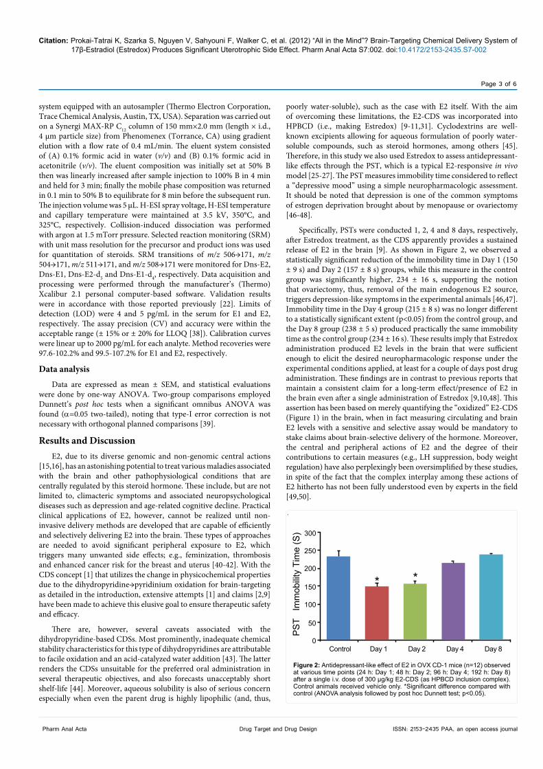
In a simple CDS, a dihydropyridine moiety is bioreversibly attached to a drug molecule (e.g., via an ester bond) [1] and, similarly to the above mentioned bioprecursor prodrug, the first step of its bioactivation involves the rapid oxidation to the corresponding pyridinium once the CDS passed the BBB from the circulation [5,7]. Then, the chemical bond between the pyridinium and drug is cleaved by appropriate enzymes (Figure 1); thus, the drug is released within the brain. It is speculated that enzymes (such as esterases) responsible for the liberation of the agent from the oxidized CDS in the brain could not
exert this action to a significant extent elsewhere in the body because the pyridinium conjugate would be rapidly eliminated when ubiquitous oxidation occurs in the periphery [1]. All these assumptions have been and are still being investigated by simplistic models [1,9-11], albeit we have used sophisticated in vivo techniques [12] to follow paradigm-shifting trends that have occurred in methods of pharmaceutical research and development by the 21st century.
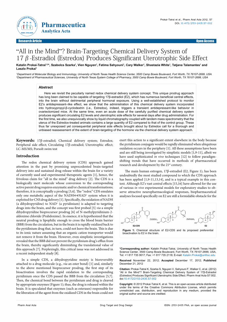
The main human estrogen, 17β-estradiol (E2, Figure 1), has been undoubtedly the most studied compound to which the CDS approach has been applied [1,9-11,13,14], and is a typical example in this con-text. Although E2’s vast central effects [15,16] have allowed for the use of various in vivo experimental models for exploratory studies to ob-serve attractive neuropharmacological responses, biopharmaceutical analyses focused specifically on E2 are still a formidable obstacle for the
*Corresponding author: Katalin Prokai-Tatrai, University of North Texas Health Science Center, 3500 Camp Bowie Boulevard, Fort Worth, TX 76107-2699, USA, Tel: +1 817 735 0617; Fax: +1 817 735 2118; E-mail: [email protected]
Received November 22, 2012; Accepted December 17, 2012; Published December 21, 2012
Citation: Prokai-Tatrai K, Szarka S, Nguyen V, Sahyouni F, Walker C, et al. (2012) “All in the Mind”? Brain-Targeting Chemical Delivery System of 17β-Estradiol (Estredox) Produces Significant Uterotrophic Side Effect. Pharm Anal Acta S7:002. doi:10.4172/2153-2435.S7-002
Copyright: © 2012 Prokai-Tatrai K, et al. This is an open-access article distributed under the terms of the Creative Commons Attribution License, which permits unrestricted use, distribution, and reproduction in any medium, provided the original author and source are credited.
AbstractHere we revisit the peculiarly named redox chemical delivery system concept. This unique prodrug approach
has long been claimed to be capable of targeting 17β-estradiol (E2), which has numerous beneficial central effects, into the brain without detrimental peripheral hormonal exposure. Using a well-established protocol to monitor E2’s antidepressant–like effect, we show that the administration of this chemical delivery system incorporated into hydroxypropyl-β-cyclodextrin (i.e., Estredox), indeed, triggers a transient antidepressant-like behavior in ovariectomized mice. At the same time, even an acute dose of the carefully purified chemical delivery system produces significant circulating E2 levels and uterotrophic side effects for several days after drug administration. For the first time, we also unequivocally show by liquid chromatography coupled with tandem mass spectrometry that the uterus of the Estredox-treated animals contains a large quantity of E2 compared to that of the control group. These thus far unexposed yet consequential peripheral side effects brought about by Estredox call for a thorough and unbiased reassessment of the extent of brain-targeting of the hormone via the chemical delivery system approach.
“All in the Mind”? Brain-Targeting Chemical Delivery System of 17β-Estradiol (Estredox) Produces Significant Uterotrophic Side EffectKatalin Prokai-Tatrai1,2*, Szabolcs Szarka1, Vien Nguyen1, Fatima Sahyouni1, Cary Walker1, Shastazia White1, Tatjana Talamantes1 and Laszlo Prokai1
1Department of Molecular Biology and Immunology, University of North Texas Health Science Center, 3500 Camp Bowie Boulevard, Fort Worth, TX 76107-2699, USA2Department of Pharmaceutical Sciences, University of North Texas System College of Pharmacy, 3500 Camp Bowie Boulevard, Fort Worth, TX 76107-2699, USA
.
HO
H
H
HO
H
H H H HH
O
O
N
O
N
HO
H
OH
O
Oxidation Hydrolysis
+
E2-CDS E2
Figure 1: Chemical structure of E2-CDS and its proposed preferential bioactivation to E2 in the brain.
Phar
mac
eutica Analytica Acta
ISSN: 2153-2435
Citation: Prokai-Tatrai K, Szarka S, Nguyen V, Sahyouni F, Walker C, et al. (2012) “All in the Mind”? Brain-Targeting Chemical Delivery System of 17β-Estradiol (Estredox) Produces Significant Uterotrophic Side Effect. Pharm Anal Acta S7:002. doi:10.4172/2153-2435.S7-002
Page 2 of 6
Drug Target and Drug DesignPharm Anal Acta ISSN: 2153-2435 PAA, an open access journal
group pursuing the development of E2-CDS [9-11]. Only an indepen-dent investigation over 20 years ago [17] has addressed the critical need for E2 pharmacokinetics and distribution in the brain using radioim-munoassays (RIA). Since then, E2 analyses by RIA were successfully done on occasion and only from plasma samples [9,10]. However, with RIAs used for E2, quality controls are sometimes difficult to achieve in a routine laboratory and these assays are also quite susceptible to artifacts caused by nonspecific binding [18]. They may also be subject to cross-reactivity and other analytical interferences. There is also of-ten very poor agreement among the results obtained by different RIAs, sometimes even with assays from the same manufacturer [18]. Similar
issues have been found for automated, chemiluminescence-based, di-rect E2 immunoassays. Method comparisons have revealed that these methods are inaccurate and prone to interferences at low analyte levels [19]. On the other hand, liquid chromatography–tandem mass spec-trometry (LC–MS/MS) has become the “gold standard” for performing quantitative bioanalyses [20,21]. Selective and sensitive assays based on this technique [22] should, therefore, be considered for bioanalytical support of projects aspiring to prove mission-critical hypotheses. In-vestigations should extend beyond an occasional assessment of merely plasma concentrations to stake claims about brain-selective delivery of drugs such as E2.
As an example highlighting the specific points expressed in the preceding paragraphs, the goal of the present work was to reassess time–dependent peripheral liability of the E2-CDS incorporated, according to the most recent practice [9,10], into (2-hydroxypropyl)-β-cyclodextrin (HPBCD) [23]. This inclusion complex is called Estredox. The assessment of peripheral effects is an apparent criterion that should be addressed by any delivery approach hoping to confine hormonal impact selectively to the brain [24]. A typical E2-responsive behavioral study with relevant clinical perspective [25-27], the Porsolt swim test (PST) [28], in ovariectomized (OVX) mice was used to confirm neuropharmacological effects. In addition, the “classical” uterotrophic response represented by the weight gain of the organ, which may be used as a biomarker of circulating estrogens [29,30], was also characterized in a temporal fashion. Ultimately, the behavioral outcomes and uterotrophic measurements were correlated with circulating E2 levels determined by an LC-MS/MS assay [22] using an experimental design that matches time-points of assessments. Collectively, these studies enabled a comprehensive and unbiased evaluation of the extent of brain-targeting of the hormone via Estredox.
Materials and MethodsChemicals and reagents
Estrone-d4 (E1-d4) and 17β-estradiol-d5 (E2-d5) internal standards with an isotopic purity of 98% were purchased from C/D/N Isotopes (Pointe-Claire, Quebec, Canada). Other reagents were obtained from Sigma-Aldrich (St. Louis, MO). Solvents used for LC-MS/MS measurements were of HPLC grade and ordered from Fisher Scientific (Atlanta, GA). E2-CDS and Estredox were prepared according to straightforward published methods [13,31,32]. Extreme care was taken for the purification of the chemically stable pyridinium precursor (“oxidized CDS”) and the E2-CDS itself by RP-HPLC [33] to ensure lack of E2 contamination. The absence of E2 in the formulated product was verified by LC-MS/MS after derivatization with dansyl (Dns) chloride (data not shown) [22].
Animals
Ovariectomized (OVX) CD-1 mice (30 ± 4 g body weight) were
purchased from Harlan Laboratories (Indianapolis, IN). All procedures were reviewed and approved by the Institutional Animal Care and Use Committee at the University of North Texas Health Science Center before initiation of the studies. In a room conditioned to 21-23°C and with a normal day/night cycle, four animals were housed per cage providing free access to food and water. Each animal was tested only once.
Porsolt swim test (PST) for the assessment of antidepressant-like activity
Mice were divided (n=6/group) into Control (vehicle), Day 1, Day 2, Day 4 and Day 8 groups. They were injected intravenously (i.v.) through the tail vein with vehicle (saline, control) and the test compound in vehicle (30 µL) at the dose of 300 µg/kg body weight. The dose was selected based on previous reports [9,10,31]. Behavioral studies for antidepressant-like activity started 24 h (Day 1), 48 h (Day 2), 96 h (Day 4) and 192 h (Day 8), respectively, after administration of the test compound (or vehicle), and were conducted according to our routinely used PST protocol [33-36]. Briefly, the immobility time (in seconds, defined as the duration of floating motionless after the cessation of struggling and making only movements necessary to keep the head above the water) was recorded for 6 min simultaneously by two trained observers who were unaware as to which animal received vehicle control or test compound.
Blood and tissue collection
The animals were anesthetized with intraperitoneal (i.p.) administration of 60 mg/kg ketamine (100 mg/mL) and 10 mg/kg xylazine (20 mg/mL) and blood was collected by cardiac puncture in a BD Vacutainer (Fisher Sci., Atlanta, GA). Blood was clotted on ice and spun at 3000 rpm for 20 min to obtain the serum sample. Animals then were perfused with cold saline through the left ventricle to avoid bias associated with E2 content originating from the blood [37]. Mice were then killed by decapitation and target tissues were collected and processed immediately. The uterus was cut above the cervical junction, visible fat was removed and the organ was weighed.
Sample preparation
Brain homogenates (20% w/v) were prepared in pH 7.4 phosphate buffer as reported before [33]. Uterus homogenates were prepared after freezing the tissue by a BioSqueezer (Biospec Products, Bartlesville, OK) then grinding to resultant powder in a Cryo-Cup Grinder (Biospec Products, Bartlesville, OK). From the collected frozen powders 20% (w/v) homogenates were made in pH 7.4 phosphate buffer. The homogenates and serum samples were spiked with 100 pg deuterium labeled internal standards (E2-d5 and E1-d4). Liquid–liquid extraction was done with methyl tert-butyl ether. The organic layer was separated by centrifugation at 2500 rpm for 10 min and carefully evaporated under a nitrogen stream at room temperature for subsequent LC-MS/MS analysis. The residue was derivatized by adding 30 µL of 1 mg/mL dansyl chloride (Dns-Cl) in acetonitrile and 20 µL of aqueous sodium bicarbonate (100 mM, pH 10.5) according to Nelson et al. [22]. The samples were vortexed and kept in a heating block at 60°C for 10 min. Afterwards, they were centrifuged at 14500 rpm for 3 min, transferred to autosampler vials, sealed, and assayed by LC-MS/MS.
LC-MS/MS
Analysis was performed using a TSQ Quantum Ultra triple-quadrupole instrument operated in positive-ion mode and interfaced via a heated-electrospray ionization (H-ESI) probe to a Surveyor LC
Citation: Prokai-Tatrai K, Szarka S, Nguyen V, Sahyouni F, Walker C, et al. (2012) “All in the Mind”? Brain-Targeting Chemical Delivery System of 17β-Estradiol (Estredox) Produces Significant Uterotrophic Side Effect. Pharm Anal Acta S7:002. doi:10.4172/2153-2435.S7-002
Page 3 of 6
Drug Target and Drug DesignPharm Anal Acta ISSN: 2153-2435 PAA, an open access journal
.
system equipped with an autosampler (Thermo Electron Corporation, Trace Chemical Analysis, Austin, TX, USA). Separation was carried out on a Synergi MAX-RP C12 column of 150 mm×2.0 mm (length × i.d., 4 µm particle size) from Phenomenex (Torrance, CA) using gradient elution with a flow rate of 0.4 mL/min. The eluent system consisted of (A) 0.1% formic acid in water (v/v) and (B) 0.1% formic acid in acetonitrile (v/v). The eluent composition was initially set at 50% B then was linearly increased after sample injection to 100% B in 4 min and held for 3 min; finally the mobile phase composition was returned in 0.1 min to 50% B to equilibrate for 8 min before the subsequent run. The injection volume was 5 µL. H-ESI spray voltage, H-ESI temperature and capillary temperature were maintained at 3.5 kV, 350°C, and 325°C, respectively. Collision-induced dissociation was performed with argon at 1.5 mTorr pressure. Selected reaction monitoring (SRM) with unit mass resolution for the precursor and product ions was used for quantitation of steroids. SRM transitions of m/z 506→171, m/z 504→171, m/z 511→171, and m/z 508→171 were monitored for Dns-E2, Dns-E1, Dns-E2-d5 and Dns-E1-d4, respectively. Data acquisition and processing were performed through the manufacturer’s (Thermo) Xcalibur 2.1 personal computer-based software. Validation results were in accordance with those reported previously [22]. Limits of detection (LOD) were 4 and 5 pg/mL in the serum for E1 and E2, respectively. The assay precision (CV) and accuracy were within the acceptable range (± 15% or ± 20% for LLOQ [38]). Calibration curves were linear up to 2000 pg/mL for each analyte. Method recoveries were 97.6-102.2% and 99.5-107.2% for E1 and E2, respectively.
Data analysis
Data are expressed as mean ± SEM, and statistical evaluations were done by one-way ANOVA. Two-group comparisons employed Dunnett’s post hoc tests when a significant omnibus ANOVA was found (a=0.05 two-tailed), noting that type-I error correction is not necessary with orthogonal planned comparisons [39].
Results and DiscussionE2, due to its diverse genomic and non-genomic central actions
[15,16], has an astonishing potential to treat various maladies associated with the brain and other pathophysiological conditions that are centrally regulated by this steroid hormone. These include, but are not limited to, climacteric symptoms and associated neuropsychological diseases such as depression and age-related cognitive decline. Practical clinical applications of E2, however, cannot be realized until non-invasive delivery methods are developed that are capable of efficiently and selectively delivering E2 into the brain. These types of approaches are needed to avoid significant peripheral exposure to E2, which triggers many unwanted side effects; e.g., feminization, thrombosis and enhanced cancer risk for the breast and uterus [40-42]. With the CDS concept [1] that utilizes the change in physicochemical properties due to the dihydropyridine→pyridinium oxidation for brain-targeting as detailed in the introduction, extensive attempts [1] and claims [2,9] have been made to achieve this elusive goal to ensure therapeutic safety and efficacy.
There are, however, several caveats associated with the dihydropyridine-based CDSs. Most prominently, inadequate chemical stability characteristics for this type of dihydropyridines are attributable to facile oxidation and an acid-catalyzed water addition [43]. The latter renders the CDSs unsuitable for the preferred oral administration in several therapeutic objectives, and also forecasts unacceptably short shelf-life [44]. Moreover, aqueous solubility is also of serious concern especially when even the parent drug is highly lipophilic (and, thus,
poorly water-soluble), such as the case with E2 itself. With the aim of overcoming these limitations, the E2-CDS was incorporated into HPBCD (i.e., making Estredox) [9-11,31]. Cyclodextrins are well-known excipients allowing for aqueous formulation of poorly water-soluble compounds, such as steroid hormones, among others [45]. Therefore, in this study we also used Estredox to assess antidepressant-like effects through the PST, which is a typical E2-responsive in vivo model [25-27]. The PST measures immobility time considered to reflect a “depressive mood” using a simple neuropharmacologic assessment. It should be noted that depression is one of the common symptoms of estrogen deprivation brought about by menopause or ovariectomy [46-48].
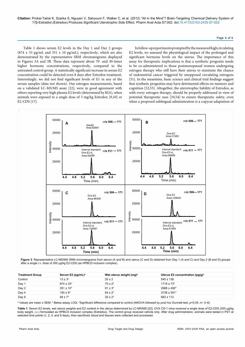
Specifically, PSTs were conducted 1, 2, 4 and 8 days, respectively, after Estredox treatment, as the CDS apparently provides a sustained release of E2 in the brain [9]. As shown in Figure 2, we observed a statistically significant reduction of the immobility time in Day 1 (150 ± 9 s) and Day 2 (157 ± 8 s) groups, while this measure in the control group was significantly higher, 234 ± 16 s, supporting the notion that ovariectomy, thus, removal of the main endogenous E2 source, triggers depression-like symptoms in the experimental animals [46,47]. Immobility time in the Day 4 group (215 ± 8 s) was no longer different to a statistically significant extent (p<0.05) from the control group, and the Day 8 group (238 ± 5 s) produced practically the same immobility time as the control group (234 ± 16 s). These results imply that Estredox administration produced E2 levels in the brain that were sufficient enough to elicit the desired neuropharmacologic response under the experimental conditions applied, at least for a couple of days post drug administration. These findings are in contrast to previous reports that maintain a consistent claim for a long-term effect/presence of E2 in the brain even after a single administration of Estredox [9,10,48]. This assertion has been based on merely quantifying the “oxidized” E2-CDS (Figure 1) in the brain, when in fact measuring circulating and brain E2 levels with a sensitive and selective assay would be mandatory to stake claims about brain-selective delivery of the hormone. Moreover, the central and peripheral actions of E2 and the degree of their contributions to certain measures (e.g., LH suppression, body weight regulation) have also perplexingly been oversimplified by these studies, in spite of the fact that the complex interplay among these actions of E2 hitherto has not been fully understood even by experts in the field [49,50].
300
250
200
150
100
50
0Control Day 1 Day 2 Day 4 Day 8
PS
T I
mm
obili
ty T
ime
(S)
* *
Figure 2: Antidepressant-like effect of E2 in OVX CD-1 mice (n=12) observed at various time points (24 h: Day 1; 48 h: Day 2; 96 h: Day 4; 192 h: Day 8) after a single i.v. dose of 300 µg/kg E2-CDS (as HPBCD inclusion complex). Control animals received vehicle only. *Significant difference compared with control (ANOVA analysis followed by post hoc Dunnett test; p<0.05).
Citation: Prokai-Tatrai K, Szarka S, Nguyen V, Sahyouni F, Walker C, et al. (2012) “All in the Mind”? Brain-Targeting Chemical Delivery System of 17β-Estradiol (Estredox) Produces Significant Uterotrophic Side Effect. Pharm Anal Acta S7:002. doi:10.4172/2153-2435.S7-002
Page 4 of 6
Drug Target and Drug DesignPharm Anal Acta ISSN: 2153-2435 PAA, an open access journal
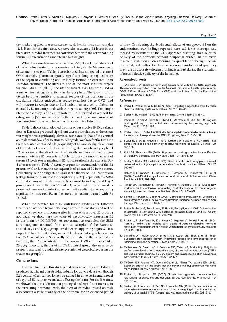
Table 1 shows serum E2 levels in the Day 1 and Day 2 groups (874 ± 33 pg/mL and 351 ± 10 pg/mL), respectively, which are also demonstrated by the representative SRM chromatograms displayed in Figures 3A and 3B. These data represent about 70- and 30-times higher hormone concentrations, respectively, compared to the untreated control group. A statistically significant increase in serum E2 concentration could be detected even 8 days after Estredox treatment. Interestingly, we did not find significant levels of E1 in any of the serum samples (data not shown). Our estrogen measurements, based on a validated LC-MS/MS assay [22], were in good agreement with others reporting very high plasma E2 levels (determined by RIA), when animals were exposed to a single dose of 3 mg/kg Estredox [9,10] or E2-CDS [17].
In follow-up experiments prompted by the measured high circulating E2 levels, we assessed the physiological impact of the prolonged and significant hormone levels on the uterus. The importance of this assay for therapeutic implications is that a synthetic progestin needs to be co-administered to those postmenopausal women undergoing estrogen therapy who still have their uterus to minimize the chance of endometrial cancer triggered by unopposed circulating estrogens [51]. In the meantime, basic science and clinical trial findings suggest that synthetic progestins may have detrimental effects on memory and cognition [52,53]. Altogether, the uterotrophic liability of Estredox, as with every estrogen therapy, should be properly addressed in view of potential therapeutic uses [10,54] to ensure therapeutic safety, even when a proposed sublingual administration is a copycat adaptation of
.
50000
25000
50000
25000
50000
25000
50000
25000
50000
25000
50000
25000
50000
25000
50000
25000
A B
DC
4.4 4.8 5.2 5.6 6.0 6.4
4.4 4.8 5.2 5.6 6.0 6.4
4.4 4.8 5.2 5.6 6.0 6.4
4.4 4.8 5.2 5.6 6.0 6.4
Time (min) Time (min)
Inte
nsity
Inte
nsity
Inte
nsity
Inte
nsity
Dns-E2Area:82661
Dns-E2Area:31283
Time (min) Time (min)
Internal standard:Dns-E2-d5Area:92099
Internal standard:Dns-E2-d5Area:109487
Internal standard:Dns-E2-d5Area:97993
Dns-E2Area:99308
Dns-E2Area:126640
m/z 506
m/z 511
m/z 506
m/z 511
m/z 506
m/z 511
m/z 506
m/z 511
171
171
171
171
171
171
171
171
Internal standard:Dns-E2-d5Area:96858
Figure 3: Representative LC-MS/MS SRM chromatograms from serum (A and B) and uterus (C and D) obtained from Day 1 (A and C) and Day 2 (B and D) groups after a single i.v. dose of 300 µg/kg E2-CDS (as HPBCD inclusion complex).
Treatment Group Serum E2 (pg/mL)a Wet uterus weight (mg)a Uterus E2 concentration (pg/g)a
Control 13 ± 3b 20 ± 2 545 ± 138Day 1 874 ± 33* 70 ± 2* 1719 ± 73*Day 2 351 ± 10* 91 ± 3* 2988 ± 492*Day 4 150 ± 9* 64 ± 2* 3738 ± 591*Day 8 48 ± 7* 32 ± 2* 683 ± 112
a Values are mean ± SEM; b Below assay LOQ; *Significant difference compared to control (ANOVA followed by post hoc Dunnett test; p<0.05, n= 3–6)
Table 1: Serum E2 levels, wet uterus weights and E2 content in the uterus determined by LC-MS/MS [22]. OVX CD-1 mice received a single dose of E2-CDS (300 µg/kg body weight, i.v.) formulated as HPBCD inclusion complex (Estredox). The control group received vehicle only. After drug administration, animals were tested in PST at selected time points (1, 2, 4, and 8 days), then sacrificed; blood and tissues were collected and processed.
Citation: Prokai-Tatrai K, Szarka S, Nguyen V, Sahyouni F, Walker C, et al. (2012) “All in the Mind”? Brain-Targeting Chemical Delivery System of 17β-Estradiol (Estredox) Produces Significant Uterotrophic Side Effect. Pharm Anal Acta S7:002. doi:10.4172/2153-2435.S7-002
Page 5 of 6
Drug Target and Drug DesignPharm Anal Acta ISSN: 2153-2435 PAA, an open access journal
the method applied to a testosterone–cyclodextrin inclusion complex [23]. Here, for the first time, we have also measured E2 levels in the uteri after Estredox treatment to correlate them with the corresponding serum E2 concentrations and uterine wet weights.
When the animals were sacrificed after PST, the enlarged uteri in all of the Estredox-treated groups were immediately visible. Measurements of wet uterine weights (Table 1) corroborated that, compared to control OVX animals, pharmacologically significant long-lasting exposure of the organ to circulating and/or locally formed E2 occurred upon Estredox treatment. The uterus is one of the most sensitive targets for circulating E2 [30,55]; the uterine weight gain has been used as a marker for estrogenic activity in the periphery. The growth of the uterus becomes sensitive to external sources of this hormone in the circulation without endogenous source (e.g., lost due to OVX) and will increase in weight due to fluid imbibition and cell proliferation elicited by E2 (or compounds with estrogenic activity) [30]. This simple uterotrophic assay is also an important EPA-approved in vivo test for estrogenicity [56] and, as such, it offers an additional and convenient screening tool to evaluate hormonal exposure after Estredox.
Table 1 shows that, adopted from previous studies [9,10], a single dose of Estredox produced significant uterus stimulation, as the uterus wet weight was significantly elevated compared to that of the control animals even 8 days after treatment. Alongside, we show for the first time that these uteri contained a large quantity of E2 (and negligible amount of E1, data not shown) further confirming that significant peripheral E2 exposure is the direct result of insufficient brain-targeting (see serum vs. uterine E2 contents in Table 1). The continuous decrease of serum E2 levels versus maximum E2 concentration in the uterus at Day 4 after treatment (Table 1) actually argues for accumulation of the E2 precursor(s) and, then, the subsequent local release of E2 in the uterus. Collectively, our findings stand against the theory of E2’s “continuous leakage from the brain into the periphery” [17,32]. Representative SRM chromatograms of the uterus extracts obtained from Day 1 and Day 2 groups are shown in Figures 3C and 3D, respectively. In any case, data presented here are in perfect agreement with earlier studies reporting significantly increased E2 in the uterus of rodents treated with E2 [57,58].
While the detailed brain E2 distribution studies after Estredox treatment have been beyond the scope of the present study and will be reported elsewhere in a comparative fashion with a novel E2 prodrug approach, we show here the value of unequivocally measuring E2 in the brain by LC-MS/MS. As representative examples, the SRM chromatograms obtained from cortical samples of the Estredox-treated Day 1 and Day 2 groups are shown in supporting Figure S1. It is important to note that endogenous E2 levels are not negligible even in the OVX rodent brain. Specifically, we estimated in the present study that, e.g., the E2 concentration in the control OVX cortex was 144 ± 28 pg/g. Therefore, tissues of an OVX control group also need to be properly analyzed to avoid misinterpretation of data obtained from the treatment group(s).
ConclusionsThe main finding of this study is that even an acute dose of Estredox
produces significant uterotrophic liability for up to 8 days even though E2’s central effect can no longer be utilized in an experimental model of a typical E2-responsive malady affecting the brain. For the first time, we showed that, in addition to a prolonged and significant increase in the circulating hormone levels, the uteri of Estredox-treated animals also contain a large quantity of the hormone for an extended period
of time. Considering the detrimental effects of unopposed E2 on the endometrium, our findings reported here call for a thorough and focused reassessment of the CDS approach asserting brain-selective delivery of the hormone without peripheral burden. In our view, reliable distribution studies focusing on quantitation through the use of an analytical method that has the necessary sensitivity and specificity to ensure an accurate estrogen profiling is a must during the evaluation of organ-selective delivery of the hormone.
Acknowledgments
We thank J.W. Simpkins for sharing his concerns with the E2-CDS approach. This work was supported in part by the National Institutes of Health (grant number AG031535 to LP and AG031421 to KPT) and the Robert A. Welch Foundation (endowment BK-0031 to LP).
References
1. Prokai L, Prokai-Tatrai K, Bodor N (2000) Targeting drugs to the brain by redox chemical delivery systems. Med Res Rev 20: 367–416.
2. Bodor N, Buchwald P (1998) All in the mind. Chem Britain 34: 36-40.
3. Pavan B, Dalpiaz A, Ciliberti N, Biondi C, Manfredini S, et al. (2008) Progress in drug delivery to the central nervous system by the prodrug approach. Molecules 13: 1035-1065.
4. Prokai-Tatrai K, Prokai L (2003) Modifying peptide properties by prodrug design for enhanced transport into the CNS. Prog Drug Res 61: 155-188.
5. Bodor N, Shek E, Higuchi T (1975) Delivery of a quaternary pyridinium salt across the blood-brain barrier by its dihydropyridine derivative. Science 190: 155-156.
6. Kokil GR, Rewatkar PV (2010) Bioprecursor prodrugs: molecular modification of the active principle. Mini Rev Med Chem 10: 1316-1330.
7. Bodor N, Roller RG, Selk SJ (1978) Elimination of a quaternary pyridinium salt delivered as its dihydropyridine derivative from brain of mice. J Pharm Sci 67: 685-687.
8. DeMar CD, Clarkson ED, Ratcliffe RH, Campbel AJ, Thangavelu SG, et al. (2010) Pro-2-PAM therapy for central and peripheral cholinesterases. Chem Biol Interact 187: 191–198.
9. Tapfer MK, Sebestyen L, Kurucz I, Horvath K, Szelenyi I, et al. (2004) New evidence for the selective, long-lasting central effects of the brain-targeted estradiol, Estredox. Pharmacol Biochem Behav 77: 423-429.
10. Sziraki I, Horvath K, Bodor N (2006) Comparative evaluation of Estredox, a brain-targeted estradiol delivery system versus traditional estrogen replacement therapy. Pharmazie 61: 140-143.
11. Patthy M, Seres G, Tóth-Sarudy E, Hazai I, Pallagi I, et al. (2008) Determination of estredox, a compound with sustained estradiol function, and its impurity profile by HPLC. Pharmazie 63: 210-216.
12. Prokai L, Prokai-Tatrai K, Zharikova AD, Nguyen V, Perjesi P, et al. (2004) Centrally acting and metabolically stable thyrotropin-releasing hormone analogues by replacement of histidine with substituted pyridinium. J Med Chem 47: 6025–6033.
13. Simpkins JW, McCornack J, Estes KS, Brewster ME, Shek E, et al. (1986) Sustained brain-specific delivery of estradiol causes long-term suppression of luteinizing hormone secretion. J Med Chem 29: 1809-1812.
14. Mullersman G, Derendorf H, Brewster ME, Estes KS, Bodor N (1998) High-performance liquid chromatographic assay of a central nervous system (CNS)-directed estradiol chemical delivery system and its application after intravenous administration to rats. Pharm Res 5: 172-177.
15. McEwen BS, Akama KT, Spencer-Segal JL, Milner TA, Waters EM (2012) Estrogen effects on the brain: actions beyond the hypothalamus via novel mechanisms. Behav Neurosci 126: 4–16.
16. Prokai L, Simpkins JW (2007) Structure-non-genomic neuroprotection relationship of estrogens and estrogen-derived compounds. Pharmacol Ther 114: 1-12.
17. Sarkar DK, Friedman SJ, Yen SS, Frautschy SA (1989) Chronic inhibition of hypothalamic-pituitary-ovarian axis and body weight gain by brain-directed delivery of estradiol-17β in female rats. Neuroendocrinology 50: 204–210.
Citation: Prokai-Tatrai K, Szarka S, Nguyen V, Sahyouni F, Walker C, et al. (2012) “All in the Mind”? Brain-Targeting Chemical Delivery System of 17β-Estradiol (Estredox) Produces Significant Uterotrophic Side Effect. Pharm Anal Acta S7:002. doi:10.4172/2153-2435.S7-002
Page 6 of 6
Drug Target and Drug DesignPharm Anal Acta ISSN: 2153-2435 PAA, an open access journal
18. Carlstrom K (1996) Low endogenous estrogen levels–analytical problems and tissue sensitivity. Acta Obstet Gynecol Scand Suppl 163: 11–15.
19. Lee CS, Smith NM, Kahn SN (1991) Analytic variability and clinical significance of different assays for serum estradiol. J Reprod Med 36: 156–160.
20. Ackermann BL, Berna MJ, Eckstein JA, Ott LW, Chaudhary AK (2008) Current applications of liquid chromatography/mass spectrometry in pharmaceutical discovery after a decade of innovation. Ann Rev Anal Chem 1: 357–396.
21. Prokai L, Zharikova A, Janaky T, Li X, Braddy AC, et al. (2001) Integration of mass spectrometry into early-phase discovery and development of central nervous system agents. J Mass Spectrom 36: 1211-1219.
22. Nelson RE, Grebe SK, OKane DJ, Singh RJ (2004) Liquid chromatography-tandem mass spectrometry assay for simultaneous measurement of estradiol and estrone in human plasma. Clin Chem 50: 373-384.
23. Stuenkel CA, Dudlet RE, Yens SS (1991) Sublingual administration of testosterone-hydroxypropyl-β-cyclodextrin inclusion complex simulates episodic androgen release in hypogonadal men. J Clin Endocrinol Metabol 72: 1054-1059.
24. Pickar JH, MacNeil T, Ohleth K (2010) SERMs: Progress and future perspectives. Maturitas 67: 129-138.
25. Walf AA, Frye CA (2009) Effects of two estradiol regimens on anxiety and depressive behaviors and trophic effects in peripheral tissues in a rodent model. Gend Med 6: 300-311.
26. Estrada-Camarena E, Fernandez-Guasti A, Lopez-Rubalcava C (2003) Antidepressant-like effect of different estrogenic compounds in the forced swimming test. Neuropsychopharmacol 28: 830–838.
27. Dhir A, Kulkarni SK (2008) Antidepressant-like effect of 17beta-estradiol: involvement of dopaminergic, serotonergic, and (or) sigma-1 receptor systems. Can J Physiol Pharmacol 86: 726-735.
28. Porsolt RD, Le Pichon M, Jalfre M (1977) Depression: A new animal model sensitive to antidepressant treatments. Nature 266: 730-732.
29. Clark JH, Mani SK (1994) Actions of ovarian steroid hormones. In: Knobil E, Neill J (Eds.), The Physiology of Reproduction, (2ndedn), Raven Press, New York, 1011–1059.
30. Owens JW, Ashby J (2002) Critical review and evaluation of the uterotrophic bioassay for the identification of possible estrogen agonists and antagonists: in support of the validation of the OECD uterotrophic protocols for the laboratory rodent. Organisation for economic co-operation and development. Crit Rev Toxicol 32: 445-520.
31. Brewster ME, Estes KS, Loftsson T, Perchalski R, Derendorf H, et al. (1988) Improved delivery through biological membranes XXXI: Solubilization and stabilization of an estradiol chemical delivery system by modified β-cyclodextrins. J Pharm Sci 77: 981–985.
32. Anderson W, Simpkins JW, Brewster ME, Bodor N (1988) Evidence for suppression of serum LH without elevation in serum estradiol or prolactin with a brain-enhanced redox delivery system for estradiol. Life Sci 42: 1493–1502.
33. Prokai-Tatrai K, Prokai L (2011) Prodrug design for brain delivery of small-and medium-sized neuropeptides. In neuropeptides: methods and protocols. Methods Mol Biol 789: 313-336.
34. Prokai-Tatrai K, Prokai L (2009) Prodrugs of thyrotropin-releasing hormone and related peptides as central nervous system agents. Molecules 14: 633-654.
35. Prokai-Tatrai K, Teixido M, Nguyen V, Zharikova AD, Prokai L (2005) A pyridinium-substituted analog of the TRH-like tripeptide pGlu-Glu-Pro-NH2 and its prodrugs as central nervous system agents. Med Chem 1: 141-152.
36. Teixido M, Prokai-Tatrai K, Wang X, Nguyen V, Prokai L (2007) Exploratory neuropharmacological evaluation of a conformationally constrained thyrotropin-releasing hormone analogue. Brain Res Bull 73: 103-107.
37. Alfinito PD, Chen X, Atherton J, Cosmi S, Deecher DC (2008) ICI 182,780 penetrates brain and hypothalamic tissue and has functional effects in the brain after systemic dosing. Endocrinol 149: 5219-5226.
38. US Department of Health and Human Services, FDA, CDER, CVM (2001) Guidance for Industry: Bioanalytical Method Validation.
39. Keppel G, Wickens TD (2004) Design and analysis: A researcher’s handbook. (4thedn), Pearson Prentice Hall, Upper Saddle River.
40. DePaolo L, Negro-Vilar A (1982) Estrogenic feminization of the LH response to orchidectomy in the rat: evidence for a hypothalamic site of action. Neuroendocrinol 34: 104-111.
41. Hsueh AJW, Peck EJ, Clark JH (1975) Progesterone antagonism of the oestrogen receptor and oestrogen-induced uterine growth. Nature 254: 337-339.
42. Koos RD (2011) Minireview: putting physiology back into estrogens’ mechanism of action. Endocrinol 152: 4481–4488.
43. Eisner U, Kuthan J (1972) The Chemistry of dihydropyridines. Chem Rev 72: 1-42.
44. Mahmoud S, Aboul-Fadl T, Sheha M, Farag H, Mouhamed AMI (2003) 1,2-Dihydroisoquinoline-N-acetic acid derivatives as new carriers for specific brain delivery I: Synthesis and estimation of oxidation kinetics using multivariate calibration method. Arch Pharm Pharm (Weinhem) 336: 573–584.
45. Gould S, Scott RC (2005) 2-Hydroxypropyl-beta-cyclodextrin (HP-beta-CD): a toxicology review. Food Chem Toxicol 43: 1451-1459.
46. Freeman EW (2010) Associations of depression with the transition to menopause. Menopause 17: 823-827.
47. Lagunas N, Calmarza-Font I, Diz-Chaves Y, Garcia-Segura LM (2010) Long-term ovariectomy enhances anxiety and depressive-like behaviors in mice submitted to chronic unpredictable stress. Horm Behav 58: 786-791.
48. Bekku N, Yoshimura H (2005) Animal model of menopausal depressive-like state in female mice: prolongation of immobility time in the forced swimming test following ovariectomy. Psychopharmacol 183: 300-307.
49. Palmer K, Gray JM (1986) Central vs. peripheral effects of estrogen on food intake and lipoprotein lipase activity in ovariectomized rats. Physiol Behav 37: 187-189.
50. Yonezawa R, Wada T, Matsumoto N, Morita M, Sawakawa K, et al. (2012) Central versus peripheral impact of estradiol on the impaired glucose metabolism in ovariectomized mice on a high-fat diet. Am J Physiol Endocrinol Metab 303: E445-E456.
51. Furness S, Roberts H, Marjoribanks J, Lethaby A, Hickey M, et al. (2012) Hormone therapy in postmenopausal women and risk of endometrial hyperplasia. Cochrane Database Syst Rev 8: CD000402.
52. Braden BB, Talboom JS, Crain ID, Simard AS, Lukas RJ, et al. (2010) Medroxyprogesterone acetate impairs memory and alters the GABAergic system in aged surgically menopausal rats. Neurobiol Learn Mem 93: 444–453.
53. Craig MC, Maki PM, Murphy DG (2005) The Women’s Health Initiative Memory Study: findings and implications for treatment. Lancet Neurol 4: 190-194.
54. Bodor N, Buchwald FP (2006) Brain-targeted delivery of estradiol: therapeutic potential and results obtained with a chemical delivery system approach. Am J Drug Deliv 3: 161-175.
55. Ayan D, Roy J, Maltais R, Poirier D (2011) Impact of estradiol structural modifications (18-methyl and/or 17-hydroxy inversion of configuration) on the in vitro and in vivo estrogenic activity. J Steroid Biochem Mol Biol 127: 324–330.
56. http://www.epa.gov/endo/pubs/uterotrophic_fs.pdf
57. Clark JH, Williams M, Upchurch S, Eriksson H, Helton E, et al. (1982) Effects of estradiol-17-alpha on nuclear occupancy of the estrogen-receptor, stimulation of nuclear type-ii sites and uterine growth. J Steroid Biochem Mol Biol 16: 323-328.
58. Wang L, Wang YD, Li DJ (2009) Differential regulation of dehydroepiandrosterone and estrogen on bone and uterus in ovariectomized mice. Osteoporos Int 20: 79-92.
This article was originally published in a special issue, Drug Target and Drug Design handled by Editor(s). Dr. Paulo Sérgio Monzani, University of North Paraná, Brazil