
A β-alanine catabolic pathway containing a highly promiscuous ω ...
32
A β-alanine catabolic pathway containing a highly promiscuous ω-transaminase from the 12- 1 aminododecanate-degrading Pseudomonas sp. Strain AAC. 2 Matthew Wilding, 1,2 Thomas S. Peat, 3 Janet Newman, 3 Colin Scott 1 3 4 1 CSIRO Land and Water Black Mountain, Canberra, Australia, 2601 5 2 CSIRO Food and Nutrition, Black Mountain, Canberra, Australia, 2601 6 3 CSIRO Manufacturing, 343 Royal Parade, Parkville, Victoria, Australia, 3052 7 8 Running title: Characterization of a β-Alanine catabolic pathway. 9 10 Correspondence should be addressed to [email protected] 11 12 AEM Accepted Manuscript Posted Online 22 April 2016 Appl. Environ. Microbiol. doi:10.1128/AEM.00665-16 Copyright © 2016 Wilding et al. This is an open-access article distributed under the terms of the Creative Commons Attribution 4.0 International license. on April 11, 2018 by guest http://aem.asm.org/ Downloaded from
Transcript of A β-alanine catabolic pathway containing a highly promiscuous ω ...
A β-alanine catabolic pathway containing a highly promiscuous ω-transaminase from the 12-1
aminododecanate-degrading Pseudomonas sp. Strain AAC. 2
Matthew Wilding,1,2 Thomas S. Peat,3 Janet Newman,3 Colin Scott1 3
4
1CSIRO Land and Water Black Mountain, Canberra, Australia, 2601 5
2CSIRO Food and Nutrition, Black Mountain, Canberra, Australia, 2601 6
3CSIRO Manufacturing, 343 Royal Parade, Parkville, Victoria, Australia, 3052 7
8
Running title: Characterization of a β-Alanine catabolic pathway. 9
10
Correspondence should be addressed to [email protected] 11
12
AEM Accepted Manuscript Posted Online 22 April 2016Appl. Environ. Microbiol. doi:10.1128/AEM.00665-16Copyright © 2016 Wilding et al.This is an open-access article distributed under the terms of the Creative Commons Attribution 4.0 International license.
on April 11, 2018 by guest
http://aem.asm
.org/D
ownloaded from
ABSTRACT 13
We previously isolated the transaminase KES23458 from Pseudomonas sp. strain AAC as a 14
promising biocatalyst for the production of 12-aminododecanoic acid, a constituent building block of 15
nylon-12. Herein, we report the subsequent characterization of this transaminase. It exhibits activity 16
with a broad substrate range which includes α, β and ω-amino acids, as well as α,ω-diamines and a 17
number of other industrially relevant compounds, and is therefore a prospective candidate for the 18
biosynthesis of a range of polyamide monomers. The crystal structure of KES23458 revealed that the 19
protein formed a dimer containing a large active site pocket and unusual phosphorylated histidine 20
residues. To infer the physiological role of the transaminase we expressed, purified and characterized 21
a dehydrogenase from the same operon, KES23460. Unlike the transaminase, the dehydrogenase 22
was shown to be quite selective, catalyzing the oxidation of malonic acid semialdehyde which was 23
formed from β-alanine transamination by KES23458. In keeping with previous reports, the 24
dehydrogenase was shown to catalyze both a CoA-dependent reaction to form acetyl-CoA, and a 25
significantly slower CoA-independent reaction to form acetate. These findings support the original 26
functional assignment of KES23458 as a β-alanine transaminase. However a seemingly well-adapted 27
active site and promiscuity towards unnatural compounds such as 12-aminododecanoic acid might 28
suggest that this enzyme performs multiple functions for Pseudomonas sp. strain AAC. 29
IMPORTANCE STATEMENT 30
The manuscript describes the characterization of an industrially relevant transaminase, able to 31
metabolize 12-aminododecanoic acid, a constituent building block of the widely used polymer Nylon-32
12, and herein we report the biochemical and structural characterization of the transaminase protein. 33
A physiological role for this highly promiscuous enzyme is proposed based on the characterization of 34
a related gene from the host organism. Molecular dynamics simulations were carried out to compare 35
the conformational changes in the transaminase protein to better understand the determinants of 36
specificity in the protein. This manuscript makes a substantial contribution that is of interest to the 37
broad biotechnology and enzymology communities, providing insight into the catalytic activity of an 38
industrially relevant biocatalyst as well as the biological function of this operon. 39
40
on April 11, 2018 by guest
http://aem.asm
.org/D
ownloaded from

KEYWORDS 41
Biocatalysis, transaminase, polyamide, nylon, aminotransferase, pyridoxal-5’-phosphate (PLP). 42
on April 11, 2018 by guest
http://aem.asm
.org/D
ownloaded from
INTRODUCTION 43
Enzymes are typically thought of as highly selective, avoiding the side reactions associated 44
with analogous (non-enzymatic) chemical reactions and producing high yields of single products. 45
However, it is perhaps underappreciated that enzymes can possess very broad substrate specificities, 46
a key requirement for their evolutionary adaptability, as it allows for the development of new catalytic 47
capabilities when required for survival or competitive advantage(1-3). For example, the ability of 48
microorganisms to degrade non-natural chemicals, such as pesticides or synthetic antibiotics, has 49
resulted in new metabolic pathways for resistance and catabolism over relatively short periods of 50
evolutionary time(4, 5). Although these proteins often evolve for catabolic purposes in the 51
microorganisms involved, these newly evolved functions represent an opportunity from a biocatalytic 52
perspective. 53
ω-Transaminases (ω-TAs) are renowned for their promiscuity and have exhibited catalytic 54
prowess in the synthesis of unnatural compounds(6). The ability of these enzymes to act on foreign 55
molecules has drawn widespread interest from both academia and industry, and whilst they are used 56
widely in the synthesis of chiral building blocks(7), they can also be used to produce more complex 57
synthetic pharmaceuticals. For example, sitagliptin, an anti-diabetic compound, can be synthesized 58
using a transaminase-mediated process which provides improvements in both yield, enantiomeric 59
excess, and productivity relative to alternative chemocatalytic methods(8). Transaminases, including 60
ω-TAs, are pyridoxal-5’-phosphate (PLP)-dependent enzymes that transfer amine groups from 61
molecules containing a primary amine (the amine donor) to acceptor molecules, aldehydes or 62
ketones(9). PLP acts as a carrier for the amine group, forming pyridoxamine-5’-phosphate (PMP) as 63
an intermediate in the reaction. The ability to install amine groups, often in an enantioselective 64
manner, is highly attractive, and when used to synthesize expensive products using relatively cheap 65
amine donors such as isopropylamine, these methods can also become more economical. Although 66
ω-transaminases are reported to have some limitations, such as low thermotolerance, substrate and 67
product inhibition, and equilibrium-controlled yields, complementary methods have been developed to 68
address these issues(7, 10-12). In addition, there are a number of X-ray structures for ω-TAs, and 69
despite having highly conserved tertiary and quaternary structures, the enzymes exhibit 70
conformational changes upon binding, and this conformational heterogeneity has been reported to 71
on April 11, 2018 by guest
http://aem.asm
.org/D
ownloaded from
make significant contributions to substrate specificity(13). Given the low homology (<30%) among the 72
class (14, 15), an understanding of the sequence-function relationship of these enzymes can be 73
challenging, and is typically only possible with a detailed understanding of the interactions between 74
the enzyme and substrate(8, 16). 75
Previously, we characterized a number of transaminases from Pseudomonas sp. strain AAC 76
(NCBI Accession number: PRJNA248604), some of which were able to metabolize 12-77
aminododecanoic acid, a non-natural compound with potential as a building block for industrial nylon-78
12 synthesis(17). Hazards associated with the synthesis of nylon-12(18), as well as global shortages 79
of the material in recent years(19) have afforded opportunities for the application of biocatalysis within 80
the materials field(20), and a number of examples have been reported to date(21, 22). However, while 81
most organisms are unable to metabolize 12-aminododecanoic acid, this bacterium contains three 82
enzymes with activity against the compound. Catalytic promiscuity is often thought to be the result of 83
an enzymatic activity which had been acquired coincidentally, and evolved under selective pressure. 84
In this case the substrate is neither toxic nor abundant in the environment, and it is unlikely that there 85
was a strong selective pressure to evolve a metabolic activity against the compound. Examples of 86
secondary reactions which provide no apparent benefit and are only encountered under laboratory 87
conditions, such as the dechlorination of chloropurines by adenosine deaminase(23), have been 88
reported, and without further evidence, it is more likely that this is the case for the Pseudomonas sp. 89
strain AAC. However, the discovery of three genes with this activity within the same organism could 90
suggest that the compound has a broader metabolic context, or that the genes co-evolved from a 91
single parent gene(24). 92
As stated above, within the Pseudomonas sp. strain AAC, three transaminases were 93
identified with 12-aminododecanoic acid activity, KES24511, KES23458 and KES23360. The most 94
promising candidate biocatalyst in terms of ease of protein production, solubility and activity was 95
KES23458, and herein we report the subsequent characterization of that protein. 96
on April 11, 2018 by guest
http://aem.asm
.org/D
ownloaded from
MATERIALS AND METHODS 97
Materials 98
A codon-optimized synthetic gene encoding KES23460 was synthesized by Life Technologies 99
(Germany). All chemicals were purchased from Sigma Aldrich Co. (USA). 100
Cloning, Expression and Purification 101
Cloning of KES23458 from genomic DNA has been described previously(17). Protein overexpression 102
was achieved by growing a culture of E. coli BL21-DE3 transformed with KES23458 inside a pETcc2 103
vector in LB media containing ampicillin (100 μg/mL) at 37°C. When the OD600 reached 0.6-1.0, the 104
culture was induced by the addition of IPTG (isopropyl β-D-1-thiogalactopyranoside; 1 mM final 105
concentration) and further incubated at 37°C for 18 hours. The cells were isolated by centrifugation in 106
a Beckman Avanti J-E centrifuge (3000 x g; 20 minutes) and the supernatant discarded. The pellet 107
was resuspended in imidazole / potassium phosphate buffer (5 mM imidazole, 10 mM phosphate, pH 108
7.5) and cell lysis was achieved by homogenization at 20 kpsi using an Avestin Emulsiflex C3. 109
Cellular debris was precipitated by centrifugation (40,000 x g, 45 minutes) and the supernatant was 110
passed over a HiTrap Chelating HP column (GE Healthcare; Little Chalfont, UK) on an Åkta FPLC 111
(Fast Protein Liquid Chromatography, GE Healthcare). Protein was eluted with an increasing 112
concentration of imidazole (5-500 mM) and the fractions containing protein were transferred into 113
potassium phosphate buffer (10 mM, pH 7.5), concentrated by centrifugation (GE Healthcare; 10k 114
MWCO) and further purified by gel filtration (Superdex 200; G.E. Healthcare) in the same phosphate 115
buffer. Purity was estimated to be >95% by SDS-PAGE. 116
A gene encoding KES23460, and codon-optimized for expression in E. coli, was ordered in the 117
expression vector pRSetB from Life Technologies (Germany). E. coli BL21-DE3 was transformed with 118
pRSetB containing the gene encoding KES23460 and overexpression and purification of KES23460 119
was achieved in the same manner as described for KES23458. 120
Protein Crystallization 121
The concentrated protein (8 mg/mL) was used to set up initial crystallization trials (JCSG+ screen, at 122
8 and 20 °C, PACT and an in-house pH vs. salt gradient screen – in total 384 experiments). The 123
on April 11, 2018 by guest
http://aem.asm
.org/D
ownloaded from
crystallization droplets consisted of 200 nL protein (8 mg/mL in 10 mM potassium phosphate buffer, 124
pH 7.5) and 200 nL crystallization cocktail, equilibrated against 50 uL of the cocktail in SD-2 sitting 125
drop plates (Molecular Dimensions, Suffolk, UK). The best crystals grew from mid-weight 126
polyethylene glycols (PEG) in the presence of magnesium (e.g. 10 % w/v PEG 4000, 0.2 M 127
magnesium chloride and 0.1 M Tris (tris(hydroxymethyl)aminomethane) hydrochloride, pH 7). 128
Crystals appeared within hours and grew to full size (70 µm x 70 µm x 200 µm) within days. Crystals 129
were selected and cryoprotected by the addition of reservoir solution supplemented with 20% 130
glycerol, and flash-cooled in N2. 131
Data Collection and Structural Determination 132
For an initial native data set, 360° of data were collected from a single crystal (1 ° oscillations) to yield 133
a complete X-ray dataset to 1.33 Å; a later data set was collected to 1.24 Å and this higher resolution 134
data set is reported in Table 2. XDS(25) was used to index the reflections and SCALA(26) was used 135
to scale the data. The structure was solved by molecular replacement using Phaser(27) and based on 136
the Omega-Amino Acid:Pyruvate Aminotransferase from Pseudomonas putida (PDB accession 137
number: 3A8U)(28). A single protomer was placed in the asymmetric unit, rebuilt manually using 138
Coot(29) and refined using Refmac(30). Details of the data collection and refinement are shown in 139
table 2. 140
All data sets were collected at the MX-2 beamline of the Australian Synchrotron. Additional 141
data sets beyond the initial native data set were processed in the same way (above) but were solved 142
using the native structure, rebuilt with Coot and refined with Refmac. The models and data have been 143
deposited in the Protein Data Bank under accession codes 4uho (1.24 Å), 4uhn (phospho-His) and 144
4uhm (1.33 Å). 145
Phosphoprotein staining 146
Sample preparation for ProQ Diamond Phosphoprotein staining was carried out following the 147
manufacturer’s protocol (Thermo Fisher Scientific). Purified KES23458 (40 μL, 5 mg/mL) ± gabaculine 148
(20 μL, 10 mg/mL), as well as crude cell lysate and BSA (20 μL, 10 mg/mL) were analysed. Staining 149
was carried out following manufacturer’s guidelines and gel visualization was carried out using a 150
on April 11, 2018 by guest
http://aem.asm
.org/D
ownloaded from
Typhoon Trio (GE Healthcare), with an excitation wavelength of 532 nm, an emission wavelength of 151
555 nm and a 500 photomultiplier tube. 152
153
Assaying for Activity 154
Activity for KES23458 was assessed using enzyme-coupled dehydrogenase assays typically 155
comprising substrate (5 mM final concentration in potassium phosphate buffer; 100 mM pH 10), co-156
substrate (pyruvate (0.5 mM final concentration) or alpha-ketoglutarate (α-KG; 0.25 mM final 157
concentration)), nicotinamide adenine dinucleotide (NAD; 1.25 mM final concentration; ε340 = 6220 M-158
1. cm-1), dehydrogenase (0.035 U of alanine dehydrogenase, ADH, where 1 U corresponds to the 159
amount of enzyme which converts 1 μmol L-alanine per minute at pH 10.0 and 30°C, or 0.925 U of 160
glutamate dehydrogenase, GDH, where 1 U will reduce 1 μmol of α-ketoglutarate to L-glutamate per 161
min at pH 7.3 at 25 °C) in potassium phosphate buffer (100 mM, pH 7-10). Following the addition of 162
the transaminase enzyme, UV absorbance at 340 nm was recorded at 28 °C for 30 minutes on a 163
SpectraMax M2 UV photospectrometer (Molecular Devices, Australia). For amination reactions, a 164
typical assay comprised substrate (5 mM), alanine (0.25 mM), NADH (0.125 mM), (NH4)2SO4 (10 165
mM), ADH (0.035 U) and transaminase. The reaction was followed by recording a hypochromic shift 166
at 340 nm in a UV spectrophotometer. 167
Thermotolerance was determined by residual activity measurements. A potassium phosphate 168
solution of the transaminase was incubated at a range of temperatures (23-68 °C) for five minutes, 169
centrifuged briefly to remove precipitate, and transferred to a UV spectrophotometer for testing using 170
the transaminase assay described above. 171
Determination of KES23460 activity was achieved using an analogous transaminase-coupled 172
assay (Figure 1B). KES23458 was added to substrate (5 mM), pyruvate (0.5 mM), cofactor (NAD or 173
NADH; 1.25 mM) and KES23460 in potassium phosphate buffer (100 mM, pH 9). Changes in 174
absorbance at 340 nm were monitored for up to thirty minutes. 175
Mass Spectrometry 176
Protein size was confirmed by mass spectrometry using an Agilent 1100 series LC with a TOF (time 177
of flight) mass spectrometer and Agilent Masshunter Workstation (v. A.02.02). A 5 μL injection of 178
on April 11, 2018 by guest
http://aem.asm
.org/D
ownloaded from
purified KES23458 (1.85 mg/mL) was injected into a mobile phase of water (+ 0.1 % formic acid) and 179
acetonitrile (+ 0.1% formic acid) (50:50 v:v; flow rate 0.8 mL/min) and analyzed in positive 180
electrospray ionization mode for two minutes. The mass spectrometer was set to detect ions with an 181
200 < m/z < 3000 with a fragmentor voltage of 225 V. The mass spectrum was deconvoluted using 182
Agilent Protein TOF confirmation software (v 1.01.00), with a limited m/z range of 600 – 3000 and a 183
signal/noise threshold of 30 to yield H+ adducts of monomeric KES23458 ± PLP. A molar excesses of 184
sodium borohydride was used to ensure complete reduction of PLP. 185
Docking Studies 186
The volume of the active site pocket was calculated using the CASTp server(31). 187
Models of KES23458 were prepared in Accelrys Discovery Studio v 3.5, using the pdb file from the 188
experimentally derived structure as a starting point, removing small molecules and water using the 189
clean protein tool. Models for the β-alanine:PLP and 12-aminododecanoic acid:PLP external aldimine 190
intermediates were created in Accelrys Discovery Studio v 3.5 and prepared for docking using the Full 191
Minimization tool in Discovery Studio v 3.5 using the default settings (CHARMm forcefield). Atomic 192
charges were calculated in Accelrys Materials Studio v8.0 using the QEq method and the substrates 193
were manually docked into the active site. The protein models were solvated in a TIP3P octahedral 194
solvent box with a 12 Å periodic boundary and charge-neutralized by the addition of Na+ ions. 195
Initial minimization on both systems was performed in AMBER (v14) over 10,000 steps under a 196
constant pressure of 1 Bar. Bonds lengths on bonds involving hydrogen were constrained in SHAKE 197
and force evaluation on these bonds was not performed. 200 ns MD simulations with a step-size of 198
0.002 ps were performed at 310 K and 1 Bar pressure with a 2 ps relaxation time. Simulations were 199
visualized in VMD (v 1.9.2) and RMSD analysis performed using the RMSD Trajectory tool. Analysis 200
was conducted on 150 ns of the simulation, removing the first 50 ns to ensure the systems and 201
equilibrated. 202
Saturation Mutagenesis Protocol 203
Mutagenesis of the DNA from Pseudomonas sp. AAC encoding KES23458 was achieved using a 204
QuikChange Lightning Multi Site-Directed Mutagenesis kit (Agilent). An annealing temperature of 60 205
°C was selected and 1.5 % DMSO was added to the reaction mixture (R414NDT Mutagenic Primer5’-206
on April 11, 2018 by guest
http://aem.asm
.org/D
ownloaded from
GAAGGAAGGCTTCTACGTGNDTTTCGGCGGCGACACCCTGCAG-3’). All other elements of the 207
protocol are described in the manufacturer’s guidelines. The CGC codon, which encodes arginine, 208
was mutated using a degenerate NDT codon(32). The resulting DNA was transformed into E. coli 209
BL21 λDE3 by electroporation and plated out on LB agar containing ampicillin (100 μg/mL). 210
Thirty-five individual colonies (> 95% coverage) were picked from the LB agar plates. An 211
additional E. coli colony containing the wild-type (WT) DNA was also picked and added as a positive 212
control to give a total of 36. Each colony was used to inoculate 200 μL LB containing ampicillin (100 213
μg/mL) in a 96 well plate. This plate (the “mother plate”) was incubated overnight at 37 °C with 214
shaking (700 rpm). 10 μL aliquots were taken from each well and added to 1 mL LBAmp in a 96-deep 215
well block (the “daughter block”). The daughter block was incubated at 37 °C for 4 hours with shaking 216
(1000 rpm), whilst the mother plate was transferred to storage at 4 °C. IPTG was added to each well 217
(1 mM final concentration) of the daughter block, which was then further incubated overnight at 37 °C 218
with shaking. 219
The daughter block was centrifuged at 3000 x g for 20 minutes and the supernatants 220
discarded. To each well, 10 μL BugBuster® (10X reagent, EMDMillipore) and 90 μL potassium 221
phosphate buffer (100 mM, pH 7.5) were added, then the block was incubated for 1 hour at 4 °C and 222
finally centrifuged at 4750 x g for 45 minutes. The 100 μL supernatant from each well was transferred 223
to the corresponding well in a new 96-well “stock” plate and stored at 4 °C. 224
For activity screening, 5 μL supernatant from each well of the stock plate was added to the standard 225
assay described previously, where the substrate was a saturated solution of 12-aminododecanoic 226
acid. UV absorbance at 340 nm was monitored for 1 hour at 1 minute intervals. Hits were identified as 227
those exhibiting a hyperchromic absorbance change. For each hit, a new 10 mL culture of LB 228
containing ampicillin (100 μg/mL) was inoculated using the corresponding culture from the mother 229
plate and incubated at 37 °C overnight with shaking. DNA was isolated from the cells using a Bioline 230
ISOLATE II Plasmid Mini Kit and DNA was sequenced by Macrogen Inc. (Seoul, S. Korea). 231
on April 11, 2018 by guest
http://aem.asm
.org/D
ownloaded from
RESULTS & DISCUSSION 232
KES23458 was previously shown to catalyze the transamination of two substrates, β-alanine and 12-233
aminododecanoic acid. Given the large difference in size between its hypothetical natural substrate 234
and the observed promiscuous substrate (β-alanine and 12-aminododecanoic acid, respectively), we 235
anticipated that the enzyme could potentially catalyze all the homologous substrates in between (i.e. 236
C4—C11) to produce a range of polyamide building blocks. As such, we sought to further 237
characterize this enzyme, both in terms of promiscuity and its physiological role with the 238
Pseudomonas sp. strain AAC. 239
Substrate Specificity and Characterization of KES23458. 240
The promiscuity of the transaminase was assessed using a well-established, dehydrogenase-coupled 241
assay (Fig. 1A) with a broad range of substrates (50 compounds; See Fig. S1). KES23458 was found 242
to be highly promiscuous, exhibiting activity with 39 of the compounds tested. These included a broad 243
range of different amines including α-, β-, and ω-amino acids, aminated cyclic substrates, and a 244
selection of diamines as well as aldehydes and ketones (substrates along with their specific and 245
relative activities are shown in Table 1). Unfortunately, as alanine dehydrogenase is strongly inhibited 246
by low millimolar concentrations of pyruvate (in the same manner as other similar 247
dehydrogenases),(33) it was not possible to obtain steady-state kinetics by this method. However it 248
was possible to obtain specific activity data for the transaminase and begin to understand its 249
substrate preferences. As anticipated, the enzyme was observed to catalyze the transamination of ω-250
aminoalkanoates ranging in length from C3-C12, with specific activities ranging from 126.1 (C12) to 251
699.7 (C8) nmol.min-1.mg-1, although no obvious correlation between substrate size and specific 252
activity was observed (see Fig. S2). In addition, the enzyme was also able to turn over a range of 253
aliphatic α/ω-diamines with specific activities ranging from 24.7 (C3) to 679.9 (C10) nmol.min-1.mg-1. 254
Like the ω-amino acids, these diamines have applications in the production of polymers, notably 6,6-255
hexamethylenediamine for the production of nylon-6,6, and as such KES23458 is a promising 256
biocatalyst for the production of a range of nylon building blocks. As with the amino acid substrates, 257
no clear correlation between the size of the diamine substrate and enzyme activity was observed 258
(See Fig. S2). 259
on April 11, 2018 by guest
http://aem.asm
.org/D
ownloaded from
The transaminase also exhibited activity with monofunctionalized aliphatic aldehydes such as 260
butyraldehyde, octanal and decanal, with the highest specific activity observed recorded for 261
butyraldehyde (295.8 nmol.min-1.mg-1). The other aldehydes were significantly slower; heptanal, 262
octanal and decanal showed specific activities of 7, 8 and 14%, respectively, relative to 263
butyraldehyde; however, this may be as a result of a lower effective concentration of these 264
compounds due to their low aqueous solubility. Again, as with the amino acids and diamines, there 265
was no clear correlation between the length of the aliphatic chain of the substrate and enzyme activity 266
(See Fig. S2). Finally, KES23458 showed activity against the biologically and industrially relevant 267
compounds, 5-hydroxymethylfurfural (HMF; specific activity 40.9 nmol.min-1.mg-1) and glyceraldehyde 268
(specific activity 101 nmol.min-1.mg-1), both of which have been identified by the US Department of 269
Energy as biorefinery product opportunities from carbohydrates(34). 270
In addition to a broad substrate range, the thermotolerance of the enzyme was assessed by assaying 271
residual activity after heat treatment. Using this method, 50% residual activity was observed after pre-272
incubation at 63 °C and only 5 % residual activity was observed after 68 °C incubation (see Figure 273
S3A). Another estimate of the thermostability of the enzyme was obtained by Differential Scanning 274
Fluorimentry (DSF), in which the fluorescence of an environmentally sensitive dye is used to follow 275
the unfolding of a protein. By this method the Tm was measured to be 58 °C (Figure S3B). . 276
Transaminase Crystallization and Structural Elucidation 277
We undertook crystallographic analysis in an effort to understand the structural basis for the broad 278
substrate specificity exhibited by the transaminase. The transaminase was expressed and purified by 279
affinity chromatography as described, then further purified by gel filtration (Figure S4). Based on its 280
elution profile the transaminase was predicted to exist as a dimer in solution (described in more detail 281
in Experimental), consistent with other known structures of ω-transaminases (13, 14). The protein was 282
concentrated to 8 mg/mL and initial crystallization trials were carried out at 8 and 20 °C. Crystals were 283
observed within hours in a number of wells containing polyethylene glycol, and although most of the 284
crystals were thin plates, chunky rectangular prisms (Figure S5), which diffracted X-rays to better than 285
1.4 Å, were observed under optimal conditions (described in Materials and Methods). Despite having 286
different morphologies, most crystals were found to be in the I222 space group with approximately the 287
same cell dimensions. The molecules in the initial, well-diffracting crystals appeared to pack in a way 288
on April 11, 2018 by guest
http://aem.asm
.org/D
ownloaded from
which would hinder access to the active site. Co-crystallization trials with 8-aminooctanoic acid, 12-289
aminododecanoic acid, D/L-3-aminoisobutyric acid, β-alanine and a common TA suicide inhibitor, 290
gabaculine in both broad (initial) screens and fine screens around the initial condition also failed to 291
yield structures with any obvious electron density for a small molecule. In addition, our lack of success 292
with substrate soaking experiments may be a result of occlusion of the active site by the crystal 293
packing. 294
The structure revealed a dimeric α/β-fold protein with a total surface area of about 37,000 Å2 and a 295
buried surface of 2,880 Å2, according to PISA(35), confirming the gel filtration results. On comparison 296
with other available ω-transaminase structures from Pseudomonas aeruginosa and Chromobacterium 297
violaceum (87% and 29% sequence identity to KES23458 respectively; PDB accession numbers 298
4B98 and 4A6T) KES23458 was observed to be similar, with backbone RMS values of 0.2 Å and 1.3 299
Å respectively (See Fig. 2C), illustrating that despite low sequence homology, overall structural 300
conservation remains high in this enzyme class. 301
The dimer contained two long active sites; each was a large, solvent accessible cavity comprised of 302
residues from both monomers (Fig. 2B) with a Connolly surface volume of 1771 Å3. This is sufficient 303
to completely enclose substrates as large as 12-aminododecanoic acid and in part rationalizes the 304
broad substrate specificity observed for the enzyme. It is unclear however why such a large active site 305
cavity has evolved when the physiological substrate and co-substrate are β-alanine and pyruvate, 306
respectively, or how selective ingress of the physiological substrate is achieved. It is possible that this 307
protein may perform more than one physiological function within the organism, which is consistent 308
with the high levels of activity observed with multiple substrates. 309
Examination of the electron density in the presumed active site showed a close analogue of 310
PLP (denoted PLP’) bound where one would expect to find the native cofactor. This density could not 311
be resolved but was atypical for a molecule of PLP. It is unclear why native PLP was not seen in 312
these very high resolution data sets as additional experiments were set up with fresh PLP added to 313
the protein and yielded the same anomalous result. Each PLP’ was coordinated to Gly120, Ser121, 314
Tyr153 Asp259, Lys288 and Thr327’ (where ‘ denotes that the residue is from the other monomer of the 315
transaminase) via hydrogen bonding and although PLP is typically bound to the protein by a covalent 316
bond (an internal or external aldimine) (36) with an active-site lysine residue, aldimine-bound PLP 317
on April 11, 2018 by guest
http://aem.asm
.org/D
ownloaded from
was not observed in the structure. However, when overlayed against other transaminase crystal 318
structures (PDB accession numbers 4B98 and 4A6T)(13), a homologous lysine residue (Lys288) lying 319
2.4 Å from the cofactor is apparent, and is likely to fulfil that role in KES23458 (See Fig. 2D). Further 320
analysis of the enzyme-cofactor complex was carried out by mass spectrometry, which confirmed the 321
identity of the full length peptide minus the N-terminal methionine (Figure S5, sequence detailed in 322
Supplementary Materials). Both the apo and holo forms of the protein were observed in the spectrum, 323
with the mass gain corresponding to aldimine bound unmodified PLP. No additional peaks which 324
could correspond to PLP analogues were observed in the spectrum. Treatment with sodium 325
borohydride was also used to reduce the aldimine and bind the cofactor irreversibly. The resultant 326
mass spectrum showed a deconvoluted peak of m/z = 50,399, corresponding to the reduced form of 327
KES23458 and PLP, supporting the presence of PLP in the protein (rather than an analogue), as well 328
as confirming aldimine bond formation. As such we would suggest that the PLP’ analogue observed in 329
the crystal structures is an artefact of the crystallization process, rather than a novel PLP-like cofactor. 330
The protein was found to contain phosphorylated histidine residues at positions 31 and 360 331
(Fig 2A). Studies of histidine phosphorylation are less extensive than for other amino acids, such as 332
serine, threonine and tyrosine as a consequence of the acid lability of the phosphate group(37). 333
However, protein phosphorylation by histidine kinases is known to be important in prokaryotic signal 334
transduction(38), and an increasing number of histidine kinase activities in higher eukaryotes have 335
been identified(39, 40). The acid lability of phosphohistidine also hindered mass spectrometry 336
characterization so phosphoprotein staining was used to confirm phosphorylation of KES23458. The 337
transaminase was treated with gabaculine to irreversibly bind the PLP and prevent reformation of an 338
internal aldimine complex with Lys288. The protein was then boiled to remove the gabaculine:PLP 339
complex, and visualization of the SDS-PAGE gel using fluorescence (detailed in Experimental 340
section) confirmed that despite removal of the phosphorylated cofactor, the protein still contained 341
phosphate, supporting the X-ray data. Although the role of this post-translational modification remains 342
unclear, and may simply be an artefact of heterologous expression, phosphorylation of histidine 343
residues at the surface of KES23458 is apparent, and under certain crystallographic conditions, these 344
unusual moieties are retained and observed. 345
Physiological Role for KES23458 346
on April 11, 2018 by guest
http://aem.asm
.org/D
ownloaded from
Interestingly, the specific activity for the proposed natural substrate, β-alanine, was only 54% of the 347
activity measured for 8-aminooctanoic acid, which was the preferred substrate under the assay 348
conditions. This observation, together with the large active site prompted further investigations to 349
probe the physiological role of the transaminase. Attempts to knockout the KES23458 gene from 350
Pseudomonas sp. strain AAC were unsuccessful (using Gene Bridges K006 kit; designed for E. coli). 351
However, analysis of the genetic organization surrounding the gene suggested it may form a 352
bicistronic operon with a putative dehydrogenase protein, KES23460. KES23460 was successfully 353
cloned, expressed and characterized. Based on the peptide sequence of the dehydrogenase, the 354
enzyme was putatively assigned the function of methylmalonate semialdehyde dehydrogenase, which 355
could be accessed by the transamination reaction of aminoisobutyrate. However because KES23458 356
had exhibited a wide substrate scope, the activity of the dehydrogenase was tested with a number of 357
substrates (shown in table S1). Substrates were selected based on prior activity with KES23458 and 358
were assayed with the dehydrogenase by coupling it to KES23458 as well as the oxidized and 359
reduced forms of both NAD and NADP cofactors. Absorbance at 340 nm was observed, anticipating 360
that the aldehydes produced by transamination would be further converted by the dehydrogenase, 361
and that the concurrent oxidation or reduction of the nicotinamide cofactor could be monitored at 340 362
nm. Unlike the transaminase, the dehydrogenase was shown to be more selective, having significant 363
activity with only one of the substrates tested, malonic acid semialdehyde, formed following the 364
transamination of β-alanine. 365
Based on previous investigations with malonate semialdehyde dehydrogenases(41-44), 366
KES23460 was further investigated to confirm its function. In keeping with previous findings for IolA 367
(44 % sequence identity to KES23460), a methylmalonate semialdehyde dehydrogenase from 368
Bacillus subtilis, KES23460 was shown to facilitate the oxidative conversion of malonic acid 369
semialdehyde both with and without CoA. In the presence of CoA, the transthioesterification step was 370
observed to be significantly faster (approx. 10-fold) than the hydrolysis when no CoA was present, 371
and this is also consistent with previous findings(44). It is plausible, based on the specificity of the 372
dehydrogenase that KES23458 and KES23460 form a β-alanine catabolic pathway in which malonic 373
acid semialdehyde is produced from β-alanine by KES23458-mediated amine transfer and then 374
converted to the common metabolic intermediate acetyl-CoA in an NAD-dependent oxidation by 375
KES23460. The reduction of acetyl-CoA by KES23460 using NADH was also tested, but could not be 376
on April 11, 2018 by guest
http://aem.asm
.org/D
ownloaded from
detected under the conditions tested and based on the rates observed, we would conclude that this 377
operon appears to form a β-alanine catabolic pathway within Pseudomonas sp. strain AAC, producing 378
acetyl-CoA. 379
380
Molecular dynamics study of KES23458 with β-alanine and 12-aminododecanoic acid 381
To improve our understanding of the structure-activity relationship of KES23458, 200 ns MD 382
simulations were carried out whereby β-alanine and 12-aminododecanoic acid were separately 383
docked in the active site of the enzyme in an external aldimine conformation. The substrates were 384
manually orientated in the active site using the electron density from PLP’ as a guide and consistent 385
with other transaminase structures, with the aromatic nitrogen of the PLP moiety in close proximity to 386
Asp259. The simulations were carried out under identical conditions in order to compare changes in 387
protein conformation and substrate binding (as described in the Experimental section). Backbone 388
RMSD analysis was performed to determine the equilibration time of the system and discarded from 389
analysis (see Supplementary Materials, Figure S7). 390
Analysis of the MD simulations exemplified the plasticity of the active site of KES23458. The 391
average backbone RMSD for each residue was calculated over 150 ns in each system. The average 392
value for each residue was then compared between the two systems and the largest observed 393
changes (> 1.5 Å) were mapped onto the original crystal structure (shown in Figure 3A). 31 residues 394
were identified whose average backbone RMSD >1.5 Å different between the two simulations, i.e. 395
these 31 residues showed the biggest differences in position when binding one substrate or the other. 396
Whilst the residues were largely located around the PLP-binding sites, loops around the entrance to 397
the active site, as well as distant loops on the surface of the protein were also observed to adopt 398
different conformations depending on the substrate bound in the system. It was also observed that 399
many of the conformational changes observed (19 of 31) were in the active site of the monomer which 400
contained PLP but no substrate. 401
On closer examination of the 12-aminododecanoic acid simulation, significant structural 402
rearrangements were apparent, with notable movements from hydrophobic residues including Phe24, 403
Phe89’, Iso166 and Asn169 (1.4 - 3.5 Å differences in RMSD between the two simulations) which 404
on April 11, 2018 by guest
http://aem.asm
.org/D
ownloaded from
combined with others to form a large surface around the substrate. These movements also acted to 405
shield the substrate from more polar residues such as Tyr153 and Arg414 (0.8 and 2.6 Å difference in 406
RMSD respectively), which rotated away from the active site (Figure 3B and C). Towards the 407
carboxylate end of the substrate, close to the surface of the protein, the active site expands and the 408
substrate appears to move randomly in the solvent accessible space. Based on the crystal structure, 409
it was initially anticipated that Lys171 may form a hydrogen bond with the α-carboxylic acid terminus of 410
the substrate, and although this loop did undergo significant conformational changes, analysis 411
revealed that this bond was only intermittently formed and no stable interactions were observed 412
between the substrate carboxylic acid and the protein. In contrast, in the β-alanine system, Arg414 413
appeared to be catalytically relevant as it was ideally positioned to stabilize the carboxylate terminus 414
of β-alanine through a stable electrostatic interaction (Figure 3C). This interaction positions the 415
substrate ideally for aldimine formation with PLP and would improve subsequent stabilization of the 416
aldimine intermediate in the active site. This supports the observation that β-alanine activity is 417
significantly higher than 12-aminododecanoic acid, as well as the notion that the active site is evolved 418
towards β-alanine catalysis. Alignment of the structure of KES23458 with other known ω-419
transaminases (PDB accession numbers 3A8U, 3FCR and 3I5T) revealed that Arg414 aligns with 420
Arg417 from other structures, the so-called “flipping arginine”, responsible for dual-substrate 421
specificity(45). To further elucidate the role of Arg414, the residue was subjected to saturation 422
mutagenesis. An NDT codon library which encodes for twelve possible amino acids was generated 423
and 35 transformants were screened (>95% coverage) for activity with 12-aminododecanoic acid. All 424
transformants exhibiting transaminase activity were recovered and DNA sequenced, and in each case 425
it was found that the protein contained an arginine residue at position 414 exclusively. A number of 426
transformants containing inactive variants were also sequenced, and were found to have had Arg414 427
replaced with Ile (ATT), Val (GTT), Gly (GGT) and Asp (GAT) residues. In addition Arg414Lys (AAA 428
codon) was also created as this was not encoded by the NDT codon, but essential to probe the 429
function of Arg414. The five variants were individually overexpressed and purified in the same manner 430
as the WT protein and tested against a small number of additional substrates (β-alanine, 1,3-431
diaminopropane, putrescine and cadaverine). Diamines were tested in order to determine whether a 432
reversal in the charge at residue 414 (Arg414Asp) could be paired with a reversed charge in the 433
substrate (carboxylic acid to amine), and longer chain amines were tested to account for the size 434
on April 11, 2018 by guest
http://aem.asm
.org/D
ownloaded from
difference between arginine and aspartic acid. All five variants were successfully purified but activity 435
was only detectable for the Arg414Lys variant, which retained activity against all of the substrates 436
tested except 12-aminododecanoic acid. In each case Arg414Lys activity was significantly lower than in 437
the WT enzyme (<20 %, see table 1c). 438
These findings are in-keeping with previous literature and indicate that Arg414 acts as a 439
“flipping-arginine” in KES23458. A positively charged group at position 414 is essential for catalysis 440
with Arg414 strongly favored at this position. Although replacement with a lysine yielded a functional 441
mutant, activity was greatly reduced for β-alanine (19.9% relative to WT KES23458) and activities with 442
other substrates were impacted more significantly (see Table 1c). 443
444
Conclusions 445
KES23458 was previously identified as a prospective industrial biocatalyst because of its ability to 446
metabolize 12-aminododecanoic acid. The subsequent analyses presented herein demonstrate that 447
KES23458 is a highly promiscuous transaminase, catalyzing amine transfers with 39 of the 50 448
substrates tested. Although this broad substrate range can be partially rationalized due to the sheer 449
size of the substrate-binding pocket, molecular dynamics simulations indicate that the enzyme is able 450
to undergo significant structural rearrangements to accommodate different substrates. As is the case 451
for the ω-transaminase family generally, the structure-function relationship of KES23458 is poorly 452
understood, and consequently, there are few examples of rationally designed ω-transaminase 453
variants with significantly improved activity towards non-physiological substrates(8, 16, 46). 454
To provide further characterization of the transaminase, additional investigations into the 455
concomitant dehydrogenase (KES23460) from the bicistronic operon containing KES23458, were 456
carried out and a physiological role for the transaminase was confirmed. In contrast to the 457
transaminase, the dehydrogenase enzyme was highly selective towards malonate semialdehyde and 458
would suggest that the transaminase in fact has a specialized function within a β-alanine catabolic 459
pathway in Pseudomonas sp. strain AAC. The dehydrogenase was also observed to facilitate both 460
CoA-dependent and independent reactions, in-keeping with previous literature findings, although 461
superior rates were observed for the CoA-dependent pathway to produce acetyl-CoA. 462
on April 11, 2018 by guest
http://aem.asm
.org/D
ownloaded from
Notwithstanding the current limitations for rational engineering of KES23458, the thorough 463
characterization of the enzyme, including an understanding of its apparent specialization towards β-464
alanine, together with its broader characterisation and a high-resolution crystal structure make it an 465
excellent candidate for biocatalyst development. KES23458 shows broad activity for a range of 466
industrially relevant amines and ω-amino acids, all of which are important precursors in polyamide 467
production. 468
469
Acknowledgements 470
MW is funded through the CSIRO Office of the Chief Executive Post-Doctoral Fellowship scheme. 471
We acknowledge the use of the CSIRO Collaborative Crystallisation Centre for crystal trials, and 472
thank the MX beamline scientists of the Australian Synchrotron for beam time. We thank Drs. Andrew 473
Warden and Chris Greening for their constructive comments on the preparation of this manuscript. 474
475
Funding Information 476
MW was supported by a CSIRO Office of the Chief Executive Post-Doctoral Fellowship. 477
478
on April 11, 2018 by guest
http://aem.asm
.org/D
ownloaded from
REFERENCES 479
1. Tawfik DS, Khersonsky O. 2010. Enzyme Promiscuity: A Mechanistic and Evolutionary 480 Perspective. Annual Review of Biochemistry 79:471-505. 481
2. Jensen RA. 1976. Enzyme recruitment in evolution of new function. Annual Reviews in 482 Microbiology 30:409-425. 483
3. Hult K, Berglund P. 2007. Enzyme promiscuity: mechanism and applications. Trends in 484 Biotechnology 25:231-238. 485
4. Lawrence PW. 2004. Evolution of Enzymes for the Metabolism of New Chemical Inputs into 486 the Environment. Journal of Biological Chemistry doi:10.1074/jbc.R400014200:41259-41262. 487
5. Janssen DB, Dinkla IJT, Poelarends GJ, Terpstra P. 2005. Bacterial degradation of 488 xenobiotic compounds: evolution and distribution of novel enzyme activities. Environmental 489 microbiology 7:1868-1882. 490
6. Bornscheuer UT, Kazlauskas RJ. 2004. Catalytic Promiscuity in Biocatalysis: Using Old 491 Enzymes to Form New Bonds and Follow New Pathways. Angew Chem, Int Ed 43:6032-492 6040. 493
7. Malik MS, Park E-S, Shin J-S. 2012. Features and technical applications of ω-494 transaminases. Applied Microbiology and Biotechnology 94:1163-1171. 495
8. Savile CK, Janey JM, Mundorff EC, Moore JC, Tam S, Jarvis WR, Colbeck JC, Krebber 496 A, Fleitz FJ, Brands J, Devine PN, Huisman GW, Hughes GJ. 2010. Biocatalytic 497 Asymmetric Synthesis of Chiral Amines from Ketones Applied to Sitagliptin Manufacture. 498 Science 329:305-309. 499
9. Ken H, Masaru G, Akihiro O, Ikuko M. 2005. Dual substrate recognition of 500 aminotransferases. The Chemical Record 5:160-172. 501
10. Höhne M, Bornscheuer UT. 2012. Application of Transaminases, p 779-820, Enzyme 502 Catalysis in Organic Synthesis doi:10.1002/9783527639861.ch19. Wiley-VCH Verlag GmbH 503 & Co. KGaA. 504
11. Shin JS, Kim BG. 1999. Asymmetric synthesis of chiral amines with ω‐transaminase. 505 Biotechnology and Bioengineering 65:206-211. 506
12. Matthias H, Uwe TB. 2009. Biocatalytic Routes to Optically Active Amines. ChemCatChem 507 1:42-51. 508
13. Sayer C, Isupov MN, Westlake A, Littlechild JA. 2013. Structural studies of Pseudomonas 509 and Chromobacterium ω-aminotransferases provide insights into their differing substrate 510 specificity. Acta crystallographica Section D, Biological crystallography 69:564-576. 511
14. Humble MS, Cassimjee KE, Håkansson M. 2012. Crystal structures of the 512 Chromobacterium violaceum ω‐transaminase reveal major structural rearrangements upon 513 binding of coenzyme PLP. The FEBS Journal 279:779-792. 514
15. Mehta PK, Hale TI. 1993. Aminotransferases: demonstration of homology and division into 515 evolutionary subgroups. European Journal of Biochemistry 214:549-561. 516
16. Byung-Kwan C, Hyung-Yeon P, Joo-Hyun S, Juhan K, Taek-Jin K, Bon-Su L, Byung-517 Gee K. 2008. Redesigning the substrate specificity of ω-aminotransferase for the kinetic 518 resolution of aliphatic chiral amines. Biotechnology and Bioengineering 99:275-284. 519
17. Wilding M, Walsh EFA, Dorrian SJ, Scott C. 2015. Identification of novel transaminases 520 from a 12-aminododecanoic acid-metabolizing Pseudomonas strain. Microbial Biotechnology 521 8:665-672. 522
18. Thiemens MH, Trogler WC. 1991. Nylon Production: An Unknown Source of Atmospheric 523 Nitrous Oxide. Science 251:932-934. 524
19. Advisen. 2013. Chemical Companies: Minimizing the Risks of Supply Chain Disruptions. 525 20. Lithner D, Larsson Å, Dave G. 2011. Environmental and health hazard ranking and 526
assessment of plastic polymers based on chemical composition. Science of The Total 527 Environment 409:3309-3324. 528
21. Schrewe M, Ladkau N, Bühler B, Schmid A. 2013. Direct Terminal Alkylamino-529 Functionalization via Multistep Biocatalysis in One Recombinant Whole-Cell Catalyst. 530 Advanced Synthesis & Catalysis 355:1693-1697. 531
22. Song J-W, Lee J-H, Bornscheuer UT, Park J-B. 2014. Microbial Synthesis of Medium-532 Chain α,ω-Dicarboxylic Acids and ω-Aminocarboxylic Acids from Renewable Long-Chain 533 Fatty Acids. Advanced Synthesis & Catalysis 356:1782-1788. 534
23. Bär HP, Drummond GI. 1966. On the mechanism of adenosine deaminase action. 535 Biochemical and biophysical research communications 24:584-587. 536
on April 11, 2018 by guest
http://aem.asm
.org/D
ownloaded from
24. Bergthorsson U, Andersson DI, Roth JR. 2007. Ohno's dilemma: Evolution of new genes 537 under continuous selection. Proceedings of the National Academy of Sciences 104:17004-538 17009. 539
25. Kabsch W. 2010. XDS. Acta Crystallographica Section D 66:125-132. 540 26. Evans P. 2006. Scaling and assessment of data quality. Acta Crystallographica Section D 541
62:72-82. 542 27. McCoy AJ, Grosse-Kunstleve RW, Adams PD, Winn MD, Storoni LC, Read RJ. 2007. 543
Phaser crystallographic software. Journal of Applied Crystallography 40:658-674. 544 28. Watanabe N, Sakabe K, Sakabe N, Higashi T, Sasaki K, Aibara S, Morita Y, Yonaha K, 545
Toyama S, Fukutani H. 1989. Crystal structure analysis of omega-amino acid:pyruvate 546 aminotransferase with a newly developed Weissenberg camera and an imaging plate using 547 synchrotron radiation. Journal of biochemistry 105:1-3. 548
29. Emsley P, Lohkamp B, Scott WG, Cowtan K. 2010. Features and development of Coot. 549 Acta Crystallographica Section D 66:486-501. 550
30. Murshudov GN, Vagin AA, Dodson EJ. 1997. Refinement of Macromolecular Structures by 551 the Maximum-Likelihood Method. Acta Crystallographica Section D 53:240-255. 552
31. Dundas J, Ouyang Z, Tseng J, Binkowski A, Turpaz Y, Liang J. 2006. CASTp: computed 553 atlas of surface topography of proteins with structural and topographical mapping of 554 functionally annotated residues. Nucleic Acids Research 34:W116-W118. 555
32. Reetz MT, Kahakeaw D, Lohmer R. 2008. Addressing the Numbers Problem in Directed 556 Evolution. ChemBioChem 9:1797-1804. 557
33. Newman J, Seabrook S, Surjadi R, Williams C, Lucent D, Wilding M, Scott C, Peat T. 558 2013. Determination of the Structure of the Catabolic N-Succinylornithine Transaminase 559 (AstC) from Escherichia coli. PloS ONE 8: e58298. 560
34. Bozell JJ, Petersen GR. 2010. Technology development for the production of biobased 561 products from biorefinery carbohydrates-the US Department of Energy's "Top 10" revisited. 562 Green Chemistry 12:539-554. 563
35. Krissinel E, Henrick K. 2007. Inference of Macromolecular Assemblies from Crystalline 564 State. Journal of Molecular Biology 372:774-797. 565
36. Oliveira EF, Cerqueira NMFSA, Fernandes PA, Ramos MJ. 2011. Mechanism of Formation 566 of the Internal Aldimine in Pyridoxal 5′-Phosphate-Dependent Enzymes. Journal of the 567 American Chemical Society 133:15496-15505. 568
37. Zu XL, Besant PG, Imhof A, Attwood PV. 2007. Mass spectrometric analysis of protein 569 histidine phosphorylation. Amino Acids 32:347-357. 570
38. Stock AM, Robinson VL, Goudreau PN. 2000. Two-Component Signal Transduction. 571 Annual Review of Biochemistry 69:183-215. 572
39. Besant PG, Attwood PV. 2005. Mammalian histidine kinases. Biochimica et Biophysica Acta 573 (BBA) - Proteins and Proteomics 1754:281-290. 574
40. Besant PG, Tan E, Attwood PV. 2003. Mammalian protein histidine kinases. The 575 International Journal of Biochemistry & Cell Biology 35:297-309. 576
41. Goodwin GW, Rougraff PM, Davis EJ, Harris RA. 1989. Purification and characterization of 577 methylmalonate-semialdehyde dehydrogenase from rat liver. Identity to malonate-578 semialdehyde dehydrogenase. The Journal of biological chemistry 264:14965-14971. 579
42. Nakamura K, Bernheim F. 1961. Studies of malonic semialdehyde dehydrogenase from 580 Pseudomonas aeruginosa. Biochimica et biophysica acta 50:147-152. 581
43. Sokatch JR, Sanders LE, Marshall VP. 1968. Oxidation of methylmalonate semialdehyde to 582 propionyl coenzyme A in Pseudomonas aeruginosa grown on valine. The Journal of biological 583 chemistry 243:2500-2506. 584
44. Stines-Chaumeil C, Talfournier F, Branlant G. 2006. Mechanistic characterization of the 585 MSDH (methylmalonate semialdehyde dehydrogenase) from Bacillus subtilis. The 586 Biochemical journal 395:107-115. 587
45. Steffen-Munsberg F, Vickers C, Thontowi A, Schätzle S, Tumlirsch T, Svedendahl 588 Humble M, Land H, Berglund P, Bornscheuer UT, Höhne M. 2013. Connecting Unexplored 589 Protein Crystal Structures to Enzymatic Function. ChemCatChem 5:150-153. 590
46. Berglund P, Humble MS, Branneby C. 2012. 7.18 C–X Bond Formation: Transaminases as 591 Chiral Catalysts: Mechanism, Engineering, and Applications A2 - Carreira, Erick M, p 390-592 401. In Yamamoto H (ed), Comprehensive Chirality doi:http://dx.doi.org/10.1016/B978-0-08-593 095167-6.00723-0. Elsevier, Amsterdam. 594
595
on April 11, 2018 by guest
http://aem.asm
.org/D
ownloaded from
Figures
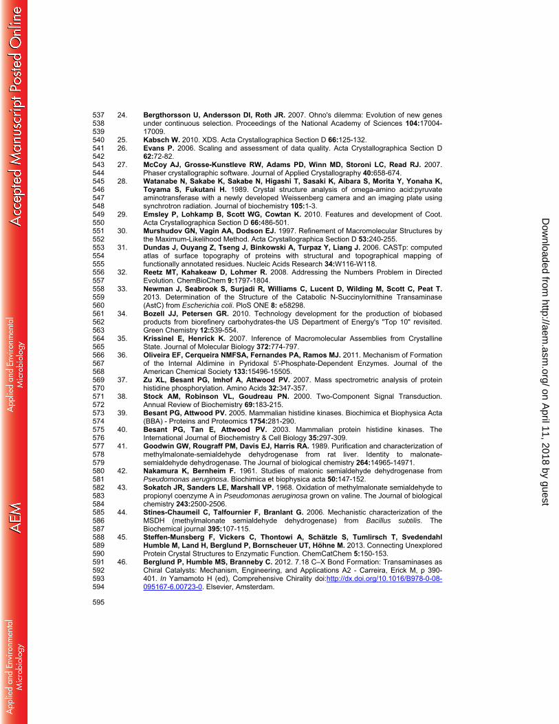
Figure 1a – Alanine dehydrogenase-coupled UV assay for transaminase activity. The transaminase catalyzes the amine transfer from the substrate to pyruvate (co-substrate), producing alanine as a co-product. The dehydrogenase then catalyzes the oxidative deamination of alanine utilizing a cofactor, NAD+ (nicotinamide adenine dinucleotide), which is concurrently reduced to NADH. Formation of NADH can be detected by UV photospectrometry as a hyperchromic shift at 340 nm.
Figure 1b – Analogous assays performed with KES23460. In this case the transaminase reaction shown is the β-alanine/pyruvate transamination reaction. If CoA is present, malonate semialdehyde produced by transamination is then converted by KES23460 to acetyl CoA in an NAD+ dependent reaction (shown in bold). Without CoA present malonate semialdehyde is converted to acetate and bicarbonate (shown in grey). Both of these reactions require the conversion of NAD+ to NADH, which can be followed by UV photospectrometry at 340 nm. Based on spectrophotometric data, and the value of Acetyl-CoA as a metabolic intermediate, the CoA-dependent pathway is likely to be the physiologically relevant reaction for these genes.
NH2
H
O
O
OHH2N
O
OHO
O
OHO
KES23458
L-ADH
340 nmNADH
X X
NH2
H
O
O
OHH2N
O
OHO
KES23458
KES23460
NADH2O
O OH O OH
a)
b)
378.3 nmol.min-1.mg-1
Acetate
KES23460
Acetyl-CoA
NADHHCO3
-518.4 nmol.min-1 .mg-
1
NAD+
CoA
NADHHCO3
-
NAD+
on April 11, 2018 by guest
http://aem.asm
.org/D
ownloaded from
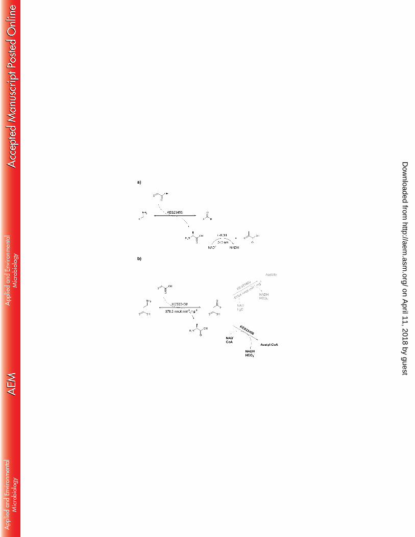
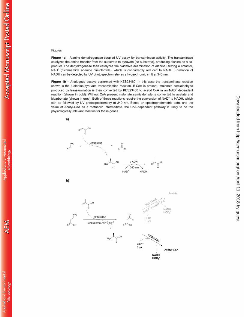
Figure 2a – 2mFo-DFc Electron density maps for the phosphohistidine groups observed in the Xray diffraction data for KES23458 at 1.5 σ. Top: His31, bottom: His360. 2b –. Dimeric form of KES23458. One monomeric subunit is shown in green, and the other in cyan, with PLP shown in each active site. 2c – Overlay. KES23458 from Pseudomonas sp. strain AAC is shown in green. 4A6T from Chromobacterium violaceum is shown in yellow. 4B98 from Pseudomonas aeruginosa is shown in magenta. 2d – KES23458 and 4A6T overlayed, with the free PLP and Lys288 from KES23458, and internal aldimine from 4A6T represented as sticks.
2a) 2b)
2c) 2d)
on April 11, 2018 by guest
http://aem.asm
.org/D
ownloaded from

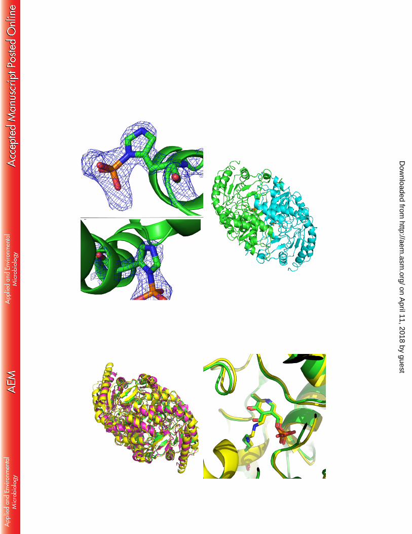
Figure 3a) The dimeric form of KES23458 with monomers shown in green and cyan. Residues which were identified as having a difference in average backbone RMSD > 1.5 Å between the two MD simulations are represented by pink dots. Note that in each case the substrate was docked into the same (green) monomeric subunit and PLP alone was docked into the cyan monomeric subunit 3b and c) – Visualization of the active site when 12-aminododecanoic acid (b) and β-alanine (c) were docked independently. In each case the substrate:PLP complex is shown in pick in a ball and stick representation. Residues which were observed to change conformation most significantly are shown in green.
3a)
3b) 3c)
on April 11, 2018 by guest
http://aem.asm
.org/D
ownloaded from
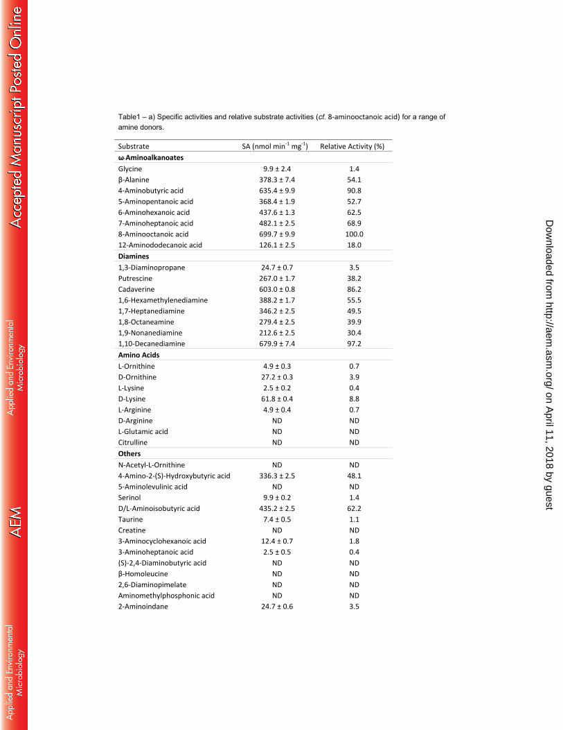
Table1 – a) Specific activities and relative substrate activities (cf. 8-aminooctanoic acid) for a range of amine donors.
Substrate SA (nmol min-1 mg-1) Relative Activity (%)
ω-Aminoalkanoates
Glycine 9.9 ± 2.4 1.4
β-Alanine 378.3 ± 7.4 54.1
4-Aminobutyric acid 635.4 ± 9.9 90.8
5-Aminopentanoic acid 368.4 ± 1.9 52.7
6-Aminohexanoic acid 437.6 ± 1.3 62.5
7-Aminoheptanoic acid 482.1 ± 2.5 68.9
8-Aminooctanoic acid 699.7 ± 9.9 100.0
12-Aminododecanoic acid 126.1 ± 2.5 18.0
Diamines
1,3-Diaminopropane 24.7 ± 0.7 3.5
Putrescine 267.0 ± 1.7 38.2
Cadaverine 603.0 ± 0.8 86.2
1,6-Hexamethylenediamine 388.2 ± 1.7 55.5
1,7-Heptanediamine 346.2 ± 2.5 49.5
1,8-Octaneamine 279.4 ± 2.5 39.9
1,9-Nonanediamine 212.6 ± 2.5 30.4
1,10-Decanediamine 679.9 ± 7.4 97.2
Amino Acids
L-Ornithine 4.9 ± 0.3 0.7
D-Ornithine 27.2 ± 0.3 3.9
L-Lysine 2.5 ± 0.2 0.4
D-Lysine 61.8 ± 0.4 8.8
L-Arginine 4.9 ± 0.4 0.7
D-Arginine ND ND
L-Glutamic acid ND ND
Citrulline ND ND
Others
N-Acetyl-L-Ornithine ND ND
4-Amino-2-(S)-Hydroxybutyric acid 336.3 ± 2.5 48.1
5-Aminolevulinic acid ND ND
Serinol 9.9 ± 0.2 1.4
D/L-Aminoisobutyric acid 435.2 ± 2.5 62.2
Taurine 7.4 ± 0.5 1.1
Creatine ND ND
3-Aminocyclohexanoic acid 12.4 ± 0.7 1.8
3-Aminoheptanoic acid 2.5 ± 0.5 0.4
(S)-2,4-Diaminobutyric acid ND ND
β-Homoleucine ND ND
2,6-Diaminopimelate ND ND
Aminomethylphosphonic acid ND ND
2-Aminoindane 24.7 ± 0.6 3.5
on April 11, 2018 by guest
http://aem.asm
.org/D
ownloaded from
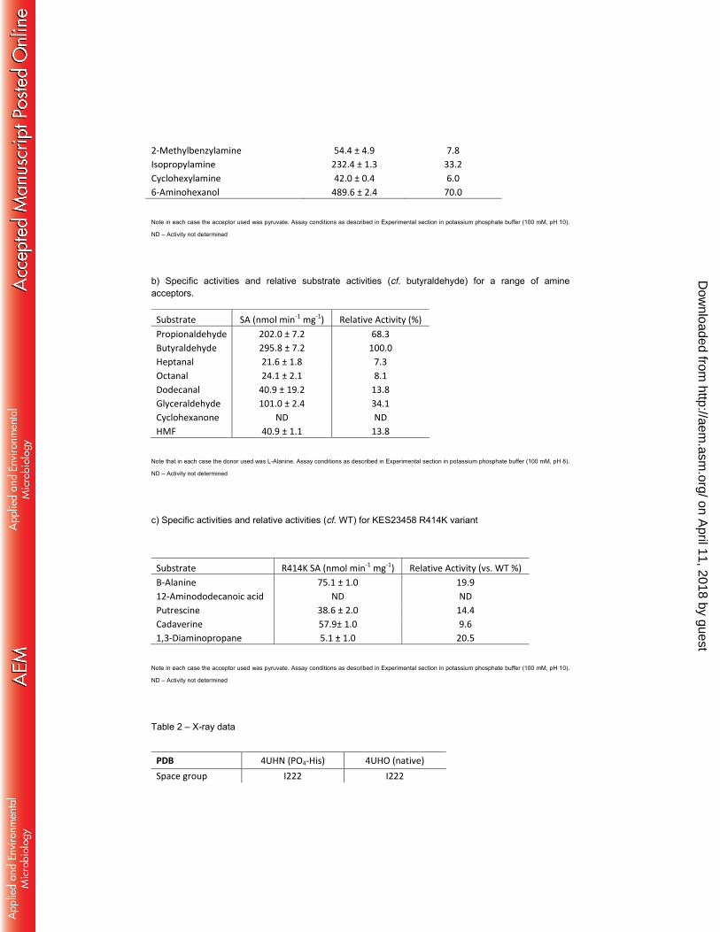
2-Methylbenzylamine 54.4 ± 4.9 7.8
Isopropylamine 232.4 ± 1.3 33.2
Cyclohexylamine 42.0 ± 0.4 6.0
6-Aminohexanol 489.6 ± 2.4 70.0
Note in each case the acceptor used was pyruvate. Assay conditions as described in Experimental section in potassium phosphate buffer (100 mM, pH 10).
ND – Activity not determined
b) Specific activities and relative substrate activities (cf. butyraldehyde) for a range of amine acceptors.
Substrate SA (nmol min-1 mg-1) Relative Activity (%)
Propionaldehyde 202.0 ± 7.2 68.3
Butyraldehyde 295.8 ± 7.2 100.0
Heptanal 21.6 ± 1.8 7.3
Octanal 24.1 ± 2.1 8.1
Dodecanal 40.9 ± 19.2 13.8
Glyceraldehyde 101.0 ± 2.4 34.1
Cyclohexanone ND ND
HMF 40.9 ± 1.1 13.8
Note that in each case the donor used was L-Alanine. Assay conditions as described in Experimental section in potassium phosphate buffer (100 mM, pH 8).
ND – Activity not determined
c) Specific activities and relative activities (cf. WT) for KES23458 R414K variant
Substrate R414K SA (nmol min-1 mg-1) Relative Activity (vs. WT %)
Β-Alanine 75.1 ± 1.0 19.9
12-Aminododecanoic acid ND ND
Putrescine 38.6 ± 2.0 14.4
Cadaverine 57.9± 1.0 9.6
1,3-Diaminopropane 5.1 ± 1.0 20.5
Note in each case the acceptor used was pyruvate. Assay conditions as described in Experimental section in potassium phosphate buffer (100 mM, pH 10).
ND – Activity not determined
Table 2 – X-ray data
PDB 4UHN (PO4-His) 4UHO (native)
Space group I222 I222
on April 11, 2018 by guest
http://aem.asm
.org/D
ownloaded from
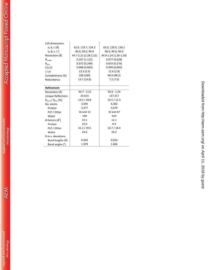
Cell dimensions
a, b, c (Å) 62.9. 119.7, 134.3 63.0, 120.0, 134.2
α, β, γ (°) 90.0, 90.0, 90.0 90.0, 90.0, 90.0
Resolution (Å) 44.7-2.21 (2.28-2.21) 44.9-1.24 (1.26-1.24)
Rmerge 0.267 (1.122) 0.077 (0.628)
Rpim 0.072 (0.299) 0.033 (0.276)
CC1/2 0.996 (0.843) 0.999 (0.845)
I / σI 13.3 (3.3) 12.6 (2.8)
Completeness (%) 100 (100) 99.9 (98.2)
Redundancy 14.7 (14.8) 7.2 (7.0)
Refinement
Resolution (Å) 44.7 - 2.21 44.9 - 1.24
Unique Reflections 24,514 137,317
Rwork / Rfree (%) 14.5 / 18.8 10.5 / 12.3
No. atoms 3,595 4,382
Protein 3,377 3,679
PLP / Other 16 and 12 16 and 67
Water 190 620
B-factors (Å2) 23.1 12.1
Protein 22.9 9.9
PLP / Other 31.2 / 39.5 10.7 / 18.0
Water 24.6 24.2
R.m.s. deviations
Bond lengths (Å) 0.020 0.010
Bond angles (°) 1.979 1.666
on April 11, 2018 by guest
http://aem.asm
.org/D
ownloaded from