
7A. Normal Anatomy, Development, and Physiology
26
Section 7 - Pancreas 353 7A. Normal Anatomy, Development, and Physiology Gia Bradley, MD Ann Scheimann, MD, MBA I. Development of the Pancreas A. Pancreatic development is influenced by both signaling and transcription factors 1. Hedgehog proteins—signaling molecules that both stimulate and inhibit pancreatic growth 2. Other factors influencing development—pancreatic duct primary cilia, fibroblast growth factor, transforming growth factor- β, vascular endothelial growth factor and homeobox transcription factor B. At 4–5 weeks’ gestation, distinct dorsal and ventral pancreatic buds arise from the endoderm of the caudal foregut (the primordial proximal duodenum) 1. The dorsal bud is larger than and slightly cranial to the ventral bud 2. Each bud communicates with the foregut through a duct C. Rotation of the duodenum causes the ventral pancreatic bud to rotate clockwise to the left of the duodenum, and brings it posterior and inferior to the dorsal pancreatic bud D. During the 7th week of gestation, the two buds fuse to form the pancreas 1. The ventral pancreatic bud forms the inferior part of the head of the pancreas and the uncinate process 2. The dorsal pancreatic bud forms the superior part of the head, the body, and the tail of the pancreas E. During the 8th week of gestation, the ductal systems of the two buds anastomose 1. The longer dorsal duct drains into the proximal part of the ventral duct to form the main pancreatic duct (duct of Wirsung), which enters the duodenum at the major duodenal papilla (ampulla of Vater) 2. If the proximal portion of the dorsal duct remains, it forms an accessory duct (duct of Santorini) that opens into a minor accessory papilla located about 2 cm above the main duct a. The accessory duct opens into a minor papilla in 33% of people and ends blindly in 8% of people, while about half of individuals do not have one F. At about 8 weeks’ of gestation, groups of endocrine cells (islets) originating from ductal epithe- lium are identifiable 1. From 10–14 weeks’ gestation, the islets form clumps and detach from the ducts G. At about 12 weeks’ gestation, exocrine cells appear along the pancreatic ducts 1. Exocrine pancreatic development continues after birth with maturation of specific diges- tive enzymes, including pancreatic amylase and lipase II. Anatomy A. Arterial supply 1. Pancreatic head—supplied by the superior pancreaticoduodenal artery (a branch of the gastroduodenal artery), and the inferior pancreaticoduodenal artery (a branch of the superior mesenteric artery) 2. Remainder of pancreas is supplied by the pancreatic branches of the splenic artery B. Venous drainage 1. Head of the pancreas is drained by superior mesenteric and portal veins 2. Body and neck of pancreas are drained by splenic vein C. Innervation 1. Acini, islets and ducts innervated by the vagus nerve 2. Blood vessels innervated by sympathetic nervous system
Transcript of 7A. Normal Anatomy, Development, and Physiology
Section 7 - Pancreas 353
7A. Normal Anatomy, Development, and Physiology
Gia Bradley, MDAnn Scheimann, MD, MBA
I. Development of the PancreasA. Pancreatic development is influenced by both signaling and transcription factors
1. Hedgehog proteins—signaling molecules that both stimulate and inhibit pancreatic growth
2. Other factors influencing development—pancreatic duct primary cilia, fibroblast growth factor, transforming growth factor-β, vascular endothelial growth factor and homeobox transcription factor
B. At 4–5 weeks’ gestation, distinct dorsal and ventral pancreatic buds arise from the endoderm of the caudal foregut (the primordial proximal duodenum)
1. The dorsal bud is larger than and slightly cranial to the ventral bud2. Each bud communicates with the foregut through a duct
C. Rotation of the duodenum causes the ventral pancreatic bud to rotate clockwise to the left of the duodenum, and brings it posterior and inferior to the dorsal pancreatic bud
D. During the 7th week of gestation, the two buds fuse to form the pancreas1. The ventral pancreatic bud forms the inferior part of the head of the pancreas and the
uncinate process2. The dorsal pancreatic bud forms the superior part of the head, the body, and the tail of
the pancreasE. During the 8th week of gestation, the ductal systems of the two buds anastomose
1. The longer dorsal duct drains into the proximal part of the ventral duct to form the main pancreatic duct (duct of Wirsung), which enters the duodenum at the major duodenal papilla (ampulla of Vater)
2. If the proximal portion of the dorsal duct remains, it forms an accessory duct (duct of Santorini) that opens into a minor accessory papilla located about 2 cm above the main duct
a. The accessory duct opens into a minor papilla in 33% of people and ends blindly in 8% of people, while about half of individuals do not have one
F. At about 8 weeks’ of gestation, groups of endocrine cells (islets) originating from ductal epithe-lium are identifiable
1. From 10–14 weeks’ gestation, the islets form clumps and detach from the ductsG. At about 12 weeks’ gestation, exocrine cells appear along the pancreatic ducts
1. Exocrine pancreatic development continues after birth with maturation of specific diges-tive enzymes, including pancreatic amylase and lipase
II. AnatomyA. Arterial supply
1. Pancreatic head—supplied by the superior pancreaticoduodenal artery (a branch of the gastroduodenal artery), and the inferior pancreaticoduodenal artery (a branch of the superior mesenteric artery)
2. Remainder of pancreas is supplied by the pancreatic branches of the splenic arteryB. Venous drainage
1. Head of the pancreas is drained by superior mesenteric and portal veins2. Body and neck of pancreas are drained by splenic vein
C. Innervation1. Acini, islets and ducts innervated by the vagus nerve2. Blood vessels innervated by sympathetic nervous system
354 The NASPGHAN Fellows Concise Review of Pediatric Gastroenterology, Hepatology and Nutrition
D. Endocrine cells1. These cells are distributed within the islets of Langerhans2. There are 4 four types:
a. A cells: produce glucagonb. B cells: produce insulinc. D cells: secrete somatostatind. F cells: secrete pancreatic polypeptide
3. These hormones enter systemic circulation via pancreatic blood flowE. Exocrine cells
1. The exocrine pancreas consists of lobules that contain acini and ductal systema. Each acinus contains ~6–8 pyramidal cells, with their apical poles facing a lumen
that empties into an intercalated ductb. Intercalated ducts fuse to form intralobular ducts that drain into interlobular
ductsc. Interlobular ducts empty into the main pancreatic duct which enters the
duodenum2. The acinus synthesizes, stores and releases pancreatic enzymes
a. The basal region of the acinar cell contains the nucleus and endoplasmic reticulum where proteins are synthesized
b. Enzymes are packaged in secretory (zymogen) granules in the Golgi complexc. Secretory granules are stored in the apical region of the cell d. Acinar basolateral membrane has multiple receptors for secretagogues
(e.g., cholecystokinin) and neurotransmitters (e.g., acetylcholine and vasoactive intestinal peptide)
F. Duct cells1. Centroacinar (proximal ductular) cells that empty into the acinar lumen and pancreatic
duct cells both modify pancreatic juice by secretion of water and bicarbonate
III. PhysiologyA. The adult human pancreas delivers ~2.5 L of fluid to the duodenum daily
1. The fluid is composed of digestive enzymes, bicarbonate to ensure an optimal pH for enzyme activity, and water
2. At rest, pancreatic secretion rate is 0.2 mL/min, with bicarbonate concentration equal to that of plasma
3. After stimulation, secretion rate increases to ~4 mL/min, and bicarbonate concentration increases to a maximum of 140 mEq/L, creating a pH of ~8.2 in pancreatic fluid
B. Regulation of pancreatic secretion1. Hormones that stimulate pancreatic fluid secretion
a. Secretin1) Major mediator of hydrogen ion-stimulated bicarbonate and water
secretion2) Released by S-type enteroendocrine cells in the proximal small intestine
in the presence of duodenal acidification (pH threshold 4.5), bile, and the products of protein and fat digestion
b. Cholecystokinin1) Major mediator of meal-stimulated enzyme secretion2) Secreted primarily by intestinal I cells in response to the products of
protein and fat digestion 2. Interdigestive pancreatic secretion
a. There is a cyclic secretion of pancreatic juice that closely follows the pattern of the migrating myoelectric complex in the intestine
b. Interdigestive secretion is important in the digestion of residual food, cellular debris and pathogens in the duodenum
c. Regulation occurs via motilin, pancreatic polypeptide and the autonomic nervous system
Section 7 - Pancreas 355
3. Postprandial pancreatic secretiona. Cephalic phase: vagus nerve mediates pancreatic secretion at the sight, smell,
taste and thought of food b. Gastric phase: distension of the stomach produces vasovagal cholinergic reflex
that causes increased pancreatic secretionc. Intestinal phase: chyme in the duodenum leads to pancreatic secretion via
secretin, cholecystokinin and the vagus nerve4. Hormones that inhibit pancreatic secretion
a. Pancreatic polypeptide: released from the islets of Langerhans in response to food and duodenal acidification
b. Peptide YY: released in response to fat in the distal ileum and colon c. Somatostatin: produced in mucosa of stomach and duodenum and in islets of
Langerhans. Released in response to fat and amino acids in the intestinal tractC. Pancreatic exocrine function (see section on Exocrine Function)
Recommended Reading
Feldman M, Friedman LS, Brandt LJ. Sleisenger and Fordtran’s Gastrointestinal and Liver Disease. 9th ed. Philadelphia, PA: Saunders Elsevier; 2010:909-930.
Kleinman RE, Goulet O-J, Mieli-Vergani G, Sanderson I, Sherman P, Shneider B. Walker’s Pediatric Gastrointes-tinal Disease. 5th ed. Hamilton, Ontario: BC Decker, Inc.;2008:1185-1201.
Wyllie R, Hyams JS. Pediatric Gastrointestinal and Liver Disease. 3rd ed. Philadelphia, PA: Saunders Elsevier; 2006:1005-1021.

356 The NASPGHAN Fellows Concise Review of Pediatric Gastroenterology, Hepatology and Nutrition
Section 7 - Pancreas 357
7B. Exocrine Function
John Pohl, MD
I. Exocrine Pancreatic DevelopmentA. The pancreas forms during the 4th week of gestation, developing from the endodermal lining
of the duodenum as ventral and dorsal outpouchings. By Week 6 of gestation, the dorsal aspect develops a nodular pattern resembling the basic acinar pancreatic anatomy, while the ventral aspect develops a connection with the early common bile duct. The ventral and dorsal elements fuse at Week 7 of gestation, and the main pancreatic duct attaches to the common bile duct (which forms from the fusion of the common bile duct, pancreatic duct and the ventral pan-creas).
B. Hedgehog proteins (signaling molecules) regulate pancreatic morphogenesis by promoting cel-lular proliferation and differentiation. Indian hedgehog protein appears to be the sole hedgehog protein involved in pancreatic growth. Pancreatic acini are present by the 3rd month of gestation. Zymogen granules appear by the 12th week of gestation. By the 20th week of gestation, zymo-gens identical to those in adult pancreas are observed. Infants have exocrine pancreatic function similar to adults at term, although zymogen size and enzyme content may differ.
II. Tests for Exocrine Pancreatic InsufficiencyA. 72-hour fecal fat collection is the gold standard indirect measure of lipolytic enzyme activity
1. Requires accurate estimate of dietary fat intake for 72 hours2. Requires complete collection of stool for 72 hours 3. Total fats are extracted from stool and weighed 4. Total fat excreted as fraction of fat intake calculated5. Normal fat excretion is <7% of intake in children over 2 years. May be higher in young
children, especially those infants younger than 6 months of age6. Very accurate measure of lipase activity, but difficult because of poor compliance with
stool collection and diet recordB. Stool trypsin/chymotrypsin measures proteolytic activity in stool
1. Indirect measure of pancreatic proteolytic enzyme secretion2. Trypsin in stool is prone to bacterial degradation, producing false low values (poor
sensitivity)C. Breath hydrogen excretion
1. Using starch as substrate, an increase in breath hydrogen excretion of 20 ppm over baseline after oral administration roughly indicates insufficiency of pancreatic amylase activity
2. Positive tests occur in patients with small bowel bacterial overgrowth and monosaccharide transport defects who may be inaccurately diagnosed with pancreatic exocrine insufficiency
D. Serum immunoreactive trypsinogen measures trypsin precursor in serum1. In true exocrine pancreatic insufficiency, serum immunoreactive trypsinogen will be very
low2. In newborns with cystic fibrosis, serum IRT is elevated because of obstruction of
pancreatic ducts and regurgitation of IRT into bloodstream3. In newborns with cystic fibrosis and distal intestinal obstruction at birth (meconium
ileus), IRT is usually low because of extensive prenatal destruction of pancreatic acini
358 The NASPGHAN Fellows Concise Review of Pediatric Gastroenterology, Hepatology and Nutrition
E. Secretin/CCK infusion test is the gold standard direct measurement of pancreatic exocrine enzyme secretion
1. IV infusion of secretin and/or cholecystokinin is followed by aspiration of duodenal contents (biliary and pancreatic secretions) by nasoduodenal tube for 15 minutes to 1 hour
2. Aspirated pancreatic secretions tested for pH, bicarbonate concentration and lipase, amylase and proteolytic activity
3. Test is expensive and invasive4. Test accuracy is increased by simultaneous perfusion of the duodenum with a
nonabsorbed marker, so that total volume of secretion following stimulation can be determined
F. Fecal elastase measures amount of pancreatic neutrophil elastase in stool1. Low fecal elastase concentration indicates pancreatic exocrine insufficiency2. Test is easy to perform3. Patients with pancreatic sufficient Shwachman-Diamond syndrome may have falsely low
fecal elastase4. Patients with short bowel syndrome have falsely low fecal elastase5. Falsely low results occur in patients with large volume diarrhea due to dilution
III. Diagnoses Associated With Exocrine Pancreatic InsufficiencyA. Acquired pancreatic exocrine insufficiency may occur as a complication of chronic pancreatitis.
Causes of chronic pancreatitis include:1. Trauma, after endoscopic retrograde cholangiopancreatogram (ERCP)2. Medication use (eg, isoniazid)3. Infections (eg, ascariasis)4. Biliary disease5. Pancreas divisum6. Metabolic causes (eg, hypercalcemia, hypertriglyceridemia, malnutrition)7. Associated with systemic diseases (hemolytic uremic syndrome or Kawasaki disease)
B. Cystic fibrosis is the most common inherited cause of exocrine pancreatic insufficiency (approximately 1 in 2,000 live births)
1. Dysfunction of the cystic fibrosis transmembrane conductance regulatory protein (CFTR)2. Most common CFTR gene mutation is ΔF5083. Measuring exocrine pancreatic function is not the recommended means of making a
diagnosis of cystic fibrosis. Knowing the extent of exocrine insufficiency may assist in management. Recommended diagnostic tests include:
a. Sweat chloride b. Nasal mucosal potential differencec. Cystic fibrosis gene (CFTR) mutation analysis
C. Second most common cause of pancreatic insufficiency is Shwachman-Diamond syndrome (1 in 75,000 live births)—exocrine pancreatic insufficiency, bone marrow abnormalities, metaphyseal dysostosis, growth retardation and immune dysfunction (see section on Shwachman-Diamond Syndrome)
D. Other causes of pancreatic exocrine insufficiency1. Hereditary pancreatitis (autosomal dominant) typically presents in 2nd decade of life,
associated with pancreatic malignancy. Can be seen with PRSS1 mutation or SPINK1 mutation affecting cationic trypsinogen gene
2. Pearson’s bone marrow syndrome: pancreatic exocrine insufficiency, sideroblastic anemia, bone marrow vacuolization
3. Congenital rubella due to cell loss, possibly from molecular mimicry/immune destruction4. Pancreatic agenesis/hypoplasia: 10 cases reported5. Isolated pancreatic enzyme defects: very rare (lipase deficiency, colipase deficiency,
enterokinase deficiency)6. Johanson-Blizzard syndrome: pancreatic exocrine insufficiency, failure to thrive, deafness,
hypothyroidism, microcephaly, abnormal hair pattern, nasal cartilage hypoplasia, small or absent permanent teeth
Section 7 - Pancreas 359
Recommended References
Beharry S, Ellis L, Corey M, et al. How useful is fecal pancreatic elastase 1 as a marker of exocrine pancreatic disease? J Pediatr. 2002;141: 84-90.
Casellas F, Guarner L, Vaquero E, et al. Hydrogen breath test with glucose in exocrine pancreatic insufficiency. Pancreas. 1998;16:481-486.
Feldman M, Friedman LS, Brandt LJ. Sleisenger and Fordtran’s Gastrointestinal and Liver Disease. 9th ed. Philadelphia, PA: Saunders Elsevier; 2010.
Pohl JF, Easley DJ. Pancreatic insufficiency in children. Pract Gastro. 2003;27(10):38-48.
Weintraub A, Blau H, Mussaffi H, et al. Exocrine pancreatic function testing in patients with cystic fibrosis and pancreatic sufficiency: a correlation study. J Pediatr Gastro Nutr. 2009;48:306-310.

360 The NASPGHAN Fellows Concise Review of Pediatric Gastroenterology, Hepatology and Nutrition
Section 7 - Pancreas 361
7C. Congenital Anomalies of the Pancreas
Maria E. Perez, DOCheryl Gariepy, MD
I. Pancreatic DivisumA. Most common congenital anomaly of the pancreasB. Failure of fusion of the dorsal and ventral pancreatic buds off the foregut (typically Weeks 7–8 of
gestation) causes formation of two separate drainage systems 1. Head of the pancreas is drained through the major papilla 2. Neck and tail of the pancreas (majority of the pancreatic tissue) is drained through the
minor papillaC. Clinical significance is controversial
1. Normal variant vs possible cause of recurrent pancreatitis2. Possible cause of recurrent pancreatitis
a. High volume of pancreatic secretions through the smaller papilla allows activated pancreatic enzymes to inflame the papilla, leading to stasis, stenosis and pancreatitis
D. Diagnosis — ERCP and MRCPE. Treatment — Endoscopic enlargement of the minor papilla, sphincteroplasty, stent placement
II. Ectopic PancreasA. Also known as pancreatic restB. Pancreatic tissue with no vascular or physical continuity with the pancreasC. Most common locations: prepyloric stomach, duodenum, Meckel diverticulumD. Clinical signs/symptoms:
1. Usually causes no symptoms2. May be the source of abdominal pain, dyspepsia, GI bleed, pyloric obstruction3. Ectopic pancreatitis is rare, but it has been documented in the adult and pediatric
population E. Diagnosis — usually an incidental finding on upper GI series or upper endoscopy; appears as a
round, slightly raised, smooth, submucosal mass with a central umbilicationF. Treatment — usually requires no treatment. Surgical resection only if the rest is thought to be
causing serious symptoms
III. Annular PancreasA. Ventral portion of the pancreas encircles the second portion of the duodenum and fuses with
the dorsal aspect of the developing pancreasB. Most common presentation is neonatal duodenal obstructionC. 75% associated with other congenital anomalies
1. Anomalies of the GI tract are most common, including malrotation; duodenal web, atresia or stenosis; TE fistula, imperforate anus and Hirschsprung disease
2. Congenital heart disease3. Increased incidence in trisomy 21
D. Clinical signs/symptoms1. Infancy: UGI obstruction, vomiting (may or may not be bilious), pancreatitis2. Adolescents/adults: duodenal obstruction, chronic pancreatitis
E. Diagnosis1. Double bubble on plain abdominal x-ray2. MRCP/ERCP provide best view
F. Treatment — surgical bypass of lesion with duodenoduodenostomy
Recommended Reading
Wyllie R, Hyams JS. Pediatric Gastrointestinal and Liver Disease. 3rd edition. Philadelphia, PA: Saunders Elsevier; 2006: Chapter 68.

362 The NASPGHAN Fellows Concise Review of Pediatric Gastroenterology, Hepatology and Nutrition
Section 7 - Pancreas 363
7D. Acute Pancreatitis
Maria E. Perez, DO Cheryl E. Gariepy, MD
I. Incidence The incidence of acute pancreatitis in children has increased in the past decade as a result of increased
physician awareness of pathophysiology and improved diagnostic methods. In adults, alcohol use and gallstones account for most cases. In children, there are multiple etiologies and many cases remain idiopathic. Management is supportive, and most patients recover without complications.
II. Three Phases in Pathophysiology of PancreatitisA. An event triggers pancreatic injury B. Acinar cell injury occurs through the activation of digestive enzymes, such as trypsinogenC. Cell injury produces a local inflammatory response with the release of inflammatory mediators
III. EtiologyA. Systemic illnesses associated with pancreatitis
1. Hemolytic uremic syndrome2. Systemic lupus3. Inflammatory bowel disease4. Sickle cell disease5. Kawasaki disease6. Shock/hypoperfusion injury7. Cystic fibrosis
B. Biliary disease—cholelithiasis, choledochal cyst, biliary sludgeC. Trauma—motor vehicle accidents or bike handle injuriesD. Medications
1. Valproic acid and L-asparaginase are drugs most commonly associated with pancreatitis at therapeutic doses
2. Azathioprine, mercaptopurine, mesalamine and metronidazole have also been associated with acute pancreatitis
E. Anatomic—pancreas divisum, annular pancreasF. Obstruction—duodenal ulcer, tumor of the papilla, duodenal Crohn diseaseG. Infections—below are major pathogens
1. Mycoplasma2. Coxsackie virus3. Mumps virus4. Varicella, HSV, CMV5. Rubeola6. Hepatitis A & B7. Influenza A & B8. HIV
H. Genetic—PRSS1 mutations, CFTR mutations, SPINK-1 mutationsI. Metabolic—hypercalcemia, hyperlipidemia, malnutritionJ. Toxins—acetaminophen overdose, organophosphates, alcohol, spider venom, heroin, amphet-
amines
IV. Clinical Manifestations/DiagnosisA. Diagnosis of acute pancreatitis requires at least two of the following three criteria:
1. Abdominal pain consistent with pancreatitis2. Elevation of serum amylase and/or lipase of at least three times the upper limit of normal3. Radiographic evidence of pancreatitis (ultrasound, CT scan, etc)
364 The NASPGHAN Fellows Concise Review of Pediatric Gastroenterology, Hepatology and Nutrition
B. Indicators of disease severity at presentation: age <7 years, lower body weight, elevated WBC, and LDH >2,000 associated with more severe disease
C. Predictors of severe disease at 48 hours1. Low serum calcium2. Low serum albumin3. Elevated BUN4. High fluid requirements
D. Signs and symptoms of acute pancreatitis are listed in Table 1. These vary based on the age of the child at presentation
1. Turner sign—bluish discoloration of the flank2. Cullen sign—bluish discoloration of the periumbilical region3. Turner and Cullen signs indicate hemorrhagic pancreatitis and are late signs in the clinical
course of pancreatitis
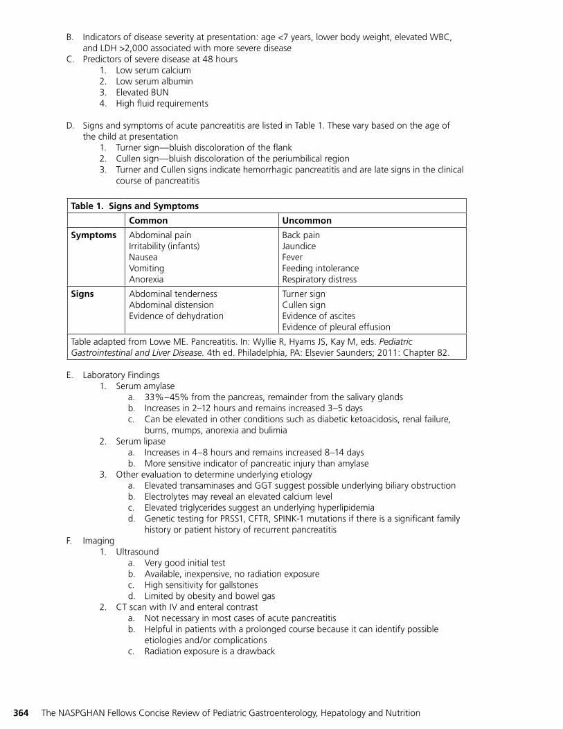
Table 1. Signs and Symptoms
Common Uncommon
Symptoms Abdominal painIrritability (infants)NauseaVomitingAnorexia
Back painJaundiceFeverFeeding intoleranceRespiratory distress
Signs Abdominal tendernessAbdominal distensionEvidence of dehydration
Turner signCullen signEvidence of ascitesEvidence of pleural effusion
Table adapted from Lowe ME. Pancreatitis. In: Wyllie R, Hyams JS, Kay M, eds. Pediatric Gastrointestinal and Liver Disease. 4th ed. Philadelphia, PA: Elsevier Saunders; 2011: Chapter 82.
E. Laboratory Findings1. Serum amylase
a. 33%–45% from the pancreas, remainder from the salivary glandsb. Increases in 2–12 hours and remains increased 3–5 daysc. Can be elevated in other conditions such as diabetic ketoacidosis, renal failure,
burns, mumps, anorexia and bulimia2. Serum lipase
a. Increases in 4–8 hours and remains increased 8–14 daysb. More sensitive indicator of pancreatic injury than amylase
3. Other evaluation to determine underlying etiologya. Elevated transaminases and GGT suggest possible underlying biliary obstructionb. Electrolytes may reveal an elevated calcium levelc. Elevated triglycerides suggest an underlying hyperlipidemiad. Genetic testing for PRSS1, CFTR, SPINK-1 mutations if there is a significant family
history or patient history of recurrent pancreatitisF. Imaging
1. Ultrasounda. Very good initial testb. Available, inexpensive, no radiation exposurec. High sensitivity for gallstonesd. Limited by obesity and bowel gas
2. CT scan with IV and enteral contrasta. Not necessary in most cases of acute pancreatitisb. Helpful in patients with a prolonged course because it can identify possible
etiologies and/or complicationsc. Radiation exposure is a drawback
Section 7 - Pancreas 365
3. Magnetic resonance cholangiopancreatography (MRCP)a. Helpful in patients with recurrent episodes of acute pancreatitisb. Can reveal anatomic abnormalities and obstructive etiologies, such as pancreatic
divisum, gallstones and choledochal cystc. Requires anesthesia in younger children
4. Endoscopic retrograde cholagiopancreatography (ERCP)a. Limited role in initial evaluation of acute pancreatitisb. Most helpful for interventional procedure (stone removal, stent placement, etc)
5. Esophagogastroduodenoscopy (EGD)a. Not routine in the evaluation of acute pancreatitisb. May reveal duodenal ulcer, tumor of the papilla or duodenal Crohn disease
6. Endoscopic ultrasounda. Not in widespread use yetb. Helpful for gallstones and microlithiasisc. Limited data in children
V. Treatment/ManagementA. Fluid Management
1. Early aggressive fluid resuscitation improves outcome and prevents severe disease in animal and human studies, by maintaining cardiovascular stability and decreasing the incidence of pancreatic necrosis
B. Pain Control1. Parenteral narcotics are preferred 2. All opiates increase sphincter of Oddi pressure, but this action does not affect outcome
or clinical courseC. Nutrition
1. Pancreatic rest (nothing by mouth) is standard in treatment of acute pancreatitis, but no clear evidence supports this practice
2. Early enteral nutrition is recommendeda. Associated with lower infection risk, reduced surgical interventions and shorter
hospital stayb. Jejunal feeds may be helpful, since there is less stimulation of the exocrine
pancreasc. No evidence-based guidelines in children. Adult studies suggest oral nutrition
be resumed when pain and nausea resolve in mild cases. In severe pancreatitis, enteral nutrition within the first 48 hours has been associated with a better outcome
3. No evidence that clear liquid or a low-fat diet improves outcome
VI. Complications/OutcomesA. Most cases resolve in 7–10 days without complicationsB. 13%–20% of children have prolonged courses with complications C. Mortality rate ranges from 2%–10%, and in children is typically associated with systemic illnessD. Peripancreatic fluid collections and pseudocyst
1. Most common complication in children (13%–16% of cases)2. Trauma often associated with pancreatic pseudocyst formation in children3. Suspect pseudocyst if acute episode is not resolving, abdominal mass develops or pan-
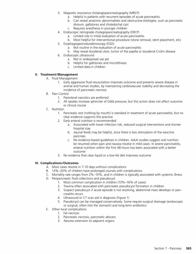
creatitis recurs4. Ultrasound or CT scan aid in diagnosis (Figure 1)5. Pseudocyst can be managed conservatively. Some require surgical drainage (endoscopic
or surgical, often into the stomach) and long-term antibiotics E. Other local complications
1. Fat necrosis2. Pancreatic necrosis, pancreatic abscess3. Abscess extension to adjacent organs
366 The NASPGHAN Fellows Concise Review of Pediatric Gastroenterology, Hepatology and Nutrition
F. Systemic complications 1. Electrolyte abnormalities2. Sepsis3. Pleural effusions4. Acute renal failure5. Coagulopathy6. Shock
Figure 1. Pseudocyst (between white arrows) near the distal body and tail of the pancreas.Figure adapted from Lowe ME. Pancreatitis. In: Wyllie R, Hyams JS, Kay M, eds. Pediatric Gastrointestinal and Liver Disease. 4th ed. Philadelphia, PA: Elsevier Saunders; 2011: Chapter 82.
Recommended Reading
Kandula L, Lowe ME. Etiology and outcome of acute pancreatitis in infants and toddlers. J Peds. 2008;152:106-110.
Lowe ME, Greer JB. Pancreatitis in Children and Adolescents. Curr Gastroenterol Reports. 2008;10:128-135.
Teh SH, Pham TH, Lee A, Stavlo PL, Hanna AM, Moir C. Pancretic pseudocyst in children: the impact of management strategies on outcome. J Pediatr Surg. 2006;41: 1889-1893.
Wyllie R, Hyams JS, Kay M, eds. Pediatric Gastrointestinal and Liver Disease. 4th ed. Philadelphia, PA: Elsevier Saunders; 2011: Chapter 82.
Section 7 - Pancreas 367
7E. Chronic Pancreatitis
Razan Alkhouri, MBBS Susan Baker, MD, PhD
I. Chronic pancreatitis Chronic pancreatitis is the continuous destruction of the pancreatic gland with irreversible scarring
of acinar and ductal cells. Each exacerbation leads to additional damage. Fortunately, the pancreas has significant reserves and is able to function with as little as 10% of the gland functioning. The etiology of chronic pancreatitis can be divided into obstructive (ductal anomalies, strictures, trauma and autoimmune), calcific (hereditary, hyperlipidemia and hypercalcemia) and idiopathic.
II. Genetic causes of chronic pancreatitis A. Hereditary pancreatitis (PRSS1) 1. Most common genetic cause of chronic pancreatitis 2. Mutation in cationic trypsinogen gene (serine protease 1) located on chromosome 7q35,
autosomal dominant 3. R112H is the most prevalent mutation followed by N291 4. Recurrent bouts of pancreatitis starting at 10–20 years of age 5. Family history usually positive for recurrent pancreatitis 6. Long-term risk of diabetes and pancreatic adenocarcinoma B. SPINK-1 gene produces pancreatic trypsin inhibitor 1. Mutations cause uninhibited activation of trypsin in pancreatic ducts 2. Mutations may be autosomal dominant, autosomal recessive or multigenic 3. Family history usually absent C. Cystic fibrosis transmembrane conductance regulator gene (CFTR) 1. Heterozygous mutations of this autosomal recessive gene found in 20%–40% of
patients with chronic pancreatitis a. Gene expressed in pancreatic ductal tissue of Type 1 is cystic fibrosis with a
CFTRsev/CFTRsev b. Type 2 is atypical cystic fibrosis with a CFTRsev/CFTRm-v c. Type 3 is CFTRsev or CFTRm-v plus a second pancreatitis modifier or
susceptibility gene in a polygenetic condition d. Type 4 is CFTRsev or CFTRm-v plus a strong environmental risk factor,
such as alcohol
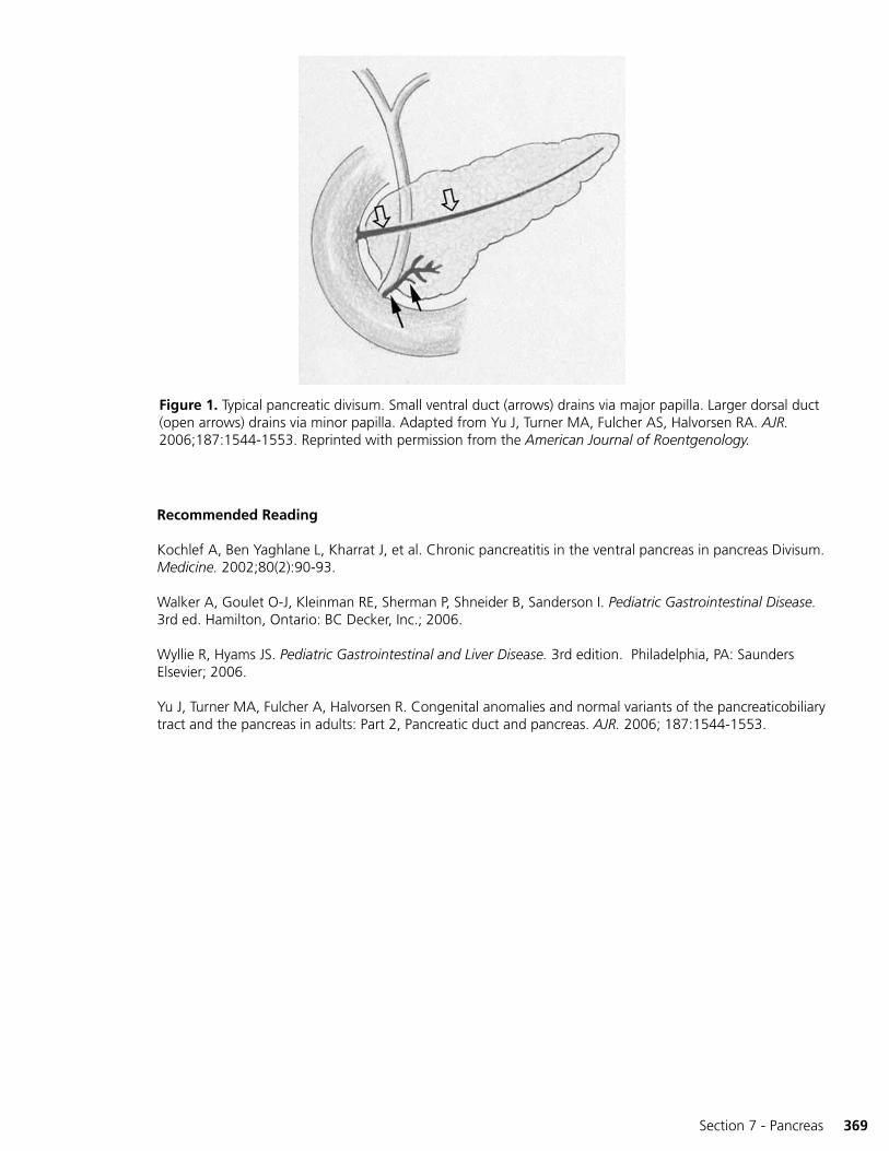
III. Causes of chronic obstructive pancreatitis A. Pancreas divisum 1. 25 % of patients with pancreatic divisum develop obstructive pancreatitis 2. Diagnosed by MRCP or ERCP (see Figure 1) 3. Optimum therapy not established a. Endoscopic sphincterotomy or surgical sphincteroplasty b. Stent of minor papilla B. Idiopathic fibrosing pancreatitis 1. Very rare 2. Presents with obstructive jaundice or abdominal pain and pancreatitis 3. Diffuse pancreatic fibrosis ± inflammation 4. Can be associated with PRSS1, SPINK-1 and CFTR 5T C. Abdominal trauma may cause obstructive pancreatitis if there is duct damage and stricture
formation D. Congenital anomalies associated with chronic pancreatitis: choledochal cyst, pancreatic ductal
duplication, anomalous pancreaticobiliary ductal union
368 The Fellows Concise Review of Pediatric Gastroenterology, Hepatology and Nutrition
E. Autoimmune pancreatitis 1. Primary: isolated pancreatic involvement may require biopsy diagnosis 2. Associated with lupus erythematosis, autoimmune hepatitis, Sjögren disease 3. Histology shows ductal and periductal infiltration by lymphocytes, plasma cells and
granulocytes with irregular narrowing of pancreatic duct and pancreatic enlargement 4. Associated increase in IgG4 (most commonly in adults) 5. Responsive to steroids
IV. Other causes of chronic pancreatitis A. Hypertriglyceridemia syndromes 1. Type I hyperlipidemia—30% develop pancreatitis 2. Type IV—15% of patients develop pancreatitis 3. Type V—30%–40% of patients develop pancreatitis 4. Common features include triglyceride levels >1,000 mg/dL B. Tropical chronic pancreatitis 1. Disease with young onset 2. Occurs in tropics 3. Associated with large intraductal calculi, steatorrhea and diabetes C. Hypercalcemia D. Organic acidemia
V. Clinical presentation of chronic pancreatitis A. Repeated bouts of pancreatitis B. Acute and chronic abdominal pain C. Chronic abdominal pain may improve in longstanding disease due to loss of pancreatic tissue D. Some patients present with diabetes, exocrine pancreatic insufficiency and obstructive jaundice
without obvious pain
VI. Diagnostic testing A. Elevated serum amylase and lipase almost always present during acute episodes but may not be
dramatic as disease progresses B. Biochemical markers of protein and fat malabsorption occur late in disease due to exocrine
insufficiency C. Pancreatic stimulation test helpful to evaluate exocrine function (see section on Pancreatic
Function Testing) D. Fecal elastase (see section Pancreatic Function Testing) is a noninvasive method to document
pancreatic exocrine insufficiency E. Functional MRCP obtained after intravenous secretin improves definition of pancreatic
duct anatomy F. Other imaging studies to clarify duct structure, cysts, masses, ductal stricture or dilation 1. Endoscopic ultrasound—head of pancreas 2. Abdominal CT 3. MRCP G. Genetic testing to clarify causation (see above)
VII. Treatment A. Treat acute episodes – (see section on Acute Pancreatitis) B. Acute and chronic pain management C. Nutrition 1. Low-fat diet, although evidence is lacking 2. Nasoduodenal feeds D. Surgical therapies 1. Puestow procedure—pancreas and main pancreatic duct are sectioned longitudinally
and oversewn with a segment of jejunum to directly drain pancreatic secretions into bowel
2. Partial pancreatectomy may relieve pain 3. Complete pancreatectomy with islet cell transplantation in recalcitrant disease 4. Celiac sympathectomy in recalcitrant disease with pain
Section 7 - Pancreas 369
Recommended Reading
Kochlef A, Ben Yaghlane L, Kharrat J, et al. Chronic pancreatitis in the ventral pancreas in pancreas Divisum. Medicine. 2002;80(2):90-93.
Walker A, Goulet O-J, Kleinman RE, Sherman P, Shneider B, Sanderson I. Pediatric Gastrointestinal Disease. 3rd ed. Hamilton, Ontario: BC Decker, Inc.; 2006.
Wyllie R, Hyams JS. Pediatric Gastrointestinal and Liver Disease. 3rd edition. Philadelphia, PA: Saunders Elsevier; 2006.
Yu J, Turner MA, Fulcher A, Halvorsen R. Congenital anomalies and normal variants of the pancreaticobiliary tract and the pancreas in adults: Part 2, Pancreatic duct and pancreas. AJR. 2006; 187:1544-1553.
Figure 1. Typical pancreatic divisum. Small ventral duct (arrows) drains via major papilla. Larger dorsal duct (open arrows) drains via minor papilla. Adapted from Yu J, Turner MA, Fulcher AS, Halvorsen RA. AJR. 2006;187:1544-1553. Reprinted with permission from the American Journal of Roentgenology.

370 The Fellows Concise Review of Pediatric Gastroenterology, Hepatology and Nutrition
Section 7 - Pancreas 371
7F. Shwachman-Diamond Syndrome
Maria E. Perez, DOCheryl E. Gariepy, MD
I. Overview/ Epidemiology A. Autosomal recessive condition characterized by the triad of exocrine pancreatic insufficiency,
bone marrow dysfunction and skeletal abnormalities B. Prevalence is 1:50,000 1. SDS the 2nd most common inherited cause of pancreatic insufficiency after cystic
fibrosis 2. SDS is the 3rd most common inherited bone marrow failure syndrome C. Variable clinical presentation with gastrointestinal and hematologic abnormalities in almost all
patients D. Median survival for all patients is 35 years. Severe bacterial infection and leukemia are the major
causes of morbidity and death
II. Pathogenesis A. Biallelic mutations in the SBDS gene on chromosome 7 occur in 90% of cases The remaining
10% have the clinical picture of SDS but no identified mutations B. The SBDS gene product seems to participate in ribosome biogenesis, RNA processing, stabilizing
the mitotic spindle and neutrophil chemotaxis C. Mutation causes failure of normal development of pancreatic acinar tissue in utero with fatty
replacement of acini D. Bone marrow abnormalities 1. Abnormal myeloid clones in bone marrow with mutations of chromosome 7 (eg,
monosomy) and dysmyelogenesis 2. Leukemia or aplastic anemia are long-term complications 3. Impaired neutrophil function E. Skeletal abnormalities 1. No correlation between genotype and skeletal phenotype 2. Delayed secondary ossification 3. Variable widening, thickening, and irregularity of the metaphyses and growth plates 4. Generalized osteopenia
III. Comparison of Pancreatic Function in CF and SDS A. Sweat chloride 1. Normal sweat chloride concentration in SDS 2. Elevated sweat chloride in CF B. Histology 1. Normal ductal elements in SDS. Fatty replacement of the acinar tissue 2. Duct obstruction, fibrosis and ectasia in CF C. Pancreatic enzyme output 1. Increased pancreatic volume and enzyme output in SDS patients over time with normal
fat absorption in approximately 50% of patients by 4 years of age 2. No increased risk of pancreatitis in SDS 3. Enzyme output in CF is dependent on genotype, whereas in patients with SDS it is NOT
dependent on genotype
372 The NASPGHAN Fellows Concise Review of Pediatric Gastroenterology, Hepatology and Nutrition
IV. Laboratory Diagnosis of SDS A. Low pancreatic secretion of lipase, amylase and trypsin in response to secretin and CCK
stimulation B. Low serum immunoreactive trypsinogen and low serum isoamylase in children <3 years of age C. Serum immunoreactive trypsinogen and fat absorption may normalize in older children/adults. D. Low serum isoamylase levels and starch malabsorption persist to adulthood E. Cyclic or persistent neutropenia. Additional cytopenias may also be present. Anemia has been
reported in 42%–66% of patients, and thrombocytopenia has been reported in 24%–60% F. Imaging 1. Abdominal CT scan or ultrasound shows a fatty pancreas of normal size 2. Skeletal x-ray findings include clinodactyly, syndactyly, genu and cubitus valgus, tooth
enamel defects, metaphyseal dysostosis
V. Clinical Manifestations A. Diarrhea, steatorrhea, failure to thrive B. Hematology 1. Recurrent neutropenia is most common abnormality (>90%) 2. Impaired neutrophil chemotaxis 3. Pancytopenia (in approximately 10%–25%) carries the worst prognosis 4. 1/3 of patients with severe chronic neutropenia develop myelodysplastic syndrome
(MDS) 5. 10%–25% of patients with severe chronic neutropenia develop acute myeloid leukemia
(AML) C. Skeletal abnormalities 1. Progressive metaphyseal dysostosis in approximately 45% of patients 2. Thoracic cage abnormalities in approximately 1/3 of patients 3. Short stature, usually remaining at less than the 5th percentile for life 4. May be at higher risk for slipped capital femoral epiphysis (SCFE) D. Others 1. Dental caries, enamel abnormalities, delayed dentition 2. Hepatomegaly and elevated transaminases are common in infancy and usually resolve by
5 years of age. Histologic abnormalities may include microvesicular and macrovesicular steatosis, periportal and portal inflammation, bridging fibrosis and glycogenosis
3. Learning difficulties including weaknesses in higher-order language skills, perceptual skills and perceptual reasoning are more common in patients with SDS compared to both healthy controls and patients with CF
4. Behavioral issues and social problems are also more common in patients with SDS compared to healthy siblings, healthy unrelated controls and patients with CF
VI. Treatment/Management A. Oral pancreatic enzyme supplementation. Steatorrhea typically resolves in approximately 50% of
patients by 4 years of age. Fecal fat measurement should be repeated to determine the need for continued supplementation
B. Fat-soluble vitamin supplementation C. Serial CBC (at least every 4–6 months) D. Bone marrow biopsy (every 1–3 years) E. Transfusions as needed F. Timely evaluation of fever and neutropenia, including physical exam, cultures and prophylactic
antibiotics G. Granulocyte colony-stimulating factor (G-CSF) for those patients with ANC <500 and with
repeated infections. The French Neutropenia Registry has documented an 80% response with G-CSF, but there has been no association between G-CSF use and the subsequent risk of AML
H. Bone marrow transplant is the only curative therapy for bone marrow failure
Section 7 - Pancreas 373
VII. Differential Diagnosis A. Cystic fibrosis: sweat chloride and genetic testing will differentiate. In addition, high
immunoreactive trypsinogen in most infants with CF B. Johanson-Blizzard syndrome 1. Exocrine pancreatic insufficiency 2. Multiple congenital abnormalities: deafness, imperforate anus, urogenital
malformations, dental anomalies 3. Endocrine abnormalities: hypothyroidism, GH deficiency, diabetes, panhypopituitarism 4. Typical facies: nasal hypoplasia with beak-shaped appearance, small misshapen teeth,
sparse hair C. Pearson Marrow-Pancreas syndrome 1. Rare mitochondrial disorder 2. Exocrine pancreatic insufficiency 3. Bone marrow involvement a. Profound sideroblastic anemia 4. Lactic acidosis is very common 5. Early death is very common D. Jeune syndrome 1. Rare autosomal-recessive disorder 2. Exocrine pancreatic insufficiency 3. Respiratory difficulties, asphyxiating thoracic dystrophy
Recommended Reading
Feldman M, Friedman LS, Brandt LJ. Sleisenger and Fordtran’s Gastrointestinal and Liver Disease. 9th ed. Philadelphia, PA: Saunders Elsevier; 2010:Chapter 57.
Ginzberg H, et al. Shwachman syndrome: Phenotypic manifestations of siblings sets and isolated cases in a large patient cohort are similar. J Peds. 1999;135:81-88.
Kerr EN, Ellis L, Dupuis A, Rommens JM, Durie PR. The behavioral phenotype of school-age children with Schwachman Diamond syndrome indicates neurocognitive dysfunction with loss of Schwachman-Bodian-Diamond syndrome gene function. J Pediatr. 2010;156:433-438.
Lacaille F, Mani TM, Brunelle F, Lallemand D, Schmitz J. Magnetic resonance imaging for diagnosis of Schwachman’s syndrome. J Pediatr Gastroenterol Nutr. 1996; 23(5):599-603.
Shimamura A. Shwachman Diamond syndrome. Semin Hematol. 200643: 178-188.Wyllie R, Hyams JS. Pediatric Gastrointestinal and Liver Disease. 3rd edition. Philadelphia, PA: Saunders Elsevier; 2006:Chapter 68.

374 The NASPGHAN Fellows Concise Review of Pediatric Gastroenterology, Hepatology and Nutrition
Section 7 - Pancreas 375
7G. Cystic FibrosisAlex Green, DO
Steve Erdman, MD
I. Description Cystic fibrosis is the most common lethal genetic disease in Caucasians. Cystic fibrosis affects many organ
systems, including the gastrointestinal tract (meconium ileus, distal intestinal obstruction syndrome), hepatobiliary system (neonatal cholestasis, steatosis, biliary cirrhosis, biliary sludge) and pancreas (exocrine dysfunction).
II. Carrier Rate/Inheritance/Diagnosis A. Autosomal recessive with carrier rate in Caucasians of 1 in 25 B. Disease prevalence 1. Caucasians 1 in 2,500 2. African Americans 1 in 15,000 3. Asians 1 in 31,000 C. Of more than 1,500 mutations now described, DF508 accounts for 66% 1. Deletion of the three nucleotides comprising the codon for phenylalanine
at position 508 D. Most cases of CF are diagnosed by newborn screen with high values of immunoreactive
trypsinogen (IRT) 1. False positive (high) IRT may be produced by: a. hypoxia, respiratory or physiologic stress b. low Apgar scores, organ damage c. trisomies 13, 18, 21 d. renal dysfunction e. hypoglycemia f. contamination of filter paper g. carrier status h. early specimen collection 2. False negative (low) IRT results seen in: a. CF genetic variant with normal pancreatic function b. Older affected infant with pancreatic insufficiency c. Meconium ileus (severe pancreatic insufficiency) E. Gold standard of CF testing remains sweat chloride. 1. Normal <40 mmol/L 2. Abnormal ≥60 mmol/L 3. Borderline readings between 40–60 require repeat sweat chloride test, CFTR mutation
analysis or nasal potential difference to confirm diagnosis 4. False positive sweat chloride test caused by a. Anorexia nervosa, Addison disease, nephrogenic diabetes insipidus and
hypothyroidism 5. False negative sweat chloride test caused by a. Edema
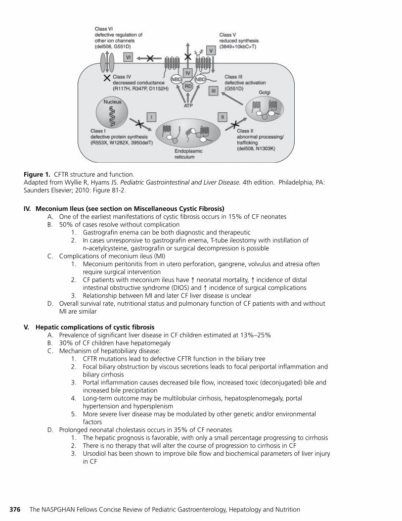
III. CFTR:Structure and Function (See Figure 1) A. Localized in the epithelial cells of the airway, pancreatic and hepatic ducts, intestine, sweat gland
and vas deferens B. CFTR opens channels in the cell membrane which transport chloride ions out of the cells C. Six classes of mutations with increasing severity 1. Mutations of lower class (I-IV) involve mutation, with effect closer to the transcription
and processing phase of the CFTR protein with complete loss of function a. More serious complications of the lower classes include pancreatic insufficiency
(PI), meconium ileus and hepatic complications 2. Mutations of higher class (V-VI) have reduced function once CFTR reaches the surface
membrane (see Figure 1)
376 The NASPGHAN Fellows Concise Review of Pediatric Gastroenterology, Hepatology and Nutrition
IV. Meconium Ileus (see section on Miscellaneous Cystic Fibrosis) A. One of the earliest manifestations of cystic fibrosis occurs in 15% of CF neonates B. 50% of cases resolve without complication 1. Gastrografin enema can be both diagnostic and therapeutic 2. In cases unresponsive to gastrografin enema, T-tube ileostomy with instillation of
n-acetylcysteine, gastrografin or surgical decompression is possible C. Complications of meconium ileus (MI) 1. Meconium peritonitis from in utero perforation, gangrene, volvulus and atresia often
require surgical intervention 2. CF patients with meconium ileus have ↑ neonatal mortality, ↑ incidence of distal
intestinal obstructive syndrome (DIOS) and ↑ incidence of surgical complications 3. Relationship between MI and later CF liver disease is unclear D. Overall survival rate, nutritional status and pulmonary function of CF patients with and without
MI are similar
V. Hepatic complications of cystic fibrosis A. Prevalence of significant liver disease in CF children estimated at 13%–25% B. 30% of CF children have hepatomegaly C. Mechanism of hepatobiliary disease: 1. CFTR mutations lead to defective CFTR function in the biliary tree 2. Focal biliary obstruction by viscous secretions leads to focal periportal inflammation and
biliary cirrhosis 3. Portal inflammation causes decreased bile flow, increased toxic (deconjugated) bile and
increased bile precipitation 4. Long-term outcome may be multilobular cirrhosis, hepatosplenomegaly, portal
hypertension and hypersplenism 5. More severe liver disease may be modulated by other genetic and/or environmental
factors D. Prolonged neonatal cholestasis occurs in 35% of CF neonates 1. The hepatic prognosis is favorable, with only a small percentage progressing to cirrhosis 2. There is no therapy that will alter the course of progression to cirrhosis in CF 3. Ursodiol has been shown to improve bile flow and biochemical parameters of liver injury
in CF
Figure 1. CFTR structure and function.Adapted from Wyllie R, Hyams JS. Pediatric Gastrointestinal and Liver Disease. 4th edition. Philadelphia, PA: Saunders Elsevier; 2010: Figure 81-2.
Section 7 - Pancreas 377
E. Hepatic steatosis is common in CF patients of all ages 1. It is a reflection of malnutrition and/or deficiency of essential fatty acids, carnitine or
choline 2. In older children, the development of diabetes can also contribute to steatosis 3. Ultrasonography and computed tomography show increased hepatic fat 4. Steatosis does not progress to serious focal biliary cirrhosis F. Focal biliary cirrhosis 1. Usually presents in teen years or later 2. CF patients are generally asymptomatic until late in the disease course 3. Hepatomegaly or splenomegaly can be present along with elevated serum transaminases 4. Elevated serum bilirubin usually a late finding 5. CF patients with high transaminases for 3–6 months should have Doppler US to assess
hepatic blood flow and possible esophageal varices 6. MRCP can be used for checking biliary tree and gallbladder 7. Liver biopsy is another option 8. Treatment includes ursodiol and nutritional improvement G. Portal hypertension 1. Pace of progression is unpredictable 2. The incidence of portal HTN and variceal bleeding in CF patients is <5% H. Cholelithiasis and biliary sludging 1. Incidence ranging from 1%–10% of CF patients 2. CF patients usually have radiolucent gallstones rich in calcium bilirubinate and proteins;
therefore, ursodiol does not help dissolve the stones 3. Symptomatic patients require cholecystectomy with intraoperative cholangiogram to as-
sess intrahepatic biliary tree I. Microgallbladder is found in 20% of CF patients 1. It is a relatively benign condition 2. Bile duct stenosis requiring dilation occurs in <1% of CF patients J. Liver transplantation 1. Almost all patients with cystic fibrosis have some evidence of liver disease, but only
3%–7% develop the degree of cirrhosis that progresses to end-stage liver disease 2. In 2002 Milkiewicz reported that among CF patients requiring liver transplant, the
average age was 13 years. Recommendation is that liver transplant be performed before lung transplant if both are needed
H. CF patients have an increased risk of malignancies including esophagus, stomach, small and large intestine, rectum, liver, biliary tract and pancreas. Greatest risk for malignancy is 20–29 years of age
VI. Nutritional Complications A. Pancreatic insufficiency (PI) 1. Exocrine pancreatic insufficiency occurs in 60% of infants and in 90% by 1 year of age 2. Protein-calorie malnutrition due to maldigestion and increased energy requirements
occurs in untreated patients 3. An acute Marasmic-Kwashiorkor-like presentation can occur with growth failure,
anemia, hypoproteinemia, edema and ascites B. Fat-soluble vitamin deficiencies occur in up to 50% CF infants by 2 months of age 1. Even when supplemented with standard ADEK, there can still be a deficiency, especially
vitamin E C. Electrolyte abnormalities 1. NaCl loss can occur with increased sweating (environmental, fever) 2. Salt supplementation at 2–4 mmol/kg/day 3. Most common presentation is hypochloremic alkalosis and dehydration 4. Very low urinary sodium is a good indicator that salt supplementation is needed
378 The NASPGHAN Fellows Concise Review of Pediatric Gastroenterology, Hepatology and Nutrition
VII. Pancreatic Exocrine Function Association With Genotypes/Abnormalities A. CFTR genotypes are more closely related to the severity of pancreatic exocrine dysfunction than
they are to the severity of lung disease B. Fecal fat excretion is more accurate than fecal elastase or serum IRT for classifying the extent of
pancreatic exocrine insufficiency C. The most accurate test of pancreatic exocrine function is direct pancreatic stimulation with
exogenous hormones (i.e., cholecystokinin or secretin) (see section on Pancreatic Function) D. Pancreatic insufficiency occurs when <10% of normal pancreatic exocrine function remains
Recommended Reading
Messick J. A 21st-century approach to cystic fibrosis: optimizing outcomes across the disease spectrum. JPGN. 2010;51:S1-S17.
Wilschanski M, Durie PR. Patterns of GI disease in adulthood associated with mutations in the CFTR gene. Gut. 2007;1153-1163.
Borowitz D, Durie PR, Clarke LL, et al. Gastrointestinal outcomes and confounders in cystic fibrosis. JPGN. 2005; 273-285.
Weintraub A, Blau H, Mussaffi H, et al. Exocrine pancreatic function testing in patients with cystic fibrosis and pancreatic sufficiency: a correlation study. JPGN. 2009;306-310.
Colombo C. Liver disease in cystic fibrosis. Curr Opin Pulmonary Med. 2007; 529-536.
Dodge JA, Turck D. Cystic fibrosis: nutritional consequences and management. Best Pract Res Clin Gastroenterol. 2006;531-546.
Wilschanski M. Patterns of gastrointestinal disease associated with mutations of CFTR. Curr Gastroenterol Rep. 2008;316-323.
Wyllie R, Hyams JS. Pediatric Gastrointestinal and Liver Disease. 4th edition. Philadelphia, PA: Saunders Elsevier; 2010.

![Glossar Deutsch- Griechisch - Praxis · C 7a Gesprächsthema, das, -themen θέμα συζήτησης C 7a laufen [εδώ] λειτουργώ C 7a Das Radio läuft. Το ράδιο](https://static.fdocument.org/doc/165x107/5fc7034600451f24d0445f37/glossar-deutsch-griechisch-praxis-c-7a-gesprchsthema-das-themen-.jpg)


![Glossar Deutsch- Griechisch - praxis.gr · 7a Jägerschnitzel, das, - σνίτσελ του κυνηγού [με μανιτάρια] 7a Kalbfleisch, das μοσχαρίσιο κρέας](https://static.fdocument.org/doc/165x107/5e06fe20a88f4c06457803b9/glossar-deutsch-griechisch-7a-jgerschnitzel-das-ff-.jpg)