510(k) Substantial Equivalence Determination Decision ...D. Type of Test: Quantitative mass...
18
1 510(k) SUBSTANTIAL EQUIVALENCE DETERMINATION DECISION SUMMARY ASSAY ONLY TEMPLATE A. 510(k) Number: k173829 B. Purpose for Submission: New device C. Measurand: Acid-β-glucocerebrosidase (ABG), acid-sphingomyelinase (ASM), acid-α-glucosidase (GAA), β-galactocerebrosidase (GALC), α-galactosidase A (GLA), α-L-iduronidase (IDUA) D. Type of Test: Quantitative mass spectrometric enzymatic activity assay E. Applicant: Wallac Oy, a subsidiary of PerkinElmer F. Proprietary and Established Names: NeoLSD MSMS kit G. Regulatory Information: 1. Regulation section: 862.1488 Lysosomal storage disorder newborn screening test system 2. Classification: II 3. Product code: PQW PQT PQU PQV
Transcript of 510(k) Substantial Equivalence Determination Decision ...D. Type of Test: Quantitative mass...
1
510(k) SUBSTANTIAL EQUIVALENCE DETERMINATION DECISION SUMMARY
ASSAY ONLY TEMPLATE
A. 510(k) Number:
k173829
B. Purpose for Submission:
New device
C. Measurand:
Acid-β-glucocerebrosidase (ABG), acid-sphingomyelinase (ASM), acid-α-glucosidase (GAA), β-galactocerebrosidase (GALC), α-galactosidase A (GLA), α-L-iduronidase (IDUA)
D. Type of Test:
Quantitative mass spectrometric enzymatic activity assay
E. Applicant:
Wallac Oy, a subsidiary of PerkinElmer
F. Proprietary and Established Names:
NeoLSD MSMS kit
G. Regulatory Information:
1. Regulation section:
862.1488 Lysosomal storage disorder newborn screening test system
2. Classification:
II
3. Product code:
PQW PQT PQU PQV
2
QCL QCM
4. Panel:
Chemistry (75)
H. Intended Use:
1. Intended use(s):
See Indication(s) for use.
2. Indication(s) for use:
The NeoLSD MSMS Kit is intended for the quantitative measurement of the activity of the enzymes acid-β-glucocerebrosidase (ABG), acid-sphingomyelinase (ASM), acid-α-glucosidase (GAA), β-galactocerebrosidase (GALC), α-galactosidase A (GLA), and α-L-iduronidase (IDUA) in dried blood spots (DBS) from newborn babies. The analysis of the enzymatic activity is intended as an aid in screening newborns for the following lysosomal storage disorders (LSD) respectively; Gaucher Disease, Niemann-Pick A/B Disease, Pompe Disease, Krabbe Disease, Fabry Disease, and mucopolysaccharidosis Type I (MPS I) Disease.
3. Special conditions for use statement(s):
This test is not intended to diagnose lysosomal storage disorders.
Test results are intended to be used in conjunction with other clinical and diagnostic findings, consistent with professional standards of practice, including confirmation by alternative methods, and clinical evaluation as appropriate.
Known causes for anomalous analytical assay results are:
- Sample not uniformly saturated with blood - Sample disk punched too close to the edge of the blood spot - Sample disk punched at the middle of the blood spot - Poorly collected specimens e.g. excessive milking or squeezing the puncture may
cause hemolysis of the specimen or a mixture of tissue fluids with the specimen. Layering successive drops of blood in the specimen may affect the measured results.
- Improperly dried specimens e.g. heating or stacking the specimen collection devices during the drying process
- Humidity and moisture or exposure to direct sunlight are detrimental to the dried blood spot sample
- Non-eluting blood spot due to deterioration of sample - Contamination of blood spot filter paper e.g. with fecal material
3
- It has been shown that female patients with the Fabry disorder may show normal GLA enzyme activities at birth but are later symptomatic
- Hematocrit levels above 65% were found to interfere with the assay by increasing the measured ABG activity that could result in false negatives for Gaucher. Refer to the interference section for complete information about this interference.
- Free (serum) hemoglobin above 16 g/dL was found to interfere with the assay by increasing the measured ABG, ASM and IDUA activity, that could result in false negative results for Gaucher, Niemann-Pick A/B, and mucopolysaccharidosis Type I (MPS I) Disease. Refer to the interference section for complete information about this interference.
The NeoLSD MSMS kit may result in;
- False negatives by not detecting Fabry disease in females - False positives by identifying pseudo deficiencies and carriers as affected for MPS I,
Gaucher, and Pompe diseases - False negative by not detecting certain late onset forms for Pompe disease - False negatives by not detecting certain late onset forms for Fabry disease - Increase rate of false positives when the specimen is exposed to high temperature
during shipping - The false negative rate is based on follow-up of subjects up to 4 years of age and on
limited data from late onset forms of the disorders since it can take several years to identify a missed late onset case.
- Certain late onset forms for Pompe disorder may have GAA enzymatic activity in the normal range and result in a false negative screening result.
- Heterozygote female Fabry patients may present with normal GLA activity and not be detected by the NeoLSD MSMS kit.
4. Special instrument requirements:
PerkinElmer TQD MSMS instrument system (k093916)
I. Device Description:
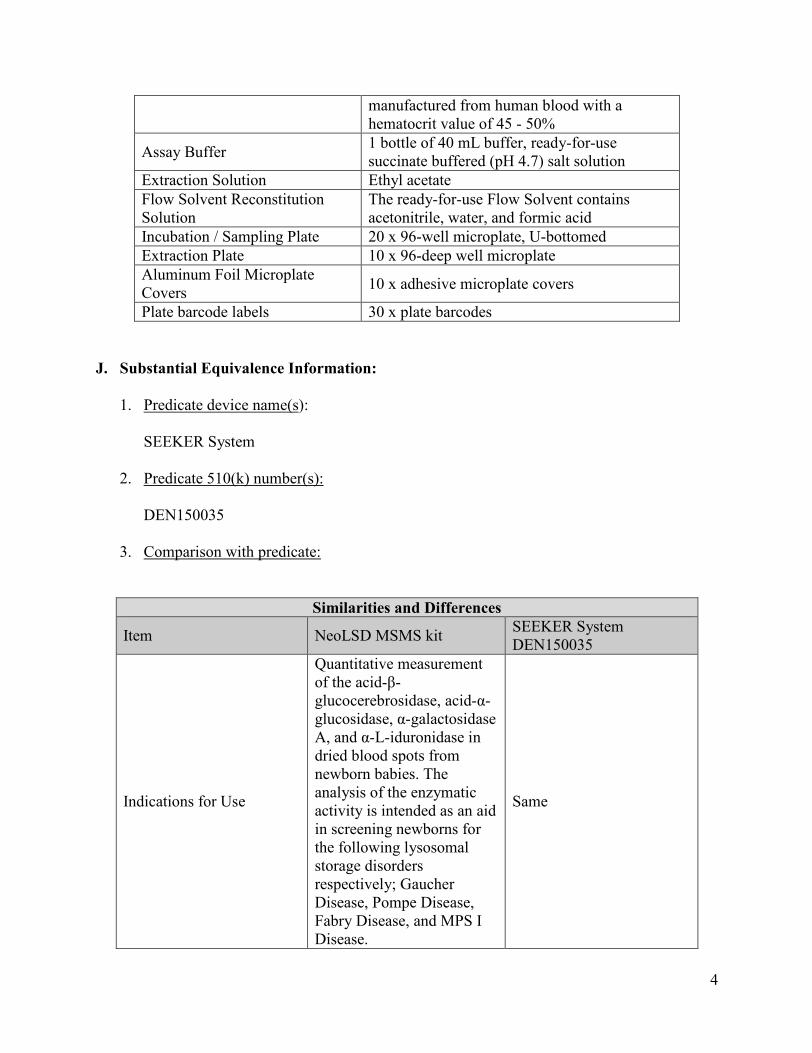
The NeoLSD MSMS kit consists of substrates, internal standards, and controls. The kit contains sufficient reagents and consumables to perform 960 assays (10 x 96-well plates). The contents of the kit are listed below:
Component Contents
Internal Standards Substrate Mix
1 vial or several vials of stable-isotope standards and designated substrates. The dried substrates and internal standards are a mixture of the 6 synthetic substrates, the corresponding 6 stable-isotope labeled internal standards, and sodium oleate
DBS Controls C1, C2, C3 control levels on DBS cassettes,
4
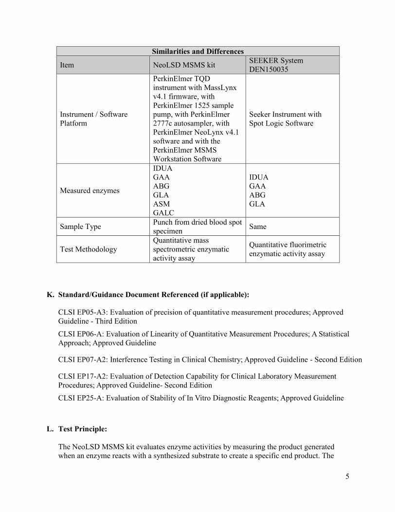
manufactured from human blood with a hematocrit value of 45 - 50%
Assay Buffer 1 bottle of 40 mL buffer, ready-for-use succinate buffered (pH 4.7) salt solution
Extraction Solution Ethyl acetate Flow Solvent Reconstitution Solution
The ready-for-use Flow Solvent contains acetonitrile, water, and formic acid
Incubation / Sampling Plate 20 x 96-well microplate, U-bottomed Extraction Plate 10 x 96-deep well microplate Aluminum Foil Microplate Covers 10 x adhesive microplate covers
Plate barcode labels 30 x plate barcodes
J. Substantial Equivalence Information:
1. Predicate device name(s):
SEEKER System
2. Predicate 510(k) number(s):
DEN150035
3. Comparison with predicate:
Similarities and Differences
Item NeoLSD MSMS kit SEEKER System DEN150035
Indications for Use
Quantitative measurement of the acid-β-glucocerebrosidase, acid-α-glucosidase, α-galactosidase A, and α-L-iduronidase in dried blood spots from newborn babies. The analysis of the enzymatic activity is intended as an aid in screening newborns for the following lysosomal storage disorders respectively; Gaucher Disease, Pompe Disease, Fabry Disease, and MPS I Disease.
Same
5
Similarities and Differences
Item NeoLSD MSMS kit SEEKER System DEN150035
Instrument / Software Platform
PerkinElmer TQD instrument with MassLynx v4.1 firmware, with PerkinElmer 1525 sample pump, with PerkinElmer 2777c autosampler, with PerkinElmer NeoLynx v4.1 software and with the PerkinElmer MSMS Workstation Software
Seeker Instrument with Spot Logic Software
Measured enzymes
IDUA GAA ABG GLA ASM GALC
IDUA GAA ABG GLA
Sample Type Punch from dried blood spot specimen Same
Test Methodology Quantitative mass spectrometric enzymatic activity assay
Quantitative fluorimetric enzymatic activity assay
K. Standard/Guidance Document Referenced (if applicable):
CLSI EP05-A3: Evaluation of precision of quantitative measurement procedures; Approved Guideline - Third Edition
CLSI EP06-A: Evaluation of Linearity of Quantitative Measurement Procedures; A Statistical Approach; Approved Guideline
CLSI EP07-A2: Interference Testing in Clinical Chemistry; Approved Guideline - Second Edition
CLSI EP17-A2: Evaluation of Detection Capability for Clinical Laboratory Measurement Procedures; Approved Guideline- Second Edition
CLSI EP25-A: Evaluation of Stability of In Vitro Diagnostic Reagents; Approved Guideline
L. Test Principle:
The NeoLSD MSMS kit evaluates enzyme activities by measuring the product generated when an enzyme reacts with a synthesized substrate to create a specific end product. The
6
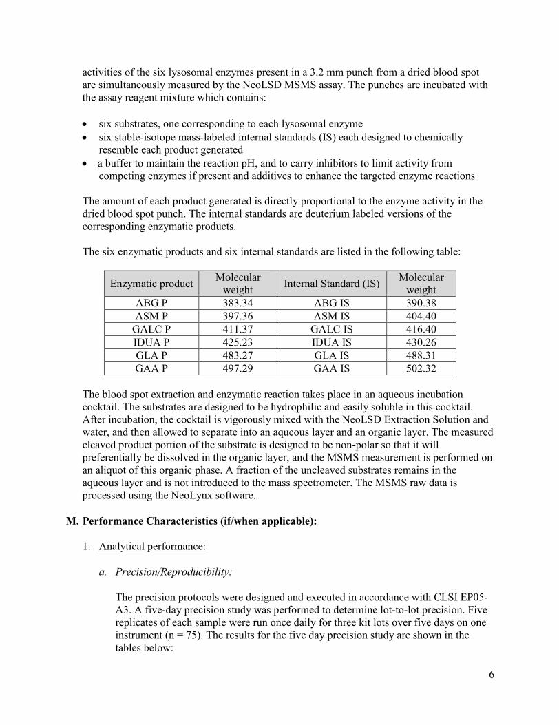
activities of the six lysosomal enzymes present in a 3.2 mm punch from a dried blood spot are simultaneously measured by the NeoLSD MSMS assay. The punches are incubated with the assay reagent mixture which contains:
· six substrates, one corresponding to each lysosomal enzyme · six stable-isotope mass-labeled internal standards (IS) each designed to chemically
resemble each product generated · a buffer to maintain the reaction pH, and to carry inhibitors to limit activity from
competing enzymes if present and additives to enhance the targeted enzyme reactions
The amount of each product generated is directly proportional to the enzyme activity in the dried blood spot punch. The internal standards are deuterium labeled versions of the corresponding enzymatic products.
The six enzymatic products and six internal standards are listed in the following table:
Enzymatic product Molecular weight Internal Standard (IS) Molecular
weight ABG P 383.34 ABG IS 390.38 ASM P 397.36 ASM IS 404.40
GALC P 411.37 GALC IS 416.40 IDUA P 425.23 IDUA IS 430.26 GLA P 483.27 GLA IS 488.31 GAA P 497.29 GAA IS 502.32
The blood spot extraction and enzymatic reaction takes place in an aqueous incubation cocktail. The substrates are designed to be hydrophilic and easily soluble in this cocktail. After incubation, the cocktail is vigorously mixed with the NeoLSD Extraction Solution and water, and then allowed to separate into an aqueous layer and an organic layer. The measured cleaved product portion of the substrate is designed to be non-polar so that it will preferentially be dissolved in the organic layer, and the MSMS measurement is performed on an aliquot of this organic phase. A fraction of the uncleaved substrates remains in the aqueous layer and is not introduced to the mass spectrometer. The MSMS raw data is processed using the NeoLynx software.
M. Performance Characteristics (if/when applicable):
1. Analytical performance:
a. Precision/Reproducibility:
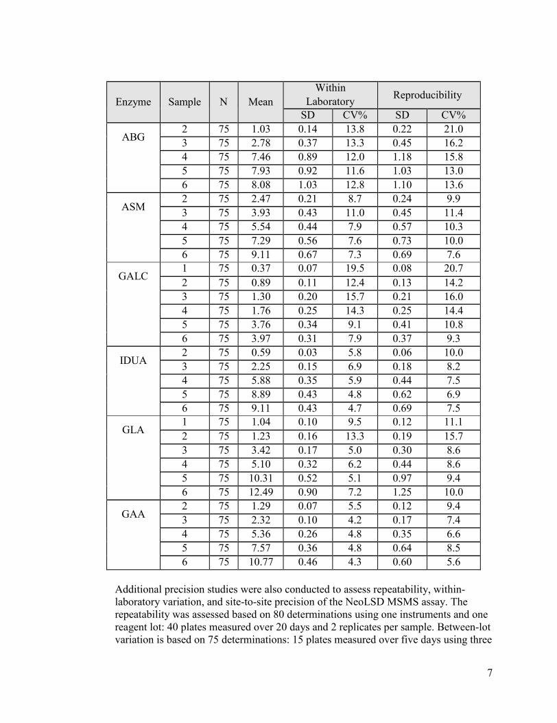
The precision protocols were designed and executed in accordance with CLSI EP05-A3. A five-day precision study was performed to determine lot-to-lot precision. Five replicates of each sample were run once daily for three kit lots over five days on one instrument (n = 75). The results for the five day precision study are shown in the tables below:
7
Enzyme Sample N Mean Within
Laboratory Reproducibility
SD CV% SD CV%
ABG 2 75 1.03 0.14 13.8 0.22 21.0 3 75 2.78 0.37 13.3 0.45 16.2 4 75 7.46 0.89 12.0 1.18 15.8 5 75 7.93 0.92 11.6 1.03 13.0 6 75 8.08 1.03 12.8 1.10 13.6
ASM 2 75 2.47 0.21 8.7 0.24 9.9 3 75 3.93 0.43 11.0 0.45 11.4 4 75 5.54 0.44 7.9 0.57 10.3 5 75 7.29 0.56 7.6 0.73 10.0 6 75 9.11 0.67 7.3 0.69 7.6
GALC 1 75 0.37 0.07 19.5 0.08 20.7 2 75 0.89 0.11 12.4 0.13 14.2 3 75 1.30 0.20 15.7 0.21 16.0 4 75 1.76 0.25 14.3 0.25 14.4 5 75 3.76 0.34 9.1 0.41 10.8 6 75 3.97 0.31 7.9 0.37 9.3
IDUA 2 75 0.59 0.03 5.8 0.06 10.0 3 75 2.25 0.15 6.9 0.18 8.2 4 75 5.88 0.35 5.9 0.44 7.5 5 75 8.89 0.43 4.8 0.62 6.9 6 75 9.11 0.43 4.7 0.69 7.5
GLA 1 75 1.04 0.10 9.5 0.12 11.1 2 75 1.23 0.16 13.3 0.19 15.7 3 75 3.42 0.17 5.0 0.30 8.6 4 75 5.10 0.32 6.2 0.44 8.6 5 75 10.31 0.52 5.1 0.97 9.4 6 75 12.49 0.90 7.2 1.25 10.0
GAA 2 75 1.29 0.07 5.5 0.12 9.4 3 75 2.32 0.10 4.2 0.17 7.4 4 75 5.36 0.26 4.8 0.35 6.6 5 75 7.57 0.36 4.8 0.64 8.5 6 75 10.77 0.46 4.3 0.60 5.6
Additional precision studies were also conducted to assess repeatability, within-laboratory variation, and site-to-site precision of the NeoLSD MSMS assay. The repeatability was assessed based on 80 determinations using one instruments and one reagent lot: 40 plates measured over 20 days and 2 replicates per sample. Between-lot variation is based on 75 determinations: 15 plates measured over five days using three
8
kit lots, with 5 replicates per sample. Site-to-site was assessed in both precision studies as well as an analysis that combined data from both studies. In all three precision studies, the NeoLSD MSMS assay showed similar precision.
b. Linearity/assay reportable range:
Linearity studies were performed following CLSI EP06-A using one lot of reagents. A high sample pool was prepared by mixing buffy coat and recombinant enzymes to obtain enzyme activities near the 95th percentile of the neonatal population, and adjusting the hematocrit to 45 - 55%. A low sample pool was prepared by washing red blood cells with saline solution then adjusting the hematocrit to 45 - 55%. Thirty levels were prepared by mixing the high sample in different proportions with the low samples, and spotted onto filter paper to generate dried blood spot specimens. Each sample was assayed in replicates of four over two runs. The degree of nonlinearity was assessed by polynomial evaluation. For any series where the polynomial fit was a better fit statistically, the maximum deviation from linearity was -12.1%.
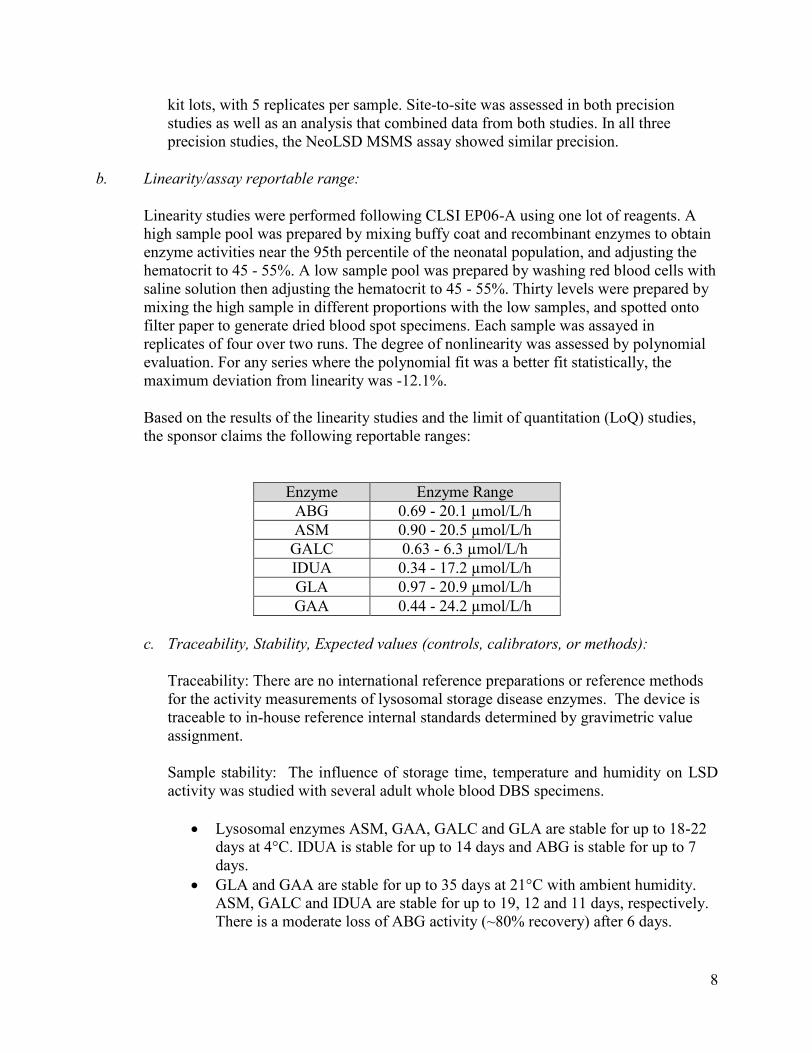
Based on the results of the linearity studies and the limit of quantitation (LoQ) studies, the sponsor claims the following reportable ranges:
Enzyme Enzyme Range ABG 0.69 - 20.1 µmol/L/h ASM 0.90 - 20.5 µmol/L/h
GALC 0.63 - 6.3 µmol/L/h IDUA 0.34 - 17.2 µmol/L/h GLA 0.97 - 20.9 µmol/L/h GAA 0.44 - 24.2 µmol/L/h
c. Traceability, Stability, Expected values (controls, calibrators, or methods):
Traceability: There are no international reference preparations or reference methods for the activity measurements of lysosomal storage disease enzymes. The device is traceable to in-house reference internal standards determined by gravimetric value assignment.
Sample stability: The influence of storage time, temperature and humidity on LSD activity was studied with several adult whole blood DBS specimens.
· Lysosomal enzymes ASM, GAA, GALC and GLA are stable for up to 18-22 days at 4°C. IDUA is stable for up to 14 days and ABG is stable for up to 7 days.
· GLA and GAA are stable for up to 35 days at 21°C with ambient humidity. ASM, GALC and IDUA are stable for up to 19, 12 and 11 days, respectively. There is a moderate loss of ABG activity (~80% recovery) after 6 days.
9
· GAA and GLA are stable at 27°C and RH 40% for up to 35 and 17 days, respectively. ASM, GALC and IDUA are stable for up to 3 days. There is a moderate loss in ASM and GALC activity (~80% recovery) after 6 days and a moderate loss in IDUA activity (~80% recovery) after 4 days. Further there is a moderate loss in ABG activity (~80% recovery) after 2 days.
· All lysosomal enzymes studied are unstable at 35°C and RH 80% with a significant loss in activity (~10-60% recovery).
In the labeling, the sponsor provides the following information:
Care should be taken in the storage and transport of the dried blood spots (DBS). Storage of specimens in an environment with elevated temperature and humidity increases the risks of false positive screening results. For long-term storage the specimens should preferably be stored at -20°C and protected from moisture. Sample exposure to high temperature and high humidity during shipping may result in increased rates of false positives.
d. Detection limit:
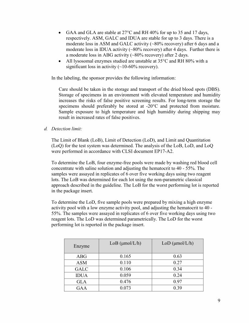
The Limit of Blank (LoB), Limit of Detection (LoD), and Limit and Quantitation (LoQ) for the test system was determined. The analysis of the LoB, LoD, and LoQ were performed in accordance with CLSI document EP17-A2.
To determine the LoB, four enzyme-free pools were made by washing red blood cell concentrate with saline solution and adjusting the hematocrit to 40 - 55%. The samples were assayed in replicates of 6 over five working days using two reagent lots. The LoB was determined for each lot using the non-parametric classical approach described in the guideline. The LoB for the worst performing lot is reported in the package insert.
To determine the LoD, five sample pools were prepared by mixing a high enzyme activity pool with a low enzyme activity pool, and adjusting the hematocrit to 40 - 55%. The samples were assayed in replicates of 6 over five working days using two reagent lots. The LoD was determined parametrically. The LoD for the worst performing lot is reported in the package insert.
Enzyme LoB (μmol/L/h) LoD (μmol/L/h)
ABG 0.165 0.63 ASM 0.110 0.27
GALC 0.106 0.34 IDUA 0.059 0.24 GLA 0.476 0.97 GAA 0.073 0.39

10
The Limit of Quantitation (LoQ) is defined as the lowest activity fulfilling the total CV% requirement of the assay. For ABG, GLA and IDUA the CV% requirement is <40%, for ASM and GAA < 30% and for GALC < 50%. If the imprecision criterion was met for activities below LoD, the LoQ was set to be equal to the LoD. The table shows the LoQ results:
Enzyme ABG ASM GAA GALC GLA IDUA LoQ
(µmol/L/h) 0.69 0.90 0.63 0.34 0.97 0.44
%CV at LoQ 21.7% 20.0% 17.5% 20.6% 17.5% 18.2%
e. Analytical specificity:
The potential interfering substances for possible mass overlaps was assessed by searching two mass spectrometry databases, NIST 14 and MassBank, and CLSI EP07-A2 for potential mass overlaps (±1 Da of the target analytes) of the six enzymatic products and six internal standards. These interfering substances were prepared as neat solutions at different concentrations in Neo MSMS Flow Solvent. The study showed that none of the potential interfering substances gave significant signal in the corresponding MRM. The potential interfering substances tested for the mass overlap study are shown in the table below:
Potential Interferent Potential
Interference MW
Corresponding NeoLSD Analyte
Corresponding NeoLSD
Exact Mass Pantoprazole 383.08 ABG P 383.34 Meropenem 383.15 ABG P 383.34 Felodipine 383.07 ABG P 383.34 Cetirizine (M2 peak) 390.15 ABG IS 390.38 S‐(5'‐Adenosyl)‐L‐methionine 398.14 ASM P 397.36
PTH‐(E‐phenylthiocarbamyl)‐ lysine
398.12 ASM P 397.36
Sulfasalazine/sulfadiazine 398.07 ASM P 397.36 Perphenazine (M2 peak) 405.15 ASM IS 404.40 Lisinopril 405.23 ASM IS 404.40 Miconazole (M2 peak) 415.98 GALC IS 416.40 Spironolactone 416.20 GALC IS 416.40 Calcitriol 416.33 GALC IS 416.40 Domperidone 425.16 IDUA P 425.23 Kanamycin 484.24 GLA P 483.27
Analytical specificity studies were performed following the recommendations in the
11
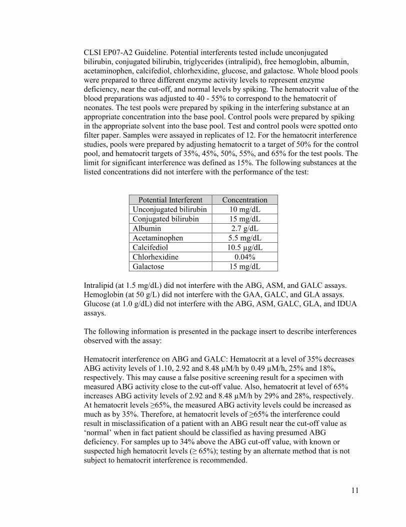
CLSI EP07-A2 Guideline. Potential interferents tested include unconjugated bilirubin, conjugated bilirubin, triglycerides (intralipid), free hemoglobin, albumin, acetaminophen, calcifediol, chlorhexidine, glucose, and galactose. Whole blood pools were prepared to three different enzyme activity levels to represent enzyme deficiency, near the cut-off, and normal levels by spiking. The hematocrit value of the blood preparations was adjusted to 40 - 55% to correspond to the hematocrit of neonates. The test pools were prepared by spiking in the interfering substance at an appropriate concentration into the base pool. Control pools were prepared by spiking in the appropriate solvent into the base pool. Test and control pools were spotted onto filter paper. Samples were assayed in replicates of 12. For the hematocrit interference studies, pools were prepared by adjusting hematocrit to a target of 50% for the control pool, and hematocrit targets of 35%, 45%, 50%, 55%, and 65% for the test pools. The limit for significant interference was defined as 15%. The following substances at the listed concentrations did not interfere with the performance of the test:
Potential Interferent Concentration Unconjugated bilirubin 10 mg/dL Conjugated bilirubin 15 mg/dL Albumin 2.7 g/dL Acetaminophen 5.5 mg/dL Calcifediol 10.5 µg/dL Chlorhexidine 0.04% Galactose 15 mg/dL
Intralipid (at 1.5 mg/dL) did not interfere with the ABG, ASM, and GALC assays. Hemoglobin (at 50 g/L) did not interfere with the GAA, GALC, and GLA assays. Glucose (at 1.0 g/dL) did not interfere with the ABG, ASM, GALC, GLA, and IDUA assays.
The following information is presented in the package insert to describe interferences observed with the assay:
Hematocrit interference on ABG and GALC: Hematocrit at a level of 35% decreases ABG activity levels of 1.10, 2.92 and 8.48 µM/h by 0.49 µM/h, 25% and 18%, respectively. This may cause a false positive screening result for a specimen with measured ABG activity close to the cut-off value. Also, hematocrit at level of 65% increases ABG activity levels of 2.92 and 8.48 µM/h by 29% and 28%, respectively. At hematocrit levels ≥65%, the measured ABG activity levels could be increased as much as by 35%. Therefore, at hematocrit levels of ≥65% the interference could result in misclassification of a patient with an ABG result near the cut-off value as ‘normal’ when in fact patient should be classified as having presumed ABG deficiency. For samples up to 34% above the ABG cut-off value, with known or suspected high hematocrit levels (≥ 65%); testing by an alternate method that is not subject to hematocrit interference is recommended.
12
Hematocrit at level of 35% increases GALC activity level of 3.38 µM/h by 22%; while at levels of 65% hematocrit decreases GALC activity levels of 0.75 and 1.02 µM/h by 24% and 20%, respectively. The decrease in GALC activity may cause a false positive screening result for a specimen with measured GALC activity close to the cut-off value. The inhibitory effect of high hematocrit values to GALC activity has been reported.
Hemoglobin interference on ABG, ASM and IDUA:
Free (serum) hemoglobin was found to interfere with the assay by increasing the measured ABG, ASM and IDUA activity, this may result in false negative results for Gaucher, Niemann-Pick A/B, and mucopolysaccharidosis Type I (MPS I) Disease.
ABG (Gaucher): Hemoglobin at a level of 16.6 g/dL increases ABG activity level near the cut-off by 27% when compared to a sample having 15.3 g/dL of hemoglobin. Hemoglobin at 18.9 g/dL increases ABG activity level of 7.68 μM/h by 30% when compared to a sample having 15.1 g/dL of hemoglobin. For samples up to 30% above the ABG cut-off value, with known or suspected high hemoglobin (>15 g/dL); testing by an alternate method that is not subject to hemoglobin interference is recommended.
ASM (Niemann-Pick A/B): Hemoglobin at a level of 17.8 g/dL hemoglobin increases ASM activity level of 1.57 μM/h by 26% when compared to a sample having 15.3 g/dL of hemoglobin. For samples up to 30% above the ASM cut-off value, with known or suspected high hemoglobin (>15 g/dL); testing by an alternate method that is not subject to hemoglobin interference is recommended.
IDUA (MPS-1): Hemoglobin at level of 18.0 g/dL increases IDUA activity level of 1.18 μM/h by 19% when compared to a sample having 15.5 g/dL of hemoglobin. For samples up to 30% above the IDUA cut-off value, with known or suspected high hemoglobin (>15 g/dL); testing by an alternate method that is not subject to hemoglobin interference is recommended.
Glucose interference on GAA: Glucose was found to interfere with the assay by decreasing the measured GAA activity. Glucose at a level of 0.50 g/dL decreases GAA activity level of 11.3 µM/h by 17%. Glucose at a level of 0.75 g/dL decreases GAA activity levels of 1.44 and 2.87 µM/h by 0.40 µM/h and 23%, respectively. Thus, glucose concentration above 0.25 g/dL with GAA may cause a false positive screening result for a specimen with measured GAA activity close to the cut-off value. However, the observed glucose concentration shown to interfere with GAA is clearly beyond the endogenous reference interval for glucose that has been reported to be for neonates (0-1 months) from 0.055 to 0.115 g/dL in whole blood. Note: Preterm infants with very-low birth weight have a high risk of hyperglycemia (blood glucose level > 0.18 g/dL) due to glucose infusion.
Triglyceride interference on GAA, GLA and IDUA: Intralipid (Triglyceride) was
13
found to interfere with the assay increasing the measured GAA, GLA and IDUA activity.
GAA: Intralipid at level of 0.23 g/dL increases GAA activity of 2.98 and 7.10 µM/h by 27% and 18%, respectively. Intralipid at level of 0.75 g/dL increases GAA activity level of 1.05 µM/h by 0.52 µM/h.
GLA: Intralipid at level of 0.30 g/dL increases GLA activity level of 7.21 µM/h by 16%; while at level 0.38 g/dL Intralipid increases GLA activity level of 3.63 µM/h by 25%. Intralipid at level of 0.75 g/dL increases GLA activity level of 1.37 µM/h by 0.65 µM/h.
IDUA: Intralipid at level of 0.38 g/dL increases IDUA activity level of 8.18 µM/h by 19% and Intralipid at level of 0.75 g/dL increases IDUA activity levels of 1.17 and 2.67 µM/h by 20%.
Note! High triglyceride concentrations (hypertriglyceridemia) in newborns due to medication effects or pathological conditions may cause a false negative screening result for a specimen with measured GAA, GLA or IDUA activity close to the cut-off value.
EDTA interference on ABG and ASM: EDTA at level 0.10 g/dL increases ABG activity level of 1.80 µM/h by 0.41 µM/h. EDTA at level of 0.04 g/dL decreases ASM activity level of 3.74 µM/h by 20%; while at level of 0.10 g/dL EDTA decreases ASM activity level of 1.18 µM/h by 16%. EDTA at a level of 0.20 g/dL decreases ASM activity level of 0.44 µM/h by 0.17 µM/h.
The concentration of EDTA found to interfere with AGB and ASM were far beyond the typical therapeutic dosage 3.4 µmol/L (0.10 mg/dL). Also, the direct application of blood from the heel-puncture to the DBS paper is intended for newborn screening eliminating the risk of contamination.
Carry-over: Carry-over was evaluated using one lot of the NeoLSD MSMS kit. Carry-over was defined as the average amount of analytes transferred from the high sample to the low sample calculated as the mean concentration difference between the first and second replicates of the low sample. Multiple enzyme concentrations were included in the study and there was no significant carry-over demonstrated by the study.
Plate Drift: The sponsor evaluated plate drift to ensure the on-board stability of processed samples for 12 and 24 hours. Two separate controls were subject to repeated testing over the course of 24 hours and showed no significant change in results over that period.
14
Filter Paper: PerkinElmer used DBS spotted onto two legally marketed dried blood spot cards. The sponsor provided information to support that results using the different filter papers were equivalent.
f. Assay cut-off:
Not applicable.
2. Comparison studies:
a. Method comparison with predicate device:
Not applicable.
b. Matrix comparison:
This device is only intended to be used with DBS specimens collected from neonates.
3. Clinical studies:
a. Clinical Sensitivity:
Not applicable.
b. Clinical specificity:
Not applicable.
c. Other clinical supportive data (when a. and b. are not applicable):
The screening performance of the NeoLSD MSMS kit was determined in a prospective clinical study of four-year-old retrospective routine samples at the Danish Section for Neonatal Screening, Department of Clinical Biochemistry and Immunology, Statens Serum Institute (SSI). 4041 newborn samples were tested. Most samples tested were from newborns ≤ 4 days old and consisted of 4011 routine samples that were 4 years old and 30 archived confirmed LSD positive newborn DBS specimens that ranged from 5.8 to 17.6 years of storage.
The 30 confirmed LSD specimens consisted of five cases of Gaucher, one case of Niemann-Pick A/B, 4 cases of Pompe, 10 cases of Krabbe, five cases of Fabry, and five cases of MPS I disease. The 4011 routine samples were tested according to the testing algorithm described in the package insert. These specimens were first tested in singlicate for all six enzymes. The specimens having at least one enzyme result below initial cut-off value were retested in duplicate to confirm the low enzyme result. The 30 confirmed positives were tested once.
15
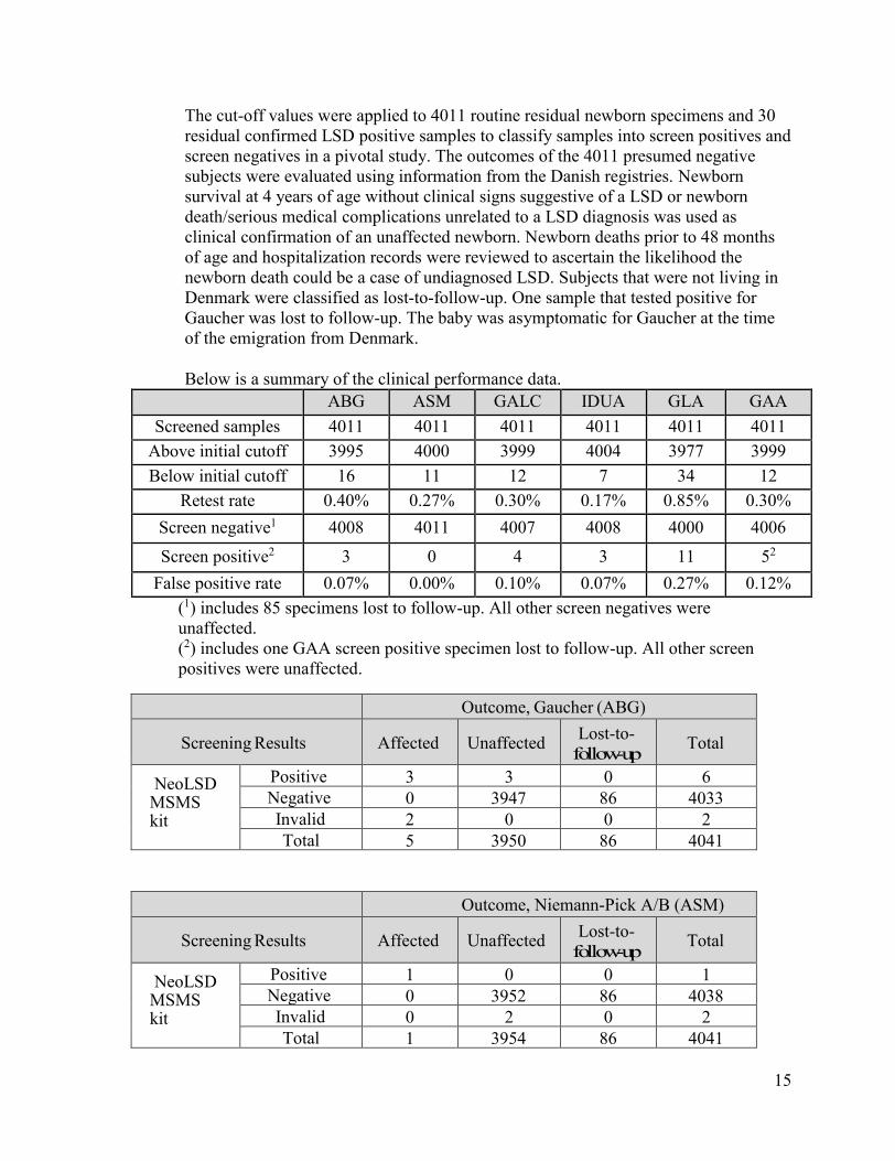
The cut-off values were applied to 4011 routine residual newborn specimens and 30 residual confirmed LSD positive samples to classify samples into screen positives and screen negatives in a pivotal study. The outcomes of the 4011 presumed negative subjects were evaluated using information from the Danish registries. Newborn survival at 4 years of age without clinical signs suggestive of a LSD or newborn death/serious medical complications unrelated to a LSD diagnosis was used as clinical confirmation of an unaffected newborn. Newborn deaths prior to 48 months of age and hospitalization records were reviewed to ascertain the likelihood the newborn death could be a case of undiagnosed LSD. Subjects that were not living in Denmark were classified as lost-to-follow-up. One sample that tested positive for Gaucher was lost to follow-up. The baby was asymptomatic for Gaucher at the time of the emigration from Denmark.
Below is a summary of the clinical performance data. ABG ASM GALC IDUA GLA GAA
Screened samples 4011 4011 4011 4011 4011 4011 Above initial cutoff 3995 4000 3999 4004 3977 3999 Below initial cutoff 16 11 12 7 34 12
Retest rate 0.40% 0.27% 0.30% 0.17% 0.85% 0.30% Screen negative1 4008 4011 4007 4008 4000 4006
Screen positive2 3 0 4 3 11 52 False positive rate 0.07% 0.00% 0.10% 0.07% 0.27% 0.12%
(1) includes 85 specimens lost to follow-up. All other screen negatives were unaffected. (2) includes one GAA screen positive specimen lost to follow-up. All other screen positives were unaffected.
Outcome, Gaucher (ABG)
Screening Results Affected Unaffected Lost-to- follow-up Total
NeoLSD MSMS kit
Positive 3 3 0 6 Negative 0 3947 86 4033 Invalid 2 0 0 2 Total 5 3950 86 4041
Outcome, Niemann-Pick A/B (ASM)
Screening Results Affected Unaffected Lost-to- follow-up Total
NeoLSD MSMS kit
Positive 1 0 0 1 Negative 0 3952 86 4038 Invalid 0 2 0 2 Total 1 3954 86 4041
16
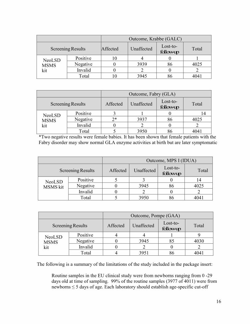
Outcome, Krabbe (GALC)
Screening Results Affected Unaffected Lost-to- follow-up Total
NeoLSD MSMS kit
Positive 10 4 0 14 Negative 0 3939 86 4025
Invalid 0 2 0 2 Total 10 3945 86 4041
Outcome, Fabry (GLA)
Screening Results Affected Unaffected Lost-to- follow-up Total
NeoLSD MSMS kit
Positive 3 11
0 14 Negative 2* 3937 86 4025 Invalid 0 2 0 2 Total 5 3950 86 4041
*Two negative results were female babies. It has been shown that female patients with the Fabry disorder may show normal GLA enzyme activities at birth but are later symptomatic
Outcome, MPS I (IDUA)
Screening Results Affected Unaffected Lost-to- follow-up Total
NeoLSD MSMS kit
Positive 5 3 0 14 Negative 0 3945 86 4025 Invalid 0 2 0 2 Total 5 3950 86 4041
Outcome, Pompe (GAA)
Screening Results Affected Unaffected Lost-to- follow-up Total
NeoLSD MSMS kit
Positive 4 4 1 9 Negative 0 3945 85 4030 Invalid 0 2 0 2 Total 4 3951 86 4041
The following is a summary of the limitations of the study included in the package insert:
Routine samples in the EU clinical study were from newborns ranging from 0 -29 days old at time of sampling. 99% of the routine samples (3977 of 4011) were from newborns ≤ 5 days of age. Each laboratory should establish age-specific cut-off
17
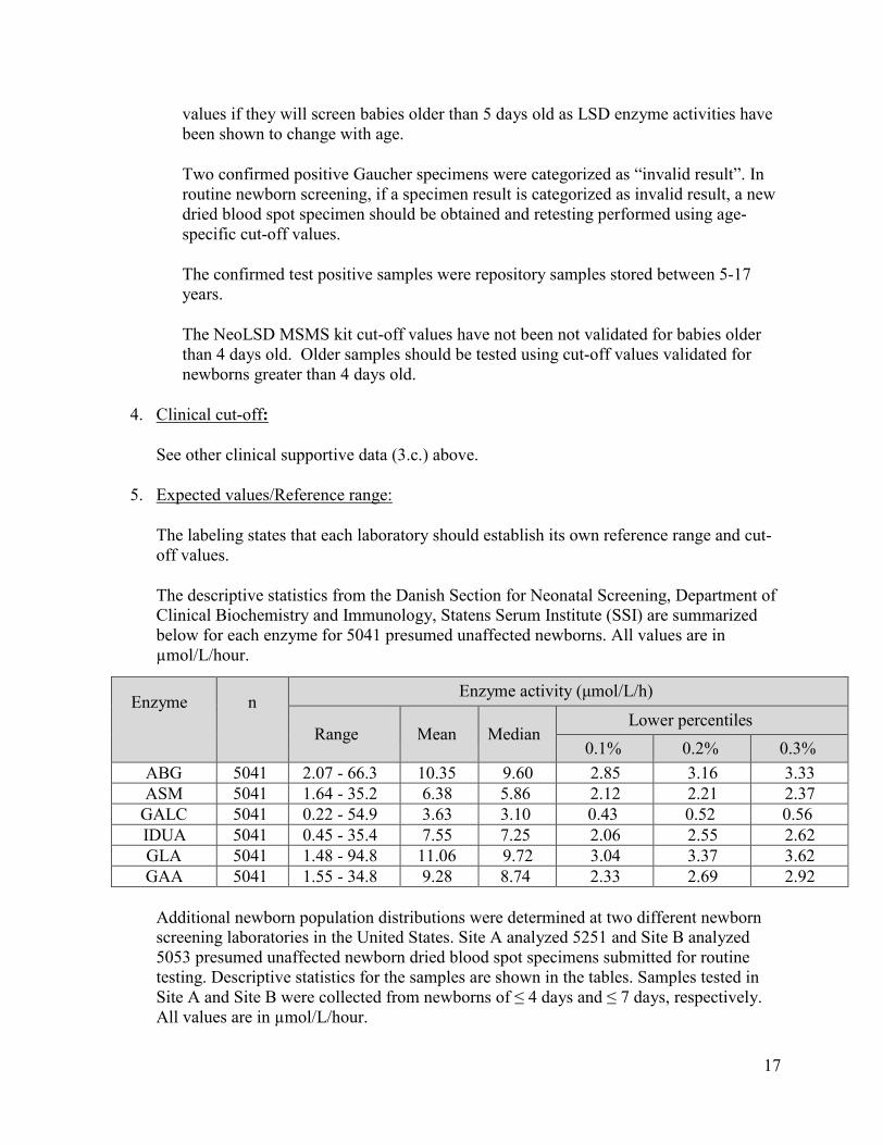
values if they will screen babies older than 5 days old as LSD enzyme activities have been shown to change with age.
Two confirmed positive Gaucher specimens were categorized as “invalid result”. In routine newborn screening, if a specimen result is categorized as invalid result, a new dried blood spot specimen should be obtained and retesting performed using age-specific cut-off values.
The confirmed test positive samples were repository samples stored between 5-17 years.
The NeoLSD MSMS kit cut-off values have not been not validated for babies older than 4 days old. Older samples should be tested using cut-off values validated for newborns greater than 4 days old.
4. Clinical cut-off:
See other clinical supportive data (3.c.) above.
5. Expected values/Reference range:
The labeling states that each laboratory should establish its own reference range and cut-off values.
The descriptive statistics from the Danish Section for Neonatal Screening, Department of Clinical Biochemistry and Immunology, Statens Serum Institute (SSI) are summarized below for each enzyme for 5041 presumed unaffected newborns. All values are in µmol/L/hour.
Enzyme n Enzyme activity (μmol/L/h)
Range Mean Median Lower percentiles
0.1% 0.2% 0.3% ABG 5041 2.07 - 66.3 10.35 9.60 2.85 3.16 3.33 ASM 5041 1.64 - 35.2 6.38 5.86 2.12 2.21 2.37
GALC 5041 0.22 - 54.9 3.63 3.10 0.43 0.52 0.56 IDUA 5041 0.45 - 35.4 7.55 7.25 2.06 2.55 2.62 GLA 5041 1.48 - 94.8 11.06 9.72 3.04 3.37 3.62 GAA 5041 1.55 - 34.8 9.28 8.74 2.33 2.69 2.92
Additional newborn population distributions were determined at two different newborn screening laboratories in the United States. Site A analyzed 5251 and Site B analyzed 5053 presumed unaffected newborn dried blood spot specimens submitted for routine testing. Descriptive statistics for the samples are shown in the tables. Samples tested in Site A and Site B were collected from newborns of ≤ 4 days and ≤ 7 days, respectively. All values are in µmol/L/hour.

18
Site A:
Enzyme n Enzyme activity (µmol/L/h)
Range Mean Median Lower percentiles
0.1% 0.2% 0.3% ABG 5251 1.39 – 77.9 10.52 9.67 2.00 2.16 2.36 ASM 5251 1.10 – 24.3 4.70 4.44 1.49 1.60 1.64
GALC 5251 0.52 – 85.0 5.49 4.63 0.86 0.96 1.02 IDUA 5251 0.09 – 18.7 6.31 6.08 0.85 1.40 1.56 GLA 5251 1.69 – 121 16.91 14.39 3.44 4.42 4.89 GAA 5251 0.75 - 43.0 10.21 9.62 2.16 2.38 2.56
Site B:
Enzyme n Enzyme activity (µmol/L/h)
Range Mean Median Lower percentiles
0.1% 0.2% 0.3% ABG 5053 1.68 – 76.5 11.4 10.4 2.94 3.23 3.33 ASM 5053 1.02 – 28.4 5.44 5.13 1.71 1.79 1.98
GALC 5053 0.53 – 33.0 4.67 4.20 0.68 0.82 0.86 IDUA 5053 0.9 – 21.8 6.65 6.39 1.59 1.98 2.09 GLA 5053 0.96 – 59.5 13.3 12.0 3.99 4.33 4.43 GAA 5053 1.11 – 36.4 11.0 10.4 2.53 2.88 3.37
N. Proposed Labeling:
The labeling is sufficient and it satisfies the requirements of 21 CFR Parts 801 and 809, as applicable and the special controls for this device type under 21 CFR 862.1488.
O. Conclusion:
The submitted information in this premarket notification is complete and supports a substantial equivalence decision.