2nd ESTRO Forum 2013 S283 - core.ac.uk · (G4 toxicity). This was associated with local disease...
2
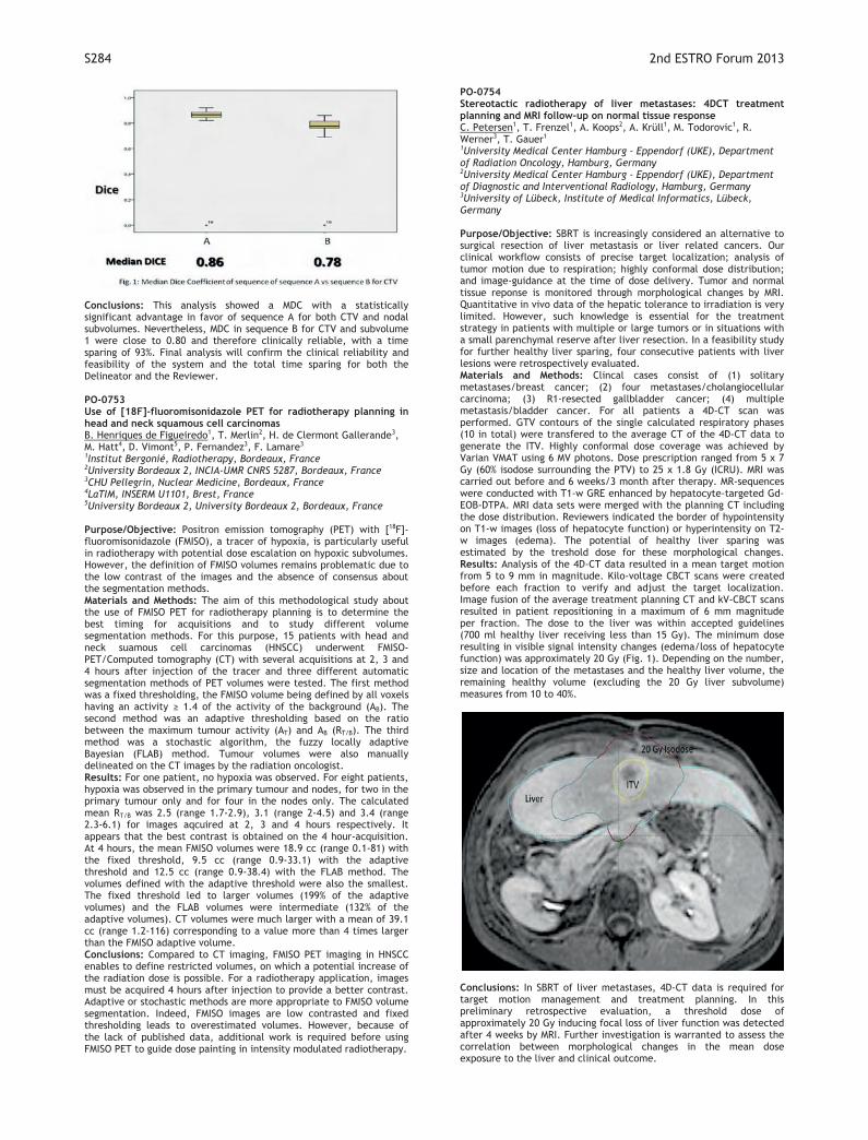
2nd ESTRO Forum 2013 S283 margin = 2.25mm (Range 1.25-3mm). Median PTV volume = 32cc (Range= 6-1879cc).Prescription dose was 30-36Gy in 3-5# (BED using α/β=10, 48-79). Dose was prescribed to the Median 64% isodose line (Range 48-78%). Median coverage was 98.3% (range 95.2-99.9%). Treatment delivery was tracked with fiducials and the Synchrony system. Results: Outcome data was available for 10 patients. Local Control rate was 100%. 4 patients (40%) progressed distantly, 2 in the IMC Node group, and 2 in the Sternum group. One of the patients that progressed distantly in the Sternal group had distant disease at the time of CyberKnife. Of those that progressed distantly, 75% had received chemotherapy as their first treatment at the time of Sternal/IMC node recurrence. Median Disease-Free Interval (DFI) in progressors was 2 years (Range 1-6 yrs), compared to a DFI in Non- progressors of 3 years (Range 1-13 yrs). Median Progression-Free Survival for the IMC node group, Sternal group, and Cohort as a whole were 6, 2 and 4 months respectively. The follow-up data was, however, less mature for the Sternal group. Overall Survival (OS) rate was 90%. The patient that died was had Sternal recurrence patient with small lung metastases at the time of CyberKnife treatment. She died of lepto-meningeal disease. Median OS for IMC node group, Sternal group and Cohort as a whole were 16, 2 and 6 months respectively. However, follow-up data is less mature for the Sternal group. Treatment was well tolerated with only 25% of the group experiencing acute toxicity (G1). Only 1 patient experienced Late toxicity (fleeting G3 symptoms), the patient had received prior rdaiotherapy. Conclusions: SBRT is a feasible treatment approach for patients with Sternal/IMC nodal recurrence of Breast Cancer. The technique is well tolerated, even in those who have received prior Radiotherapy. The outcome data is still immature, but the early Local Control rates (100%), and Overall Survival rates (90%) at a Median follow-up of 11 months are encouraging. PO-0751 Stereotactic body radiation therapy (SBRT) for abdominopelvic re- irradiation: early results K. Aitken 1 , H. Taylor 1 , E. Wells 2 , D. Tait 1 , N. van As 1 , A. Taylor 1 , S. Lalondrelle 1 1 Royal Marsden NHS Foundation Trust, Radiotherapy, London, United Kingdom 2 Royal Marsden NHS Foundation Trust, Radiotherapy Physics, London, United Kingdom Purpose/Objective: The management of disease recurrence within a previously irradiated pelvic field is a challenging clinical problem. The ability to re-irradiate the pelvis to a therapeutically meaningful dose is limited by the cumulative dose received by adjacent pelvic organs at risk (OAR). The Cyberknife radiosurgery system enables highly conformal dosimetry with rapid dose fall off outside of the target. This, in combination with the use of small treatment margins facilitated by tumour tracking, make it a potentially attractive delivery platform for re-irradiation. The aim of this retrospective study was to review the toxicity and early efficacy data of patients receiving SBRT with the Cyberknife platform for pelvic re-irradiation at our institution. Materials and Methods: Patients receiving Cyberknife SBRT to abdominopelvic targets sited within a previously irradiated field were retrospectively identified. Details on primary site histology, prior external beam radiotherapy (EBRT), re-irradiation dose and indication for re-treatment were obtained from the patient’s electronic notes. Radiotherapy toxicity was graded using CTCAE v4.0. Follow up consisted of clinical examination and radiological assessment. In field local control and time to progression were calculated from the day of completing SBRT until the date of radiological evidence of progression using standard RECIST criteria. Results: 16 patients were treated between 01.08.11-01.11.12. Median follow up was 6 months (range 1-15). 8 patients had primary gynaecological malignancies, 7 colorectal and 1 melanoma. Median age was 59 years (range 27-85). Indications for re-irradiation were nodal recurrence (6), soft tissue recurrence (7) and positive margins following surgery (3). Median time from previous EBRT was 20 months (range 1-156). Median previous EBRT dose was 50.4 Gy (range 30-54) in 28 fractions (range 10-39). 3 patients with gynaecological primaries had also received prior brachytherapy. Median re-irradiation dose was 30 Gy (range 18-35) in 3 fractions. 44% of patients had no recordable toxicity. 3 patients with pelvic side wall recurrences experienced G2 pain during treatment, which resolved shortly after completion of SBRT. One patient developed a colovesical fistula 2 months after SBRT (G4 toxicity). This was associated with local disease recurrence therefore the aetiology was unclear. At last follow up, local control is 94% (15/16 patients). A single patient had an in field recurrence 2 months after SBRT. 6/16 patients (38%) have relapsed at distant sites. Median time to relapse at any site was 3 months (range 2-11). Conclusions: At this early time point of data analysis, minimal toxicity has been observed to date. Cyberknife SBRT is a promising method for pelvic re-irradiation and warrants further investigation. POSTER: CLINICAL TRACK: TARGET AND VOLUME DEFINITION AND IMAGING PO-0752 Reliability and feasibility of automatic segmentation in rectal cancer: a perspective study. M.A. Gambacorta 1 , C. Valentini 1 , N. Dinapoli 1 , L. Boldrini 1 , M.C. Barba 1 , G.C. Mattiucci 1 , D. Pasini 1 , S. Manfrida 1 , N. Caria 2 , V. Valentini 1 1 Università Cattolica del S. Cuore, Radiotherapy and Oncology, Roma, Italy 2 Varian Medical Systems, Clinical Solution, Palo Alto, USA Purpose/Objective: The use of autosegmentation computed systems in clinical setting for locally advanced rectal cancer is time sparing with an acceptable Dice Coefficient for CTV, Mean Dice Coefficient (MDC) of 0.70, while it does not reach an adequate value for small nodal subvolumes (MDC=0.67) (1) . In this phase II study the objective is to verify in clinical setting the time sparing and the Dice Coefficient in bigger volumes (CTV and grouped nodal subvolumes) in locally advanced rectal cancer using the Smart Segmentation Knowledge Based Contouring (SS-KBC)® research program. (1) Clinical validation of atlas-based auto-segmentation of pelvic volumes and normal tissue in rectal tumors using autosegmentation computed system. Gambacorta et al, Acta Oncologica, in press Materials and Methods: 29 consecutive patients were selected between June and September 2012; images of 14 patients as atlas, 15 for validation. To test the reliability of the system for bigger volumes, the nodal subvolumes were grouped according to clinical stage and site of the tumor (tab1). According to our ongoing QA program two operators were involved: a Delineator and a Reviewer. CTV and pelvic grouped nodal subsites were contoured by Delineator in 2 different sequences (A-manual vs B-autosegmentation) of contouring using the same planning CT; all of them underwent to Indipendent Check by Reviewer. To improve the reliability of the system many anthropometric characteristics where analyzed (including BMI, sex, age, fertility state, sacro-coccigeal distance and the most anterior distance between upper iliac crests). The analysis was conducted to test the reliability of the system using Dice Coefficient and the total time spared by the Delineator to complete the 2 different sequences. Results: In clinical practice the time spared by operator 1 to complete sequence A and B was of 14min vs 1min respectively; there was a statistically significant better MDC in favor of sequence A vs sequence B: -CTV: sequence A (MDC=0.86) vs sequence B (MDC=0.78), p=0.001 (fig.1) -Subvolume 1: sequence A (MDC=0.86) vs sequence B (MDC=0.77), p=0.001 -Subvolume 2a: sequence A (MDC=0.76) vs sequence B (MDC=0.66), p=0.001
Transcript of 2nd ESTRO Forum 2013 S283 - core.ac.uk · (G4 toxicity). This was associated with local disease...
2nd ESTRO Forum 2013 S283
margin = 2.25mm (Range 1.25-3mm). Median PTV volume = 32cc (Range= 6-1879cc).Prescription dose was 30-36Gy in 3-5# (BED using α/β=10, 48-79). Dose was prescribed to the Median 64% isodose line (Range 48-78%). Median coverage was 98.3% (range 95.2-99.9%). Treatment delivery was tracked with fiducials and the Synchrony system. Results: Outcome data was available for 10 patients. Local Control rate was 100%. 4 patients (40%) progressed distantly, 2 in the IMC Node group, and 2 in the Sternum group. One of the patients that progressed distantly in the Sternal group had distant disease at the time of CyberKnife. Of those that progressed distantly, 75% had received chemotherapy as their first treatment at the time of Sternal/IMC node recurrence. Median Disease-Free Interval (DFI) in progressors was 2 years (Range 1-6 yrs), compared to a DFI in Non-progressors of 3 years (Range 1-13 yrs). Median Progression-Free Survival for the IMC node group, Sternal group, and Cohort as a whole were 6, 2 and 4 months respectively. The follow-up data was, however, less mature for the Sternal group. Overall Survival (OS) rate was 90%. The patient that died was had Sternal recurrence patient with small lung metastases at the time of CyberKnife treatment. She died of lepto-meningeal disease. Median OS for IMC node group, Sternal group and Cohort as a whole were 16, 2 and 6 months respectively. However, follow-up data is less mature for the Sternal group. Treatment was well tolerated with only 25% of the group experiencing acute toxicity (G1). Only 1 patient experienced Late toxicity (fleeting G3 symptoms), the patient had received prior rdaiotherapy. Conclusions: SBRT is a feasible treatment approach for patients with Sternal/IMC nodal recurrence of Breast Cancer. The technique is well tolerated, even in those who have received prior Radiotherapy. The outcome data is still immature, but the early Local Control rates (100%), and Overall Survival rates (90%) at a Median follow-up of 11 months are encouraging. PO-0751 Stereotactic body radiation therapy (SBRT) for abdominopelvic re-irradiation: early results K. Aitken1, H. Taylor1, E. Wells2, D. Tait1, N. van As1, A. Taylor1, S. Lalondrelle1 1Royal Marsden NHS Foundation Trust, Radiotherapy, London, United Kingdom 2Royal Marsden NHS Foundation Trust, Radiotherapy Physics, London, United Kingdom Purpose/Objective: The management of disease recurrence within a previously irradiated pelvic field is a challenging clinical problem. The ability to re-irradiate the pelvis to a therapeutically meaningful dose is limited by the cumulative dose received by adjacent pelvic organs at risk (OAR). The Cyberknife radiosurgery system enables highly conformal dosimetry with rapid dose fall off outside of the target. This, in combination with the use of small treatment margins facilitated by tumour tracking, make it a potentially attractive delivery platform for re-irradiation. The aim of this retrospective study was to review the toxicity and early efficacy data of patients receiving SBRT with the Cyberknife platform for pelvic re-irradiation at our institution. Materials and Methods: Patients receiving Cyberknife SBRT to abdominopelvic targets sited within a previously irradiated field were retrospectively identified. Details on primary site histology, prior external beam radiotherapy (EBRT), re-irradiation dose and indication for re-treatment were obtained from the patient’s electronic notes. Radiotherapy toxicity was graded using CTCAE v4.0. Follow up consisted of clinical examination and radiological assessment. In field local control and time to progression were calculated from the day of completing SBRT until the date of radiological evidence of progression using standard RECIST criteria. Results: 16 patients were treated between 01.08.11-01.11.12. Median follow up was 6 months (range 1-15). 8 patients had primary gynaecological malignancies, 7 colorectal and 1 melanoma. Median age was 59 years (range 27-85). Indications for re-irradiation were nodal recurrence (6), soft tissue recurrence (7) and positive margins following surgery (3). Median time from previous EBRT was 20 months (range 1-156). Median previous EBRT dose was 50.4 Gy (range 30-54) in 28 fractions (range 10-39). 3 patients with gynaecological primaries had also received prior brachytherapy. Median re-irradiation dose was 30 Gy (range 18-35) in 3 fractions. 44% of patients had no recordable toxicity. 3 patients with pelvic side wall recurrences experienced G2 pain during treatment, which resolved shortly after completion of SBRT. One patient developed a colovesical fistula 2 months after SBRT (G4 toxicity). This was associated with local disease recurrence therefore the aetiology was unclear. At last follow up, local control is 94% (15/16 patients). A single patient had an in field recurrence 2
months after SBRT. 6/16 patients (38%) have relapsed at distant sites. Median time to relapse at any site was 3 months (range 2-11). Conclusions: At this early time point of data analysis, minimal toxicity has been observed to date. Cyberknife SBRT is a promising method for pelvic re-irradiation and warrants further investigation.
POSTER: CLINICAL TRACK: TARGET AND VOLUME DEFINITION AND IMAGING
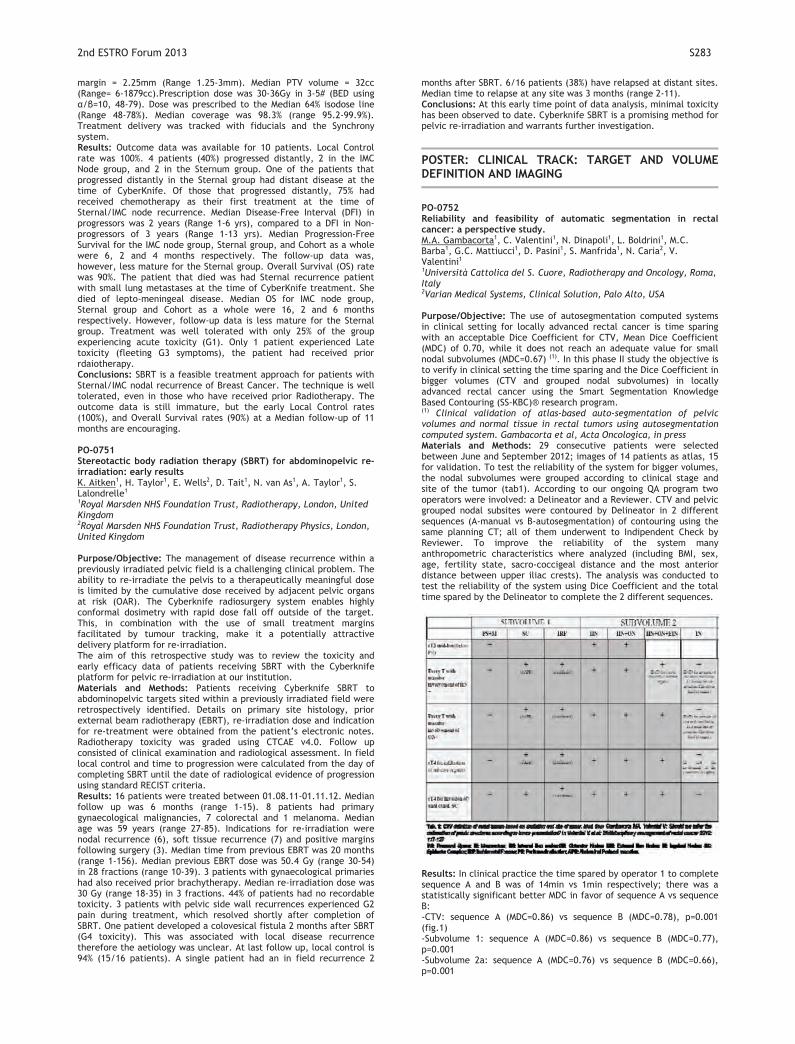
PO-0752 Reliability and feasibility of automatic segmentation in rectal cancer: a perspective study. M.A. Gambacorta1, C. Valentini1, N. Dinapoli1, L. Boldrini1, M.C. Barba1, G.C. Mattiucci1, D. Pasini1, S. Manfrida1, N. Caria2, V. Valentini1 1Università Cattolica del S. Cuore, Radiotherapy and Oncology, Roma, Italy 2Varian Medical Systems, Clinical Solution, Palo Alto, USA Purpose/Objective: The use of autosegmentation computed systems in clinical setting for locally advanced rectal cancer is time sparing with an acceptable Dice Coefficient for CTV, Mean Dice Coefficient (MDC) of 0.70, while it does not reach an adequate value for small nodal subvolumes (MDC=0.67) (1). In this phase II study the objective is to verify in clinical setting the time sparing and the Dice Coefficient in bigger volumes (CTV and grouped nodal subvolumes) in locally advanced rectal cancer using the Smart Segmentation Knowledge Based Contouring (SS-KBC)® research program. (1) Clinical validation of atlas-based auto-segmentation of pelvic volumes and normal tissue in rectal tumors using autosegmentation computed system. Gambacorta et al, Acta Oncologica, in press Materials and Methods: 29 consecutive patients were selected between June and September 2012; images of 14 patients as atlas, 15 for validation. To test the reliability of the system for bigger volumes, the nodal subvolumes were grouped according to clinical stage and site of the tumor (tab1). According to our ongoing QA program two operators were involved: a Delineator and a Reviewer. CTV and pelvic grouped nodal subsites were contoured by Delineator in 2 different sequences (A-manual vs B-autosegmentation) of contouring using the same planning CT; all of them underwent to Indipendent Check by Reviewer. To improve the reliability of the system many anthropometric characteristics where analyzed (including BMI, sex, age, fertility state, sacro-coccigeal distance and the most anterior distance between upper iliac crests). The analysis was conducted to test the reliability of the system using Dice Coefficient and the total time spared by the Delineator to complete the 2 different sequences.
Results: In clinical practice the time spared by operator 1 to complete sequence A and B was of 14min vs 1min respectively; there was a statistically significant better MDC in favor of sequence A vs sequence B: -CTV: sequence A (MDC=0.86) vs sequence B (MDC=0.78), p=0.001 (fig.1) -Subvolume 1: sequence A (MDC=0.86) vs sequence B (MDC=0.77), p=0.001 -Subvolume 2a: sequence A (MDC=0.76) vs sequence B (MDC=0.66), p=0.001
S284 2nd ESTRO Forum 2013
Conclusions: This analysis showed a MDC with a statistically significant advantage in favor of sequence A for both CTV and nodal subvolumes. Nevertheless, MDC in sequence B for CTV and subvolume 1 were close to 0.80 and therefore clinically reliable, with a time sparing of 93%. Final analysis will confirm the clinical reliability and feasibility of the system and the total time sparing for both the Delineator and the Reviewer. PO-0753 Use of [18F]-fluoromisonidazole PET for radiotherapy planning in head and neck squamous cell carcinomas B. Henriques de Figueiredo1, T. Merlin2, H. de Clermont Gallerande3, M. Hatt4, D. Vimont5, P. Fernandez3, F. Lamare3 1Institut Bergonié, Radiotherapy, Bordeaux, France 2University Bordeaux 2, INCIA-UMR CNRS 5287, Bordeaux, France 3CHU Pellegrin, Nuclear Medicine, Bordeaux, France 4LaTIM, INSERM U1101, Brest, France 5University Bordeaux 2, University Bordeaux 2, Bordeaux, France Purpose/Objective: Positron emission tomography (PET) with [18F]-fluoromisonidazole (FMISO), a tracer of hypoxia, is particularly useful in radiotherapy with potential dose escalation on hypoxic subvolumes. However, the definition of FMISO volumes remains problematic due to the low contrast of the images and the absence of consensus about the segmentation methods. Materials and Methods: The aim of this methodological study about the use of FMISO PET for radiotherapy planning is to determine the best timing for acquisitions and to study different volume segmentation methods. For this purpose, 15 patients with head and neck suamous cell carcinomas (HNSCC) underwent FMISO-PET/Computed tomography (CT) with several acquisitions at 2, 3 and 4 hours after injection of the tracer and three different automatic segmentation methods of PET volumes were tested. The first method was a fixed thresholding, the FMISO volume being defined by all voxels having an activity ≥ 1.4 of the activity of the background (AB). The second method was an adaptive thresholding based on the ratio between the maximum tumour activity (AT) and AB (RT/B). The third method was a stochastic algorithm, the fuzzy locally adaptive Bayesian (FLAB) method. Tumour volumes were also manually delineated on the CT images by the radiation oncologist. Results: For one patient, no hypoxia was observed. For eight patients, hypoxia was observed in the primary tumour and nodes, for two in the primary tumour only and for four in the nodes only. The calculated mean RT/B was 2.5 (range 1.7-2.9), 3.1 (range 2-4.5) and 3.4 (range 2.3-6.1) for images aqcuired at 2, 3 and 4 hours respectively. It appears that the best contrast is obtained on the 4 hour-acquisition. At 4 hours, the mean FMISO volumes were 18.9 cc (range 0.1-81) with the fixed threshold, 9.5 cc (range 0.9-33.1) with the adaptive threshold and 12.5 cc (range 0.9-38.4) with the FLAB method. The volumes defined with the adaptive threshold were also the smallest. The fixed threshold led to larger volumes (199% of the adaptive volumes) and the FLAB volumes were intermediate (132% of the adaptive volumes). CT volumes were much larger with a mean of 39.1 cc (range 1.2-116) corresponding to a value more than 4 times larger than the FMISO adaptive volume. Conclusions: Compared to CT imaging, FMISO PET imaging in HNSCC enables to define restricted volumes, on which a potential increase of the radiation dose is possible. For a radiotherapy application, images must be acquired 4 hours after injection to provide a better contrast. Adaptive or stochastic methods are more appropriate to FMISO volume segmentation. Indeed, FMISO images are low contrasted and fixed thresholding leads to overestimated volumes. However, because of the lack of published data, additional work is required before using FMISO PET to guide dose painting in intensity modulated radiotherapy.
PO-0754 Stereotactic radiotherapy of liver metastases: 4DCT treatment planning and MRI follow-up on normal tissue response C. Petersen1, T. Frenzel1, A. Koops2, A. Krüll1, M. Todorovic1, R. Werner3, T. Gauer1 1University Medical Center Hamburg - Eppendorf (UKE), Department of Radiation Oncology, Hamburg, Germany 2University Medical Center Hamburg - Eppendorf (UKE), Department of Diagnostic and Interventional Radiology, Hamburg, Germany 3University of Lübeck, Institute of Medical Informatics, Lübeck, Germany Purpose/Objective: SBRT is increasingly considered an alternative to surgical resection of liver metastasis or liver related cancers. Our clinical workflow consists of precise target localization; analysis of tumor motion due to respiration; highly conformal dose distribution; and image-guidance at the time of dose delivery. Tumor and normal tissue reponse is monitored through morphological changes by MRI. Quantitative in vivo data of the hepatic tolerance to irradiation is very limited. However, such knowledge is essential for the treatment strategy in patients with multiple or large tumors or in situations with a small parenchymal reserve after liver resection. In a feasibility study for further healthy liver sparing, four consecutive patients with liver lesions were retrospectively evaluated. Materials and Methods: Clincal cases consist of (1) solitary metastases/breast cancer; (2) four metastases/cholangiocellular carcinoma; (3) R1-resected gallbladder cancer; (4) multiple metastasis/bladder cancer. For all patients a 4D-CT scan was performed. GTV contours of the single calculated respiratory phases (10 in total) were transfered to the average CT of the 4D-CT data to generate the ITV. Highly conformal dose coverage was achieved by Varian VMAT using 6 MV photons. Dose prescription ranged from 5 x 7 Gy (60% isodose surrounding the PTV) to 25 x 1.8 Gy (ICRU). MRI was carried out before and 6 weeks/3 month after therapy. MR-sequences were conducted with T1-w GRE enhanced by hepatocyte-targeted Gd-EOB-DTPA. MRI data sets were merged with the planning CT including the dose distribution. Reviewers indicated the border of hypointensity on T1-w images (loss of hepatocyte function) or hyperintensity on T2-w images (edema). The potential of healthy liver sparing was estimated by the treshold dose for these morphological changes. Results: Analysis of the 4D-CT data resulted in a mean target motion from 5 to 9 mm in magnitude. Kilo-voltage CBCT scans were created before each fraction to verify and adjust the target localization. Image fusion of the average treatment planning CT and kV-CBCT scans resulted in patient repositioning in a maximum of 6 mm magnitude per fraction. The dose to the liver was within accepted guidelines (700 ml healthy liver receiving less than 15 Gy). The minimum dose resulting in visible signal intensity changes (edema/loss of hepatocyte function) was approximately 20 Gy (Fig. 1). Depending on the number, size and location of the metastases and the healthy liver volume, the remaining healthy volume (excluding the 20 Gy liver subvolume) measures from 10 to 40%.
Conclusions: In SBRT of liver metastases, 4D-CT data is required for target motion management and treatment planning. In this preliminary retrospective evaluation, a threshold dose of approximately 20 Gy inducing focal loss of liver function was detected after 4 weeks by MRI. Further investigation is warranted to assess the correlation between morphological changes in the mean dose exposure to the liver and clinical outcome.


![Edition 2015 - Eureka 3D · PDF fileArchimedes’ Challenge was an ... was the derivation of an accurate approximation of pi ... archimedes‘ challenge archimedes‘ challenge [2]](https://static.fdocument.org/doc/165x107/5a9434457f8b9a8b5d8c73fb/edition-2015-eureka-3d-challenge-was-an-was-the-derivation-of-an-accurate.jpg)