2 Androgen Action
23
23 From: Contemporary Endocrinology: Androgens in Health and Disease Edited by: C. Bagatell and W. J. Bremner © Humana Press Inc., Totowa, NJ INTRODUCTION Androgen action in human male subjects is predominantly the result of the biological activities of testosterone and 5α-dihydrotestosterone (DHT) (see Fig. 1). Although the former steroid is the predominant androgen present in the peripheral circulation at a 10- fold to 12-fold greater concentration, the local tissue concentration of DHT may be greater, as is its biological potency. Testosterone is synthesized within Leydig cells and secreted by the testes, whereas DHT is formed primarily as a metabolic product in peripheral tissues expressing steroid 5α-reductase activity. The equilibrium kinetic properties of lipophilic steroids suggest that testosterone can enter cells by passive diffusion across the cell membrane. However, only 1–2% of the testosterone present in the blood is free to diffuse into tissues, because the vast majority of the steroid is bound to sex hormone-binding globulin (SHBG; 40–50%) and to albumin (50–60%). Normal physiologic levels of circulating testosterone are necessary for adequate androgen bio- logic activity; however, some actions of androgens within tissues require the local con- version of testosterone to its more biologically active metabolite, DHT. A single molecular form of the androgen receptor (AR) exists within androgen target cells. The inactive cytoplasmic AR is a member of a large macromolecular chaperone complex, which dissociates upon the binding of testosterone or DHT to its receptor. The binding of steroid produces a conformational (or allosteric) change to an activated receptor complex that translocates into the nucleus and binds with high affinity as homodimers 2 Androgen Action Terry R. Brown, PhD CONTENTS INTRODUCTION ANDROGENS AND ANTIANDROGENS SEX HORMONE-BINDING GLOBULIN ANDTROGEN METHBOLISM STEROID 5α-REDUCTASE ENZYME ANDROGEN RECEPTOR ANDROGEN RECEPTOR AND HUMAN PATHOLOGY SUMMARY REFERENCES
Transcript of 2 Androgen Action
Chapter 2/Androgen Action 23
23
From: Contemporary Endocrinology: Androgens in Health and DiseaseEdited by: C. Bagatell and W. J. Bremner © Humana Press Inc., Totowa, NJ
INTRODUCTION
Androgen action in human male subjects is predominantly the result of the biologicalactivities of testosterone and 5α-dihydrotestosterone (DHT) (see Fig. 1). Although theformer steroid is the predominant androgen present in the peripheral circulation at a 10-fold to 12-fold greater concentration, the local tissue concentration of DHT may begreater, as is its biological potency. Testosterone is synthesized within Leydig cells andsecreted by the testes, whereas DHT is formed primarily as a metabolic product inperipheral tissues expressing steroid 5α-reductase activity. The equilibrium kineticproperties of lipophilic steroids suggest that testosterone can enter cells by passivediffusion across the cell membrane. However, only 1–2% of the testosterone present inthe blood is free to diffuse into tissues, because the vast majority of the steroid is boundto sex hormone-binding globulin (SHBG; 40–50%) and to albumin (50–60%). Normalphysiologic levels of circulating testosterone are necessary for adequate androgen bio-logic activity; however, some actions of androgens within tissues require the local con-version of testosterone to its more biologically active metabolite, DHT. A singlemolecular form of the androgen receptor (AR) exists within androgen target cells. Theinactive cytoplasmic AR is a member of a large macromolecular chaperone complex,which dissociates upon the binding of testosterone or DHT to its receptor. The bindingof steroid produces a conformational (or allosteric) change to an activated receptorcomplex that translocates into the nucleus and binds with high affinity as homodimers
2 Androgen Action
Terry R. Brown, PhD
CONTENTS
INTRODUCTION
ANDROGENS AND ANTIANDROGENS
SEX HORMONE-BINDING GLOBULIN
ANDTROGEN METHBOLISM
STEROID 5α-REDUCTASE ENZYME
ANDROGEN RECEPTOR
ANDROGEN RECEPTOR AND HUMAN PATHOLOGY
SUMMARY
REFERENCES
02/Brow/023-044/F 04/11/2003, 12:14 PM23
24 Brown
to specific binding sites on nuclear chromatin adjoining androgen-responsive genes.ARs function as transcription factors and form larger complexes via interactions withadditional nuclear proteins that serve as transcriptional coactivators or adaptors, someof which possess intrinsic kinase, methyltransferase, or histone acetyltransferase activi-ties. These multifactorial transcriptional complexes activate (or repress) androgen-regu-lated gene transcription by RNA polymerase, which alters the levels of specific mRNAs.The translation of mRNAs on cytoplasmic ribosomes directs the synthesis of proteinsresponsible for androgen-induced changes in cell function, growth, or differentiation.
It is important to remember that testosterone also serves as a precursor for estrogenbiosynthesis. Therefore, estrogenic actions can occur in men following aromatization oftestosterone to estradiol and binding of the latter steroid to nuclear estrogen receptors.
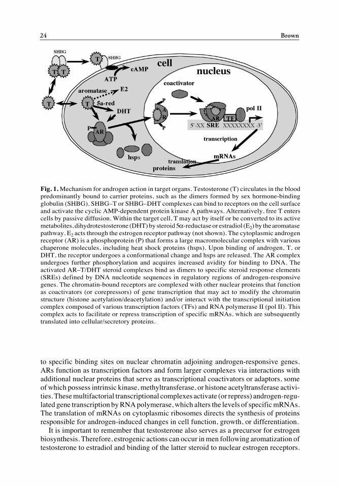
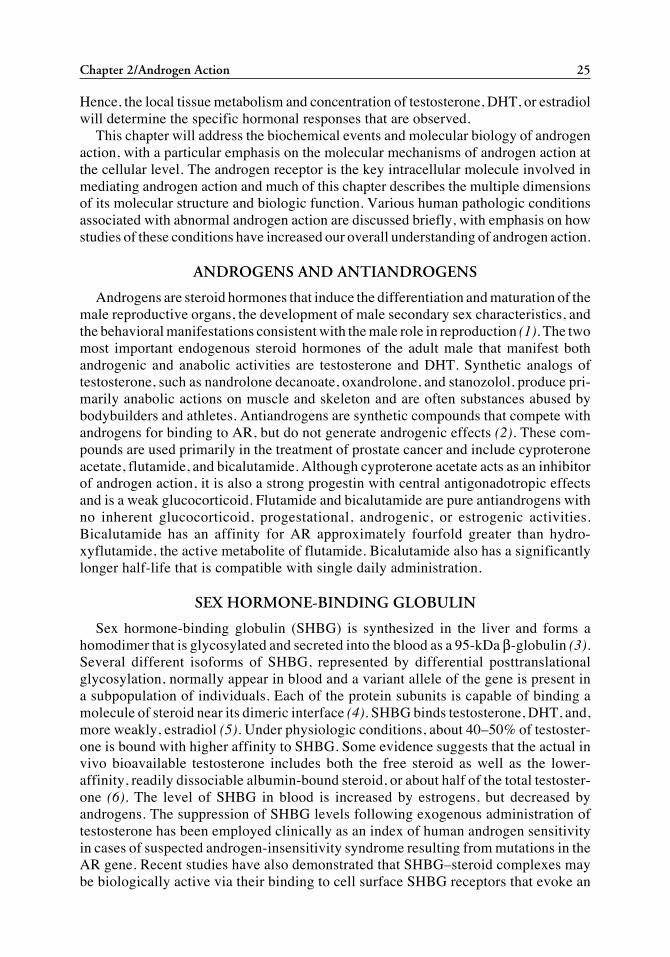
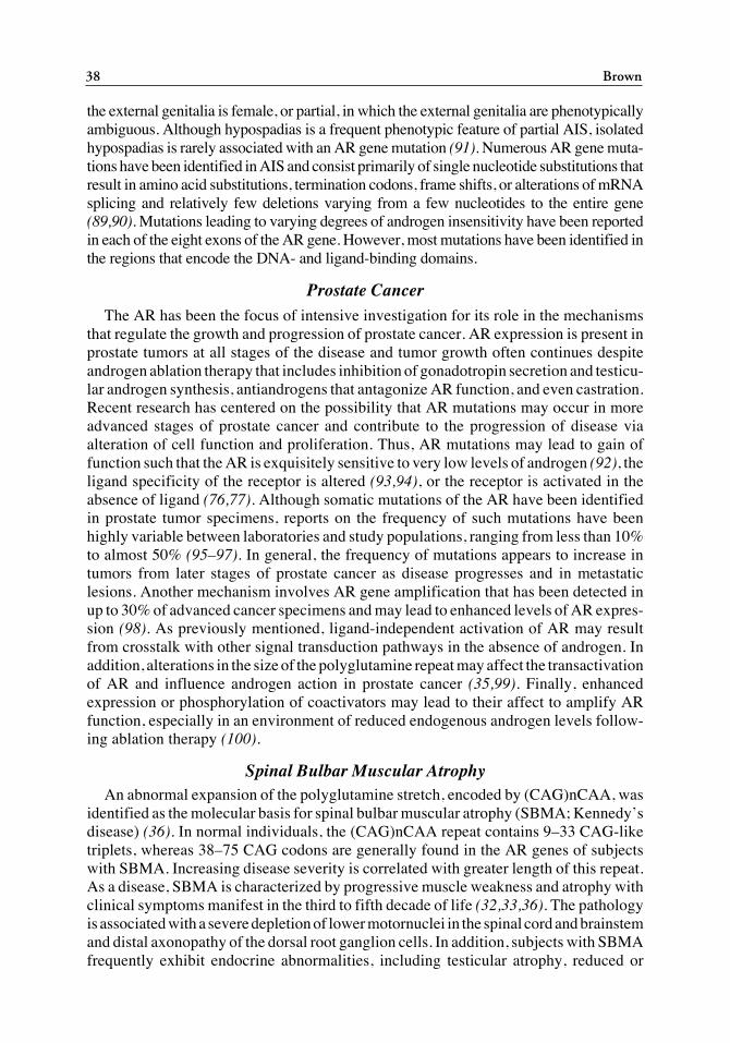
Fig. 1. Mechanism for androgen action in target organs. Testosterone (T) circulates in the bloodpredominantly bound to carrier proteins, such as the dimers formed by sex hormone-bindingglobulin (SHBG). SHBG–T or SHBG–DHT complexes can bind to receptors on the cell surfaceand activate the cyclic AMP-dependent protein kinase A pathways. Alternatively, free T enterscells by passive diffusion. Within the target cell, T may act by itself or be converted to its activemetabolites, dihydrotestosterone (DHT) by steroid 5α-reductase or estradiol (E2) by the aromatasepathway. E2 acts through the estrogen receptor pathway (not shown). The cytoplasmic androgenreceptor (AR) is a phosphoprotein (P) that forms a large macromolecular complex with variouschaperone molecules, including heat shock proteins (hsps). Upon binding of androgen, T, orDHT, the receptor undergoes a conformational change and hsps are released. The AR complexundergoes further phosphorylation and acquires increased avidity for binding to DNA. Theactivated AR–T/DHT steroid complexes bind as dimers to specific steroid response elements(SREs) defined by DNA nucleotide sequences in regulatory regions of androgen-responsivegenes. The chromatin-bound receptors are complexed with other nuclear proteins that functionas coactivators (or corepressors) of gene transcription that may act to modify the chromatinstructure (histone acetylation/deacetylation) and/or interact with the transcriptional initiationcomplex composed of various transcription factors (TFs) and RNA polymerase II (pol II). Thiscomplex acts to facilitate or repress transcription of specific mRNAs, which are subsequentlytranslated into cellular/secretory proteins.
02/Brow/023-044/F 04/11/2003, 12:14 PM24
Chapter 2/Androgen Action 25
Hence, the local tissue metabolism and concentration of testosterone, DHT, or estradiolwill determine the specific hormonal responses that are observed.
This chapter will address the biochemical events and molecular biology of androgenaction, with a particular emphasis on the molecular mechanisms of androgen action atthe cellular level. The androgen receptor is the key intracellular molecule involved inmediating androgen action and much of this chapter describes the multiple dimensionsof its molecular structure and biologic function. Various human pathologic conditionsassociated with abnormal androgen action are discussed briefly, with emphasis on howstudies of these conditions have increased our overall understanding of androgen action.
ANDROGENS AND ANTIANDROGENS
Androgens are steroid hormones that induce the differentiation and maturation of themale reproductive organs, the development of male secondary sex characteristics, andthe behavioral manifestations consistent with the male role in reproduction (1). The twomost important endogenous steroid hormones of the adult male that manifest bothandrogenic and anabolic activities are testosterone and DHT. Synthetic analogs oftestosterone, such as nandrolone decanoate, oxandrolone, and stanozolol, produce pri-marily anabolic actions on muscle and skeleton and are often substances abused bybodybuilders and athletes. Antiandrogens are synthetic compounds that compete withandrogens for binding to AR, but do not generate androgenic effects (2). These com-pounds are used primarily in the treatment of prostate cancer and include cyproteroneacetate, flutamide, and bicalutamide. Although cyproterone acetate acts as an inhibitorof androgen action, it is also a strong progestin with central antigonadotropic effectsand is a weak glucocorticoid. Flutamide and bicalutamide are pure antiandrogens withno inherent glucocorticoid, progestational, androgenic, or estrogenic activities.Bicalutamide has an affinity for AR approximately fourfold greater than hydro-xyflutamide, the active metabolite of flutamide. Bicalutamide also has a significantlylonger half-life that is compatible with single daily administration.
SEX HORMONE-BINDING GLOBULIN
Sex hormone-binding globulin (SHBG) is synthesized in the liver and forms ahomodimer that is glycosylated and secreted into the blood as a 95-kDa β-globulin (3).Several different isoforms of SHBG, represented by differential posttranslationalglycosylation, normally appear in blood and a variant allele of the gene is present ina subpopulation of individuals. Each of the protein subunits is capable of binding amolecule of steroid near its dimeric interface (4). SHBG binds testosterone, DHT, and,more weakly, estradiol (5). Under physiologic conditions, about 40–50% of testoster-one is bound with higher affinity to SHBG. Some evidence suggests that the actual invivo bioavailable testosterone includes both the free steroid as well as the lower-affinity, readily dissociable albumin-bound steroid, or about half of the total testoster-one (6). The level of SHBG in blood is increased by estrogens, but decreased byandrogens. The suppression of SHBG levels following exogenous administration oftestosterone has been employed clinically as an index of human androgen sensitivityin cases of suspected androgen-insensitivity syndrome resulting from mutations in theAR gene. Recent studies have also demonstrated that SHBG–steroid complexes maybe biologically active via their binding to cell surface SHBG receptors that evoke an
02/Brow/023-044/F 04/11/2003, 12:14 PM25
26 Brown
increase in intracellular cAMP levels (7). Androgen-binding protein (ABP) is a prod-uct of the same gene that is expressed in testicular Sertoli cells and secreted into theseminiferous tubules, where it binds a significant proportion of the testosterone and/or DHT present in the intratubular fluid of the testes and epididymides. This mayexplain, in part, the apparent requirement for high intratesticular levels of testosteronein the maintenance of spermatogenesis.
ANDROGEN METABOLISM
Testosterone is, itself, an active steroid with both androgenic actions to promotespermatogenesis in the testes and anabolic actions to increase tissue mass in muscle. Insexual tissues, such as the skin and prostate, testosterone is irreversibly converted pri-marily to the active metabolite DHT (8). DHT can be further reduced to 5α-androstane-3α-, 17β-diol (3α-diol) and subsequently conjugated with glucuronide to form 3α-diolglucuronide (9). These latter reactions are reversible such that 3α-diol, or its glucu-ronide, can be converted back to DHT. Most of the DHT, 3α-diol, and their glucuronidesthat appear in blood are derived from extrasplanchnic metabolism. Inactivation of tes-tosterone occurs primarily in the liver and involves the formation of 17-ketosteroids viaoxidation of the 17-OH group, reduction of the A ring, and reduction of the 3-keto groupwith the formation of polar metabolites that include diols, triols, and their conjugates(10). Testosterone and its synthetic precursor androstenedione can be aromatized toestradiol and estrone, respectively, by the cytochrome P450 aromatase enzyme, desig-nated CYP19 (11). The enzyme complex catalyzes a multistep reaction leading toremoval of the methyl group as formic acid and the rearrangement of the steroid A ringto an aromatic structure. This reaction requires NADPH as a cofactor for NADPH–cytochrome P450 reductase to enable the transfer of reducing equivalents to the enzyme,with 3 mol of oxygen being consumed in the sequence of hydroxylation reactions.Approximately 60–70 µg of estradiol are formed each day in normal men, primarilywithin adipose tissue, and this serves to further diversify the biological actions derivedfrom androgens at the individual tissue level.
STEROID 5α-REDUCTASE ENZYME
The conversion of testosterone to a variety of 5α- and 5β-reduced metabolites wasknown prior to the discovery that the 5α-reduced metabolite DHT was the principalintracellular androgen concentrated within the nuclei of many androgen-responsive tar-get tissues, such as the prostate (8). DHT proved to be twice as potent as testosterone inbioassays. Its physiologic importance was confirmed in human subjects with abnormalmale sex differentiation and decreased serum concentrations of DHT resulting fromgenetic deficiency of steroid 5α-reductase enzyme activity (12). Russell and Wilson (8)discovered that steroid 5α-reductase was present as two isoforms encoded by differentgenes, each containing 5 exons and having 50% identity of their nucleotide sequences.The 28- to 29-kDa enzyme proteins are localized to the endoplasmic reticulum andnuclear membranes, bind testosterone as a substrate, and require NADPH as a cofactor.The human type 1 isoform is present at low levels in the prostate but is the predominantisozyme expressed in the skin and liver. It is encoded by a gene on the short arm ofchromosome 5, has an optimal activity across a broad pH range from 6.5 to 8.0, has a highKm (1–5 µM) for testosterone, and is relatively insensitive (Ki = 300–500 nM) to the
02/Brow/023-044/F 04/11/2003, 12:14 PM26
Chapter 2/Androgen Action 27
4-azasteroid inhibitor finasteride. The type 2 isozyme is encoded by its gene on the shortarm of chromosome 2, has an acidic pH (5.0) optimum, and a low Km (0.1–1.0 µM) fortestosterone and is sensitive to inhibition by finasteride (Ki = 3–5 nM). As discussed ina later chapter, molecular defects in the type 2 isozyme are responsible for the reducedserum and tissue DHT concentrations and inadequate virilization of the urogenital sinusand external genitalia observed in some infants with male pseudohermaphroditismbecause of steroid 5α-reductase enzyme deficiency.
ANDROGEN RECEPTOR
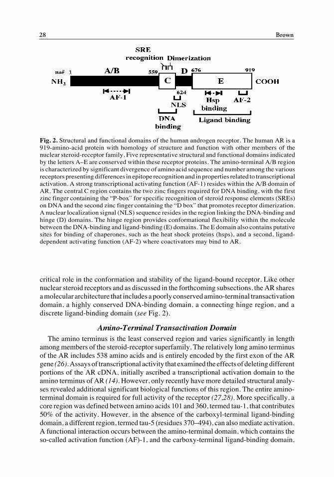
The AR is a member of the larger superfamily of nuclear ligand-dependent transcrip-tion factors that includes all of the steroid receptors among its members (13). Thus, theAR gene and protein share homology and conservation of structure and function with theother steroid receptors (14) (see Fig. 2). The human AR gene locus resides on the Xchromosome between q11 and q12 and is estimated to span a region of approx 90 kb(15,16). The gene encodes eight exons and transcription is initiated from one of two siteswithin a 13-bp region of a single promoter that lacks a TATA or a CCAAT box (17). TheAR promoter contains a GC box near the initiation site and an adjacent upstreamhomopurine/homopyrimidine stretch. Transcription from the initiation site at +13, butnot +1, is dependent on binding of Sp1 to the GC box. The purine/pyrimidine region canbind Sp1 in its normal double-stranded B-DNA conformation, but a novel single-strandbinding protein may also be involved (18). Relative to the rather large size of the ARgene, the predominant mRNA transcript is 10.6 kb, with a minor 8.5-kb transcript alsodetected in various tissues. The 10.6-kb transcript includes a 1.1-kb 5' untranslatedregion (UTR), the 2.7-kb open reading frame (ORF) and a relatively long 3' UTR of 6.8kb, whereas the shorter, alternative 8.5-kb mRNA transcript arises from differentialsplicing in the 3' UTR. Recently, unique androgen-responsive regulatory regions withinexons 4 and 5 have been postulated to mediate upregulation of AR mRNA transcriptionthat occurs in several cell types in response to androgen (19). A portion of the 5' UTRof AR mRNA is capable of forming a stem-loop secondary structure that has beensuggested to play a role in the induction of AR translation (20).
Variations in the coding region of the cDNA clones for the human AR arise as a resultof polymorphisms in two stretches of the nucleotide sequence that encode amino acidpolymeric repeats for glutamine and glycine within the amino terminus of the approx110-kDa protein. Hence, the number of amino acids reported in the literature for AR varybetween 910 and 919. All numeric references to amino acid residues in this chapter arederived from the original AR cDNA cloned by Lubahn et al. (21) and have been adoptedas the international reference standard for the Human Androgen Receptor MutationDatabase maintained at McGill University (22).
Posttranslational modification of AR occurs via phosphorylation on several serineresidues and results in a shift in the apparent molecular weight (MW) to 112–114 kDa(23,24). The translation of an 87-kDa (AR-A) minor isoform of the receptor has beenreported to occur from an alternative initiation methionine codon at position 189, but thisisoform generally represents less than 20% of the total AR protein expressed in tissues(26). Interestingly, cell transfection studies showed that transcriptional activation by theamino truncated AR-A isoform could not be distinguished from the full-length AR-Bisoform. More recent studies, however, suggest that the entire amino terminus plays a
02/Brow/023-044/F 04/11/2003, 12:14 PM27
28 Brown
critical role in the conformation and stability of the ligand-bound receptor. Like othernuclear steroid receptors and as discussed in the forthcoming subsections, the AR sharesa molecular architecture that includes a poorly conserved amino-terminal transactivationdomain, a highly conserved DNA-binding domain, a connecting hinge region, and adiscrete ligand-binding domain (see Fig. 2).
Amino-Terminal Transactivation DomainThe amino terminus is the least conserved region and varies significantly in length
among members of the steroid-receptor superfamily. The relatively long amino terminusof the AR includes 538 amino acids and is entirely encoded by the first exon of the ARgene (26). Assays of transcriptional activity that examined the effects of deleting differentportions of the AR cDNA, initially ascribed a transcriptional activation domain to theamino terminus of AR (14). However, only recently have more detailed structural analy-ses revealed additional significant biological functions of this region. The entire amino-terminal domain is required for full activity of the receptor (27,28). More specifically, acore region was defined between amino acids 101 and 360, termed tau-1, that contributes50% of the activity. However, in the absence of the carboxyl-terminal ligand-bindingdomain, a different region, termed tau-5 (residues 370–494), can also mediate activation.A functional interaction occurs between the amino-terminal domain, which contains theso-called activation function (AF)-1, and the carboxy-terminal ligand-binding domain,
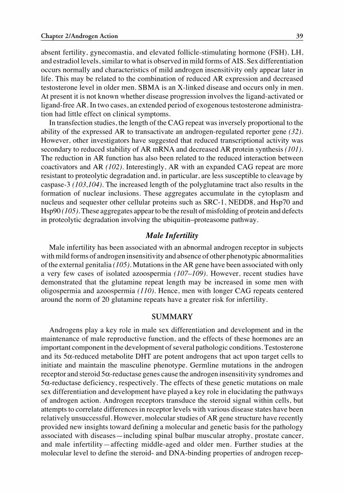
Fig. 2. Structural and functional domains of the human androgen receptor. The human AR is a919-amino-acid protein with homology of structure and function with other members of thenuclear steroid-receptor family. Five representative structural and functional domains indicatedby the letters A–E are conserved within these receptor proteins. The amino-terminal A/B regionis characterized by significant divergence of amino acid sequence and number among the variousreceptors presenting differences in epitope recognition and in properties related to transcriptionalactivation. A strong transcriptional activating function (AF-1) resides within the A/B domain ofAR. The central C region contains the two zinc fingers required for DNA binding, with the firstzinc finger containing the “P-box” for specific recognition of steroid response elements (SREs)on DNA and the second zinc finger containing the “D box” that promotes receptor dimerization.A nuclear localization signal (NLS) sequence resides in the region linking the DNA-binding andhinge (D) domains. The hinge region provides conformational flexibility within the moleculebetween the DNA-binding and ligand-binding (E) domains. The E domain also contains putativesites for binding of chaperones, such as the heat shock proteins (hsps), and a second, ligand-dependent activating function (AF-2) where coactivators may bind to AR.
02/Brow/023-044/F 04/11/2003, 12:14 PM28
Chapter 2/Androgen Action 29
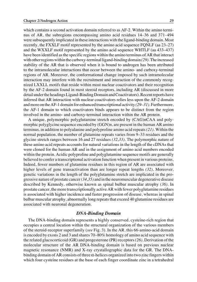
which contains a second activation domain referred to as AF-2. Within the amino termi-nus of AR, the subregions encompassing amino acid residues 14–36 and 371–494were subsequently implicated in these interactions with the ligand-binding domain. Mostrecently, the FXXLF motif represented by the amino acid sequence FQNLF (aa 23–27)and the WXXLF motif represented by the amino acid sequence WHTLF (aa 433–437)have been identified as the specific regions within the amino terminus of AR that interactwith other regions within the carboxy-terminal ligand-binding domain (29). The increasedstability of the AR that is observed when it is bound to androgen has been attributedto the intramolecular interactions that occur between the amino- and carboxy-terminalregions of AR. Moreover, the conformational change imposed by such intramolecularinteraction may interfere with the recruitment and interaction of the commonly recog-nized LXXLL motifs that reside within most nuclear coactivators and their recognitionby the AF-2 domain found in most steroid receptors, including AR (discussed in moredetail under the headings Ligand-Binding Domain and Coactivators). Recent reports haveinferred that AR interaction with nuclear coactivators relies less upon the AF-2 domainand more on the AF-1 domain for enhanced transcriptional activity (29–31). Furthermore,the AF-1 domain to which coactivators binds appears to be distinct from the regioninvolved in the amino- and carboxy-terminal interaction within the AR protein.
A unique, polymorphic polyglutamine stretch encoded by (CAG)nCAA and poly-morphic polyglycine sequence encoded by (GGN)n, are present in the human AR aminoterminus, in addition to polyalanine and polyproline amino acid repeats (21). Within thenormal population, the number of glutamine repeats varies from 9–33 residues and theglycine stretch ranges between 16 and 27 residues (32,33). The polymorphic nature ofthese amino acid repeats accounts for natural variations in the length of the cDNAs thatwere cloned for the human AR and in the assignment of amino acid numbers encodedwithin the protein. Acidic polyproline and polyglutamine sequence motifs are generallybelieved to confer a transcriptional activation function when present in various proteins.Indeed, fewer numbers of glutamine residues in this region of AR are associated withhigher levels of gene transactivation than are longer repeat lengths (32). Moreover,genetic variations in the length of the polyglutamine stretch are implicated in the pro-gressive nature of prostate cancer (34,35) and in the neuromuscular degenerative diseasedescribed by Kennedy, otherwise known as spinal bulbar muscular atrophy (36). Inprostate cancer, the more transcriptionally active AR with fewer polyglutamine residuesis associated with higher incidence and faster progression of disease, whereas in spinalbulbar muscular atrophy, abnormally long repeats that exceed 40 glutamine residues areassociated with neuronal degeneration.
DNA-Binding DomainThe DNA-binding domain represents a highly conserved, cysteine-rich region that
occupies a central location within the structural organization of the various membersof the steroid-receptor superfamily (see Fig. 3). In the AR, this 66-amino-acid domainis encoded by exons 2 and 3 and shares 70–80% homology of amino acid sequence withthe related glucocorticoid (GR) and progesterone (PR) receptors (26). Derivation of themolecular structure of the AR DNA-binding domain is based on previous nuclearmagnetic resonance (NMR) and X-ray crystallographic data for the GR. The DNA-binding domain of AR consists of three α-helices organized into two zinc fingers withinwhich four cysteine residues at the base of each finger coordinate zinc in a tetrahedral
02/Brow/023-044/F 04/11/2003, 12:14 PM29
30 Brown
array (37). The first zinc finger incorporates a perpendicularly oriented α-helix wherethe amino acid residues gly577, ser578, and val581 at its carboxy terminus form theproximal (P) box, which is conserved in AR, GR, and PR. Mutational analyses showedthat this conserved sequence is responsible for the common recognition and binding ofthese three receptors to a similar DNA steroid response element (SRE) consisting of theconsensus palindromic nucleotide sequence, -AGAACAnnnTGTTCT-. The α-helixcontaining the P box is positioned in the major groove of DNA where the three con-served amino acid residues make base-specific contacts with DNA. The second zincfinger has two α-helices, the first of which forms the distal (D) box, consisting of fiveamino acids (ala-ser-arg-asn-asp) that form a symmetric interface for homodimerizationof the receptor that accompanies its binding to a palindromic SRE, as previously dem-onstrated for GR. The first and third helices within the DNA-binding domain are per-pendicular to each other and form numerous hydrogen bonds.
The critical features of protein–DNA binding that differentially determine the speci-ficity of AR, GR, and PR binding to DNA regulatory sequences, and hence their dis-criminatory function in steroid-specific gene regulation, are now under scrutiny. Recentexperiments have identified genes that are specifically responsive to androgens andanalysis of the AR-binding sites in their promoters has revealed SRE motifs that resembledirect nucleotide repeat sequences rather than palindromic, inverted nucleotide repeatsequences (38) (see Table 1). Further analysis of the binding of AR to these direct-repeatnucleotide binding sites suggested that the dimerization interface within the ARhomodimers differed from that observed on inverted-repeat DNA-binding sites. Thesefindings predicted that dimerization of the AR occurred in a head-to-tail orientation ofthe monomeric AR molecules and that the intermolecular interactions at the dimeriza-tion interface involved amino acids in the second zinc finger and the adjoining hingeregion (38). Subsequent mutation analysis revealed that three specific amino acids,thr585, gly610, and leu617, are involved in the recognition of the direct nucleotide repeat
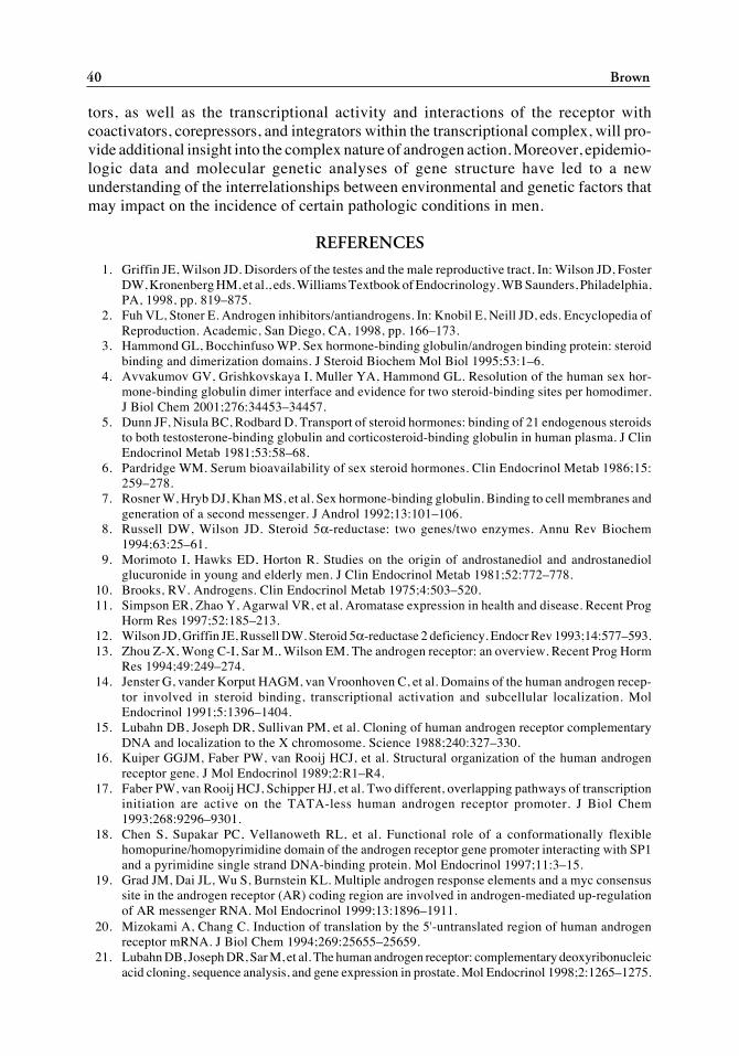
Fig. 3. Amino acid sequence of the human androgen-receptor DNA-binding domain. The single-letter amino acid code is shown for the sequence within the two zinc fingers formed by tetrahedralcoordination of zinc by each of four cysteines in the androgen receptor DNA-binding domain.The proximal “P box,” comprised of the amino acids G, S, and V, for specific recognition of theandrogen response element on DNA is indicated in the first finger and the distal “D box,”composed of A, S, R, N, and D, involved in dimerization is indicated in the second finger. Threehelices are formed within the structure of the DNA-binding domain, with helices 1 and 3 arrangedin perpendicular plane to the DNA.
02/Brow/023-044/F 04/11/2003, 12:14 PM30
Chapter 2/Androgen Action 31
that serves as an AR-specific binding site in the rat probasin gene promoter. Furthersupport for this hypothesis will evolve as the AR-binding sites are identified within thepromoters of other androgen-responsive genes. In addition, the proximity of bindingsites on the DNA of gene promoters for other transcription factors can also modify thespecificity for AR binding to adjacent SREs; hence, the transcriptional activity of AR isalso dependent on the context of individual promoter elements (39).
Nuclear Localization Signal and the Hinge RegionIn the absence of ligand, the AR is primarily localized to the cytoplasm, whereas the
addition of androgen induces its rapid translocation to the nucleus (14). A bipartitenuclear targeting signal sequence encoded at the junction between exons 3 and 4 func-
Table 1Classification of Androgen Response Elements
A. High affinity, nonspecificGRE177 GTTACA aac TGTTCTC3(1) ARE AGTACT tga TGTTCTGRE2 TAT TGTACA gga TGTTCTPSA ARE1 AGCACT tgc TGTTCTSLP-HRE-3 GAAACA gcc TGTTCT
B. High affinity, AR specificPB-ARE-2 GGTTCT tgg AGTACTSLP-HRE-2 TGGTCA ggc AGTTCTSC ARE1.2 GGCTCT ttc AGTTCT
C. Low affinity, nonspecificPB-ARE-1 ATAGCA tct TGTTCTMVDP pARE TGAAGT tcc TGTTCTGPX5 ATCCTA tgt TGTTCTCRP2 AGAACA aaa TGTACA
D. Low affinity, AR specificSC ARE AGCAGG ctg TGTCCC
Note: Four classes of androgen response elements have been definedby Claessens et al. (38) based on gel-shift assays of protein–DNA bindingwith AR or GR and by transfection experiments in which transcription ofan ARE-reporter gene is activated by either AR and/or GR. The androgenresponse elements (AREs) and glucocorticoid response elements (GREs)are derived from the promoter sequences: GRE177 from the MMTV-LTR; C3(1) ARE from the C3 subunit of the rat prostate prostatein gene;GRE TAT from the mouse liver tyrosine aminotransferase gene; PSAARE1 from the human prostate-specific antigen gene; SLP-HRE-2 and-3 from the mouse sex-limited protein gene; PB-ARE-1 and -2 from therat prostate probasin gene; SC ARE1.2 from the human secretorycomponent gene; VDP pARE from the mouse vas deferens protein gene;GPX5 from the mouse epididymal glutathione peroxidase 5 gene; andCRP2 from the cystatin-related protein gene.
02/Brow/023-044/F 04/11/2003, 12:14 PM31
32 Brown
tions to shuttle the AR through nuclear pores. The signal sequence consists of 2 clustersof basic amino acids, separated by 10 amino acids, that reside within the region that joinsthe DNA-binding domain and the hinge region (40). Amino acid substitutions or dele-tions in this region bounded by residues 617–633 cause an almost complete cytoplasmiclocalization of the receptor.
Recent advances in imaging techniques and the in vivo expression of chimeric ARtagged with a modified version of green fluorescent protein have revealed a number ofimportant properties associated with ligand-dependent nuclear translocation of thereceptor in live cells (41). First, in the absence of ligand, the AR is predominantlylocalized to the cellular cytoplasm. Second, upon the addition of androgen, such as DHT,the nuclear translocation of AR is apparent within minutes and is nearly complete within30 min. The apparent rate and extent of nuclear import is reduced with ligands thatdisplay reduced affinity for the receptor or antagonist activity. In the presence of andro-gens that act as agonists and activate transcription, the nuclear AR appears tightly asso-ciated with chromatin and organized within distinct subnuclear compartments. Bycomparison, when cells are exposed to antagonists, such as the antiandrogens flutamideor bicalutamide, the AR exhibits ligand-dependent migration into the nucleus but doesnot display a pattern of subnuclear compartmentalization as observed in the presence ofandrogen. Furthermore, androgen withdrawal releases the receptor from its chromatinassociation and exports it back into the cytoplasmic compartment for recycling when thesteroid is reintroduced. The small nuclear ring finger protein SNURF, which interactswith the AR through a region overlapping the bipartite nuclear localization signal, facili-tates AR nuclear import and retards its export upon hormone withdrawal (42). Furtherdetails regarding the processes of nuclear import and export are likely to become appar-ent from additional experiments using sophisticated imaging techniques.
The hinge region has been associated with conformational flexibility induced by thebinding of ligand to the carboxy-terminal domain and the binding of the ligand-activatedreceptor to DNA. Interestingly, a repressor activity is associated with the hinge regionin AR. Deletion of amino acid residues 628–646 from the hinge region of AR created amutant receptor with a twofold higher ligand-dependent transactivation activity than thewild-type receptor (43). Moreover, interaction of the mutant receptor with the p160coactivator TIF2 was significantly enhanced, as was its effect on AR transactivation.Mutations of the AR involving single-amino-acid substitutions within the hinge regionalso occur in prostate cancer. Recently, several of these mutant ARs were shown to haveincreased transcriptional activity when compared with the wild-type AR (44).
Ligand-Binding DomainAndrogen agonists and antagonists bind to the carboxy-terminal ligand-binding
domain of AR encoded by exons 4–8 (26). In the absence of ligand, some steroid recep-tors actually repress gene transactivation by occupying nuclear chromatin sites. Bycontrast, the AR is predominantly localized to the cytoplasm in the absence of ligand andexists within a large macromolecular complex of chaperone proteins that includes sev-eral members of the heat shock family (45). In this state, the AR is maintained in aninactive conformation that is not capable of binding to DNA. Deletion of the ligand-binding domain relieves its repressive function such that the truncated AR protein is ableto bind to DNA and is constitutively active as a result of expression of its intrinsic AF-1 activation domain (14). Alternatively, the binding of an androgen agonist causes the
02/Brow/023-044/F 04/11/2003, 12:14 PM32
Chapter 2/Androgen Action 33
inactive macromolecular chaperone complex to dissociate and promotes the nucleartranslocation and DNA binding of AR (46). Agonist binding also activates thetransactivation function of AF-2 and this coincides with alteration of the AR molecularconformation. The active conformation of the receptor is stabilized by the presence ofandrogen and AR homodimers bind in the nucleus to androgen response elements (AREs)on DNA (47). Androgens bind to either the nonphosphorylated or phosphorylated formsof AR. Although the significance of posttranslational modification of AR by phospho-rylation has not been clearly defined, its role in ligand-independent activation via alter-native signal transduction pathways has been inferred from a number of recent studies.
A series of 12 conserved helices form the ligand-binding domain of steroid receptorsas revealed by X-ray crystallographic analyses of these receptors conducted in the pres-ence and absence of agonists and antagonists (48,49). The AR ligand-binding-domaincrystal structure in the presence of the synthetic ligand, R1881, contains nine α-helices,two 310-helices, and four short β-strands associated in two antiparallel β-sheets (50,51)(see Fig. 4). The AR, like PR, has no helix 2, but its helix 12 is longer than those of someother steroid receptors and has an extended carboxy-terminal amino acid chain. Thehelices are arranged in a “helical sandwich” pattern and helices 4 and 5, and 10 and 11,are contiguous. In the case of AR, helices 10 and 11 are interrupted by a proline residueat position 868 which causes a kink between the helices. As in PR, there is a very shortβ-sheet formed by the loop between helices 8 and 9 and the carboxy-terminus, whichholds helix 12 in the closed agonist conformation, close to and capping the ligand-binding site (see Fig. 5). The androgen ligand DHT interacts with helices 3, 5, and 11within the ligand-binding domain. The AF-2 region resides within helix 12 of the AR andother receptors. When the receptor is occupied by a steroid agonist, helix 12 closes overthe ligand-binding pocket to form a surface for interaction with transcriptionalcoactivators; by contrast, binding of antagonists prevents this interface from forming.Conformational changes that accompany binding of steroid agonists also promote theintramolecular interactions that occur between the amino-terminal transactivation (AF-1)and carboxy-terminal ligand-binding (AF-2) domains in AR, interactions that are criticalfor receptor transactivation (29). The relatively weaker transactivation effect of the AF-2domain in AR has been associated with a competition between the internal AF-1 domainand coactivators for interaction with AF-2. It has been suggested that the p160 family ofcoactivators interact with AR by forming a bridge that links the AF-1 and AF-2 domains(28–31,52).
Steroid Response Elements in DNASteroid response elements minimally contain a core recognition motif of six nucle-
otide basepairs, generally consisting of two core motifs (half-sites) separated by a spacerof variable length (53,54) (see Table 1). The nucleotide sequence of the core motif isrelatively specific for subgroups of receptors; the AR, GR, and PR all bind to hexamerhalf-sites with the consensus sequence TGTTCT. However, an ARE sequence differsfrom that for the estrogen receptor, TGACCT, at positions 3 and 4, which determinesreceptor-specific recognition. The consensus SRE for AR is a 15-bp sequence,AGAACAnnnTGTTCT, organized as an inverted or palindromic repeat of core motifs,but which fails in most circumstances to discriminate among AR, GR, and PR. Morerecently, the identification of synthetic and naturally occurring direct repeats that specifybinding of AR, rather than GR, or PR, have suggested that regulatory regions in the
02/Brow/023-044/F 04/11/2003, 12:14 PM33
34 Brown
promoters of androgen-responsive genes may utilize these sequence-specific bindingsites (38,55) (see Table 1). As mentioned previously, the dimerization of AR monomersin an antiparallel configuration when bound to DNA may also direct AR-specifictransactivation (38).
Transcriptional ActivationSequence-specific DNA-binding transcription factors, such as AR, interact with other
general transcription factors in the control of gene activation. These general factors, inturn, interact with the core promoter elements to induce basal transcription. RNA poly-merase II and the general transcription factors form the transcription initiation complex
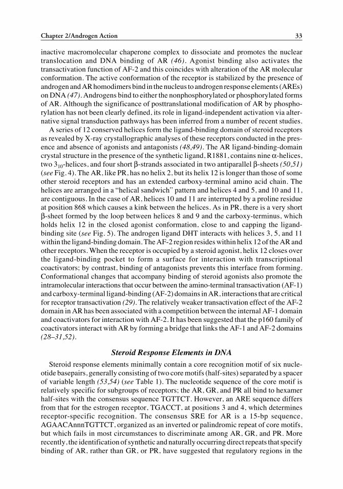
Fig. 4. Amino acid primary sequence of the human androgen-receptor ligand-binding domainand its homology with the ligand-binding domains of the human progesterone and glucocorticoidreceptors. The single-amino-acid code is shown for each of the receptors and the amino acidresidue number is indicated (*) for the androgen receptor. The positions of each of the conserved11 α-helices (H1–H12) and the 4 β-strands in the AR secondary structure are indicated by thesolid and dashed lines, respectively, based on the recently published crystal structures of theandrogen receptor. (From refs. 50 and 51.)
02/Brow/023-044/F 04/11/2003, 12:14 PM34
Chapter 2/Androgen Action 35
in conjunction with the TATA-box binding proteins, TATA-binding protein (TBP) andTBP-associated factors (TAFIIs). Steroid receptors may enhance basal transcription,either by direct interaction with the basal transcription machinery or with TAFIIs (56).For example, interaction of the amino terminus of AR with TFIIF has been reported (57).Most such interactions between AR and the transcriptional machinery imply directbinding of AR to chromatin. However, AR may not necessarily bind to DNA but rathermay bind directly with other sequence specific DNA-binding transcription factors, suchas AP-1 and nuclear factor (NF)-κB to modulate gene transcription (58,59).
Coactivators and CorepressorsCoactivators are cellular proteins that interact with the agonist-activated steroid
receptor complexes to enhance transactivation of target genes, whereas, in most cases,corepressors have an inverse effect through interaction with antagonist-bound or ligand-free receptors (60–62). A general model for coactivator and corepressor function hasevolved. Initially, the repressor protein associates with the receptor on chromatin tomaintain a transcriptionally inactive structure by tethering of histone deacetylases to theDNA at sites close to the responsive element for the receptor. Subsequently, binding ofhormone by the receptor causes release of the corepressor and recruitment ofacetyltransferases, which disrupt the chromatin template by acetylation of histones.Finally, interaction between the activation domains of both the receptor and the recruitedcoactivator with the basal transcription factors results in gene transcription. Whereas theaforementioned mechanisms may be generally applicable, the ligand-free AR existspredominantly in the cytoplasm in a complex of chaperone proteins and is probably notinvolved in interactions with corepressors that possess histone deacetylase activity as
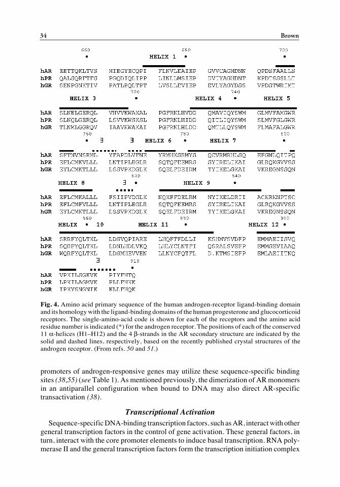
Fig. 5. Tertiary ribbon structure of the human AR ligand-binding domain in the presence of itsligand based on its crystal structure. The ligand, methyltrienolone (R1881), which makes contactwith helices 3, 5, and 11 is shown within the binding pocket of the AR, which is capped by helix12 in the presence of androgen agonists. The AR crystal structure was published by Matias et al.(ref. 50) and can be viewed at the internet website http://ww.rcsb.org/pdb/ in the Protein DataBank (PDB) Structure Explorer under the molecule reference number 1E3G.
02/Brow/023-044/F 04/11/2003, 12:14 PM35

36 Brown
part of an inactive chromatin-bound complex. However, when steroid antagonists bindto receptors, these complexes can bind to DNA, yet they fail to activate gene transcrip-tion. This effect may be the result of failure of coactivators to interact with the antago-nist-bound form of the receptor or to the hypothetical binding of a corepressor andassociated histone deacetylase. Coactivators may have several roles, including expres-sion of intrinsic histone acetyltransferase (HAT) activity, recruitment of other proteinswith HAT activity, and as integrators that enable regulatory molecules to be recruitedand assembled at sites of transcriptional activity (60,61).
A number of coactivators are either directly or indirectly involved in chromatin remod-eling (60,61). The so-called p160 family of coactivators have been subdivided into threesubgroups, represented by SRC-1 (steroid-receptor coactivator-1), SRC-2 which includesTIF2 (transcriptional intermediary factor 2) and GRIP-1 (glucocorticoid-receptor inter-acting protein 1) and SRC-3, which includes TRAM-1 (thyroid-receptor activator mol-ecule 1), ACTR, and AIB1 (amplified in breast cancer 1). In general, these coactivatorsinteract with the ligand-activated AF-2 regions of different steroid receptors to enhancetranscriptional activity. However, for AR, the interaction of these coactivators appears tobe predominantly with the AF-1 region and much less with the AF-2 region (30,31). Thecoactivators possess one or more receptor interaction domains represented by the aminoacid sequence motif LXXLL. The coactivators like SRC-1 also interact with integratorssuch as p300/CBP (cAMP-response element binding [CREB]-binding protein) and pCAF(p300/CBP-associated factor), proteins that possess intrinsic HAT activity, with the over-all effect being synergistic for transcriptional activation (60,61).
Other proteins have been identified as coactivators or effectors of transcriptionalactivity through their specific interaction with AR. Among these are a series of proteinsof varying molecular weight that have been reported by Chang et al. The most well-characterized of these proteins is ARA-70 (AR-associated protein, 70 kDa), whichinteracts in a ligand-dependent manner with AR to increase its transcriptional activity(63). Other members of this group include ARA24, ARA54, ARA55, and ARA160 (64–66). The interaction between AR and other putative coregulator proteins has beenreported (67–75). These proteins include ARIP3 (androgen-receptor interacting pro-tein)/PIAS 1 (protein inhibitor of activated STAT), SNURF, ANPK (androgen nuclearprotein kinase), BRCA1 (breast cancer susceptibility), BAG-1, Smad3, cyclin E, Ubc9(ubiquitin E2-conjugating enzyme), and HBO1 (MYST family, human origin recogni-tion complex). The encyclopedia of proteins continues to grow, but the biologicalrelevance of individual protein interactions with AR will require further investigation.
By contrast to molecules that function as coactivators, the corepressors act to repressbasal promoter activity in the absence of hormone. Studies of the corepressors, NCo-R(nuclear-receptor corepressor), SMRT (silencing mediator for retinoids and thyroidhormone), and RIP140 (receptor interacting protein, 140 kDa) have provided somegeneral insights into the activities of these molecules (62). The relative activity andinteraction of general corepressors on AR transcriptional activity have not been reported,but the PIASy isoform functions in transrepression of AR activity (75).
Ligand-Independent ActivationSteroid receptors, including AR, can also be stimulated to activate gene transcription
in the absence of ligand by growth factors, such as insulin-like growth factor I (IGF-I),keratinocyte growth factor (KGF), epidermal growth factor (EGF), luteinizing hor-
02/Brow/023-044/F 04/11/2003, 12:14 PM36
Chapter 2/Androgen Action 37
mone-releasing hormone (LHRH), neuropeptides, HER-2/neu, interleukin-6 (IL-6) andother agents that directly or indirectly increase intracellular kinase activity or diminishphosphatase activity (76,77). Although the AR is a phosphoprotein, it is unclear whetherpeptide-activated kinases directly phosphorylate the AR or whether this results in recep-tor activation, possibly through enhanced binding to DNA. In addition to the AR itself,phosphorylation of coactivators and basal transcription factors could explain the ARligand-independent transcriptional activity. Importantly, peptide activation of AR activ-ity is synergistic with minimal levels of androgens, thus lowering the androgen thresholdconcentration required for full AR function. These observations are particularly relevantfor androgen-deprivation therapy of patients with prostate cancer, in whom the level ofandrogen is decreased but not completely ablated, and in androgen-independent tumors,where AR expression is present.
Protein kinase A (PKA) can induce prostate-specific antigen (PSA) mRNA andandrogen-responsive reporters in prostate cancer cells, an effect that is blocked byantiandrogens (78). Forskolin directly activates AR transcriptional activity (79). Inter-estingly, the binding of AR to ARE sequences is increased when nuclear extracts areprepared from forskolin-treated cells, despite the observation that nuclear levels of ARare 10-fold higher in androgen-treated cells. Both cAMP and the activator of proteinkinase C (PKC), TPA, act synergistically to enhance androgen-stimulated AR tran-scriptional activity (80,81). 8-Bromo-cAMP has also been shown to induce phospho-rylation of SRC-1 and to facilitate ligand-independent activation (82). IL-6 activatesAR in prostate cancer cells through a mechanism that involves crosstalk between ARand PKA, PKC, and/or MAPK via ErbB2 (83). AP-1 is a complex of transcriptionfactors encoded by c-fos and c-jun protooncogenes and has been implicated in cellgrowth, differentiation, and development with its activity modulated by growth factors,cytokines, oncogenes, and activation of PKC by tumor promoters. The fact that c-juninhibits AR action in some prostate cells and activates it in others emphasizes possiblecell- and/or promoter-specific responses that may be explained by differences in therequirements of a particular promoter for coactivators or by differences between cellsin the availability of required coactivators (78,84,85). The HER2/Neu protooncogenehas been linked to proliferation of prostate cancer cells and can induce AR trans-activation of the PSA gene by the mitogen-activated protein (MAP) kinase-dependentphosphorylation of AR (86,87). Inactivation of the PTEN tumor suppressor gene inprostate cancer allows the AKT/PKB pathway to be constitutively active. Alterna-tively, activation of PI3 kinase by IGF-I can lead to activation of AKT/PKB (88). AKTbinds to and phosphorylates AR. Taken together, these results show that multiplekinase-dependent phosphorylation pathways can be activated in prostate cancer cells,suggesting that phosphorylation events affecting AR, related transcription factors, orcoactivators may lead to ligand-independent activation of AR.
ANDROGEN RECEPTOR AND HUMAN PATHOLOGY
Androgen Insensitivity Syndrome
The AR is required for normal male sex differentiation and development. Much of ourknowledge regarding AR function is derived from studies of human subjects with mutationsof the AR gene and defects in the biological action of androgens that underlie the androgen-insensitivity syndrome (AIS) (89,90). AIS can be either complete in which the phenotype of
02/Brow/023-044/F 04/11/2003, 12:14 PM37
38 Brown
the external genitalia is female, or partial, in which the external genitalia are phenotypicallyambiguous. Although hypospadias is a frequent phenotypic feature of partial AIS, isolatedhypospadias is rarely associated with an AR gene mutation (91). Numerous AR gene muta-tions have been identified in AIS and consist primarily of single nucleotide substitutions thatresult in amino acid substitutions, termination codons, frame shifts, or alterations of mRNAsplicing and relatively few deletions varying from a few nucleotides to the entire gene(89,90). Mutations leading to varying degrees of androgen insensitivity have been reportedin each of the eight exons of the AR gene. However, most mutations have been identified inthe regions that encode the DNA- and ligand-binding domains.
Prostate CancerThe AR has been the focus of intensive investigation for its role in the mechanisms
that regulate the growth and progression of prostate cancer. AR expression is present inprostate tumors at all stages of the disease and tumor growth often continues despiteandrogen ablation therapy that includes inhibition of gonadotropin secretion and testicu-lar androgen synthesis, antiandrogens that antagonize AR function, and even castration.Recent research has centered on the possibility that AR mutations may occur in moreadvanced stages of prostate cancer and contribute to the progression of disease viaalteration of cell function and proliferation. Thus, AR mutations may lead to gain offunction such that the AR is exquisitely sensitive to very low levels of androgen (92), theligand specificity of the receptor is altered (93,94), or the receptor is activated in theabsence of ligand (76,77). Although somatic mutations of the AR have been identifiedin prostate tumor specimens, reports on the frequency of such mutations have beenhighly variable between laboratories and study populations, ranging from less than 10%to almost 50% (95–97). In general, the frequency of mutations appears to increase intumors from later stages of prostate cancer as disease progresses and in metastaticlesions. Another mechanism involves AR gene amplification that has been detected inup to 30% of advanced cancer specimens and may lead to enhanced levels of AR expres-sion (98). As previously mentioned, ligand-independent activation of AR may resultfrom crosstalk with other signal transduction pathways in the absence of androgen. Inaddition, alterations in the size of the polyglutamine repeat may affect the transactivationof AR and influence androgen action in prostate cancer (35,99). Finally, enhancedexpression or phosphorylation of coactivators may lead to their affect to amplify ARfunction, especially in an environment of reduced endogenous androgen levels follow-ing ablation therapy (100).
Spinal Bulbar Muscular AtrophyAn abnormal expansion of the polyglutamine stretch, encoded by (CAG)nCAA, was
identified as the molecular basis for spinal bulbar muscular atrophy (SBMA; Kennedy’sdisease) (36). In normal individuals, the (CAG)nCAA repeat contains 9–33 CAG-liketriplets, whereas 38–75 CAG codons are generally found in the AR genes of subjectswith SBMA. Increasing disease severity is correlated with greater length of this repeat.As a disease, SBMA is characterized by progressive muscle weakness and atrophy withclinical symptoms manifest in the third to fifth decade of life (32,33,36). The pathologyis associated with a severe depletion of lower motornuclei in the spinal cord and brainstemand distal axonopathy of the dorsal root ganglion cells. In addition, subjects with SBMAfrequently exhibit endocrine abnormalities, including testicular atrophy, reduced or
02/Brow/023-044/F 04/11/2003, 12:15 PM38
Chapter 2/Androgen Action 39
absent fertility, gynecomastia, and elevated follicle-stimulating hormone (FSH), LH,and estradiol levels, similar to what is observed in mild forms of AIS. Sex differentiationoccurs normally and characteristics of mild androgen insensitivity only appear later inlife. This may be related to the combination of reduced AR expression and decreasedtestosterone level in older men. SBMA is an X-linked disease and occurs only in men.At present it is not known whether disease progression involves the ligand-activated orligand-free AR. In two cases, an extended period of exogenous testosterone administra-tion had little effect on clinical symptoms.
In transfection studies, the length of the CAG repeat was inversely proportional to theability of the expressed AR to transactivate an androgen-regulated reporter gene (32).However, other investigators have suggested that reduced transcriptional activity wassecondary to reduced stability of AR mRNA and decreased AR protein synthesis (101).The reduction in AR function has also been related to the reduced interaction betweencoactivators and AR (102). Interestingly, AR with an expanded CAG repeat are moreresistant to proteolytic degradation and, in particular, are less susceptible to cleavage bycaspase-3 (103,104). The increased length of the polyglutamine tract also results in theformation of nuclear inclusions. These aggregates accumulate in the cytoplasm andnucleus and sequester other cellular proteins such as SRC-1, NEDD8, and Hsp70 andHsp90 (105). These aggregates appear to be the result of misfolding of protein and defectsin proteolytic degradation involving the ubiquitin–proteasome pathway.
Male InfertilityMale infertility has been associated with an abnormal androgen receptor in subjects
with mild forms of androgen insensitivity and absence of other phenotypic abnormalitiesof the external genitalia (105). Mutations in the AR gene have been associated with onlya very few cases of isolated azoospermia (107–109). However, recent studies havedemonstrated that the glutamine repeat length may be increased in some men witholigospermia and azoospermia (110). Hence, men with longer CAG repeats centeredaround the norm of 20 glutamine repeats have a greater risk for infertility.
SUMMARY
Androgens play a key role in male sex differentiation and development and in themaintenance of male reproductive function, and the effects of these hormones are animportant component in the development of several pathologic conditions. Testosteroneand its 5α-reduced metabolite DHT are potent androgens that act upon target cells toinitiate and maintain the masculine phenotype. Germline mutations in the androgenreceptor and steroid 5α-reductase genes cause the androgen insensitivity syndromes and5α-reductase deficiency, respectively. The effects of these genetic mutations on malesex differentiation and development have played a key role in elucidating the pathwaysof androgen action. Androgen receptors transduce the steroid signal within cells, butattempts to correlate differences in receptor levels with various disease states have beenrelatively unsuccessful. However, molecular studies of AR gene structure have recentlyprovided new insights toward defining a molecular and genetic basis for the pathologyassociated with diseases—including spinal bulbar muscular atrophy, prostate cancer,and male infertility—affecting middle-aged and older men. Further studies at themolecular level to define the steroid- and DNA-binding properties of androgen recep-
02/Brow/023-044/F 04/11/2003, 12:15 PM39
40 Brown
tors, as well as the transcriptional activity and interactions of the receptor withcoactivators, corepressors, and integrators within the transcriptional complex, will pro-vide additional insight into the complex nature of androgen action. Moreover, epidemio-logic data and molecular genetic analyses of gene structure have led to a newunderstanding of the interrelationships between environmental and genetic factors thatmay impact on the incidence of certain pathologic conditions in men.
REFERENCES
1. Griffin JE, Wilson JD. Disorders of the testes and the male reproductive tract. In: Wilson JD, FosterDW, Kronenberg HM, et al., eds. Williams Textbook of Endocrinology. WB Saunders, Philadelphia,PA, 1998, pp. 819–875.
2. Fuh VL, Stoner E. Androgen inhibitors/antiandrogens. In: Knobil E, Neill JD, eds. Encyclopedia ofReproduction. Academic, San Diego, CA, 1998, pp. 166–173.
3. Hammond GL, Bocchinfuso WP. Sex hormone-binding globulin/androgen binding protein: steroidbinding and dimerization domains. J Steroid Biochem Mol Biol 1995;53:1–6.
4. Avvakumov GV, Grishkovskaya I, Muller YA, Hammond GL. Resolution of the human sex hor-mone-binding globulin dimer interface and evidence for two steroid-binding sites per homodimer.J Biol Chem 2001;276:34453–34457.
5. Dunn JF, Nisula BC, Rodbard D. Transport of steroid hormones: binding of 21 endogenous steroidsto both testosterone-binding globulin and corticosteroid-binding globulin in human plasma. J ClinEndocrinol Metab 1981;53:58–68.
6. Pardridge WM. Serum bioavailability of sex steroid hormones. Clin Endocrinol Metab 1986;15:259–278.
7. Rosner W, Hryb DJ, Khan MS, et al. Sex hormone-binding globulin. Binding to cell membranes andgeneration of a second messenger. J Androl 1992;13:101–106.
8. Russell DW, Wilson JD. Steroid 5α-reductase: two genes/two enzymes. Annu Rev Biochem1994;63:25–61.
9. Morimoto I, Hawks ED, Horton R. Studies on the origin of androstanediol and androstanediolglucuronide in young and elderly men. J Clin Endocrinol Metab 1981;52:772–778.
10. Brooks, RV. Androgens. Clin Endocrinol Metab 1975;4:503–520.11. Simpson ER, Zhao Y, Agarwal VR, et al. Aromatase expression in health and disease. Recent Prog
Horm Res 1997;52:185–213.12. Wilson JD, Griffin JE, Russell DW. Steroid 5α-reductase 2 deficiency. Endocr Rev 1993;14:577–593.13. Zhou Z-X, Wong C-I, Sar M., Wilson EM. The androgen receptor: an overview. Recent Prog Horm
Res 1994;49:249–274.14. Jenster G, vander Korput HAGM, van Vroonhoven C, et al. Domains of the human androgen recep-
tor involved in steroid binding, transcriptional activation and subcellular localization. MolEndocrinol 1991;5:1396–1404.
15. Lubahn DB, Joseph DR, Sullivan PM, et al. Cloning of human androgen receptor complementaryDNA and localization to the X chromosome. Science 1988;240:327–330.
16. Kuiper GGJM, Faber PW, van Rooij HCJ, et al. Structural organization of the human androgenreceptor gene. J Mol Endocrinol 1989;2:R1–R4.
17. Faber PW, van Rooij HCJ, Schipper HJ, et al. Two different, overlapping pathways of transcriptioninitiation are active on the TATA-less human androgen receptor promoter. J Biol Chem1993;268:9296–9301.
18. Chen S, Supakar PC, Vellanoweth RL, et al. Functional role of a conformationally flexiblehomopurine/homopyrimidine domain of the androgen receptor gene promoter interacting with SP1and a pyrimidine single strand DNA-binding protein. Mol Endocrinol 1997;11:3–15.
19. Grad JM, Dai JL, Wu S, Burnstein KL. Multiple androgen response elements and a myc consensussite in the androgen receptor (AR) coding region are involved in androgen-mediated up-regulationof AR messenger RNA. Mol Endocrinol 1999;13:1896–1911.
20. Mizokami A, Chang C. Induction of translation by the 5'-untranslated region of human androgenreceptor mRNA. J Biol Chem 1994;269:25655–25659.
21. Lubahn DB, Joseph DR, Sar M, et al. The human androgen receptor: complementary deoxyribonucleicacid cloning, sequence analysis, and gene expression in prostate. Mol Endocrinol 1998;2:1265–1275.
02/Brow/023-044/F 04/11/2003, 12:15 PM40
Chapter 2/Androgen Action 41
22. Gottlieb B, Beitel LK, Trifiro MA. Variable expressivity and mutation databases: the androgenreceptor gene mutations database. Hum Mutat 2001;17:382–388.
23. Kemppainen JA, Lane MV, Sar M, Wilson EM. Androgen receptor phosphorylation, turnover,nuclear transport, and transcriptional activation. J Biol Chem 1992;267:968–974.
24. Blok LJ de Ruiter PE, Brinkmann AO. Androgen receptor phosphorylation. Endocr Res 1996;58:569–575.25. Gao T, McPhaul MJ. Functional activities of the A and B forms of the human androgen receptor in
response to androgen receptor agonists and antagonists. Mol Endocrinol 1998;12:654–663.26. Lubahn DB, Brown TR, Simental JA, et al. Sequence of the intron/exon junctions of the coding
region of the human androgen receptor gene and identification of a point mutation in a family withcomplete androgen insensitivity. Proc Natl Acad Sci USA 1989;86:9534–9538.
27. Doesburg P, Kuil CW, Berrevoets CA, et al. Functional in vivo interaction between the amino-terminal, transactivation domain and the ligand binding domain of the androgen receptor. Biochem-istry 1997;36:1052–1064.
28. He B, Kemppainen JA, Voegel JJ, et al. Activation function 2 in the human androgen receptor ligandbinding domain mediates interdomain communication with the NH2-terminal domain. J Biol Chem1999;274:37219–37225.
29. He B, Kemppainen JA, Wilson EM. FXXLF and WXXLF sequences mediate the NH2- terminal inter-action with the ligand binding domain of the androgen receptor. J Biol Chem 2000;275:22986–22994.
30. Alen P, Claessens F, Verhoeven G. et al. The androgen receptor amino- terminal domain plays a keyrole in p160 coactivator-stimulated gene transcription. Mol Cell Biol 1999;19:6085–6097.
31. Bevan Cl, Hoare S, Claessens F, et al. The AF1 and AF2 domains of the androgen receptor interactwith distinct regions of SRC1. Mol Cell Biol 1999;19:8383–8392.
32. Trifiro MA, Kazemi-Esfarjani P, Pinsky L. X-Linked muscular atrophy and the androgen receptor.Trends Endocrinol Metab 1994;5:416–421.
33. Choong CS, Wilson EM. Trinucleotide repeats in the human androgen receptor: a molecular basisfor disease. J Mol Endocrinol 1998;21:2235–2257.
34. Irvine RA, Yu MC, Ross RK, Coetzee GA. The CAG and GGC microsatellites of the androgen receptorgene are in linkage disequilibrium in men with prostate cancer. Cancer Res 1995;55:1937–1940.
35. Giovannucci E, Stampfer MJ, Krithivas K, et al. The CAG repeat within the androgen receptor geneand its relationship to prostate cancer. Proc Natl Acad Sci USA 1997;94:3320–3323.
36. LaSpada AR, Wilson EM, Lubahn DB. Androgen receptor gene mutations in X-linked spinal andbulbar muscular atrophy. Nature 1991;352:77–79.
37. Freedman LP. Anatomy of the steroid receptor zinc finger region. Endocr Rev 1992;3:29–145.38. Claessens F. Verrijdt G, Schoenmakers E, et al. Selective DNA binding by the androgen receptor as
a mechanism for hormone-specific gene regulation. J Steroid Biochem Mol Biol 2001;76:23–30.39. Gonzales MI, Robins D. Oct-1 preferentially interacts with androgen receptor in a DNA- dependent
manner that facilitates recruitment of SRC-1. J Biol Chem 2001;276:6420–6428.40. Zhou Z-X, Sar M, Simental JA, et al. Ligand-dependent bipartite nuclear targeting signal in the
human androgen receptor. J Biol Chem 1994;269:13115–13123.41. Tyagi RK, Lavrovsky Y, Ahn SC, et al. Dynamics of intracellular movement and nucleocytoplasmic
recycling of the ligand-activated androgen receptor in living cells. Mol Endocrinol 2000;14:1162–1174.42. Poukka H, Karvonen U, Yoshikawa N, et al. The RING finger protein SNURF modulates nuclear
trafficking of the androgen receptor. J Cell Sci 2000;113:2991–3001.43. Wang Q, Lu J, Yong EL. Ligand- and coactivator-mediated transactivation function (AF2) of the
androgen receptor ligand-binding domain are inhibited by the cognate hinge region. J Biol Chem2001;276:7493–7499.
44. Buchanan G, Yan M, Harris JM, et al. Mutations at the boundary of the hinge and ligand bindingdomain of the androgen receptor confer increased transactivation function. Mol Endocrinol2001;15:46–56.
45. Pratt WB. Role of heat-shock proteins in steroid receptor function. In: Parker M, ed. Frontiers inMolecular Biology: Steroid Hormone Action. IRL, Oxford, 1993, pp. 64–93.
46. Wong CI, Zhou Z-X, Sar M, Wilson EM. Steroid requirement for androgen receptor dimerizationand DNA binding. J Biol Chem 1993;268:19004–19012.
47. Langley E, Zhou Z-X, Wilson EM. Evidence for an anti-parallel orientation of the ligand- activatedhuman androgen receptor dimer. J Biol Chem 1994;270:29983–29990.
48. Brzozowski AM, Pike ACW, Dauter Z, et al. Molecular basis of agonism and antagonism in theoestrogen receptor. Nature 1997;389:753–758.
02/Brow/023-044/F 04/11/2003, 12:15 PM41
42 Brown
49. Williams SP, Sigler PB. Atomic structure of progesterone complexed with its receptor. Nature1998;393:392–395.
50. Matias PM, Donner P, Coelho R, et al. Structural evidence for ligand specificity in the bindingdomain of the human androgen receptor. J Biol Chem 2000;275: 26164–26171.
51. Sack JS, Kish KF, Wang C, et al. Crystallographic structures of the ligand-binding domains of theandrogen receptor and its T877A mutant complexed with the natural agonist dihydrotestosterone.Proc Natl Acad Sci USA 2001;98:4904–4909.
52. Berrevoets CA, Doesburg P, Sketetee K, et al. Functional interactions of the AF-2 domain coreregion of the human androgen receptor with the amino-terminal domain and with the transcriptionalcoactivator TIF-2 (transcriptional intermediary factor-2). Mol Endocrinol 1998;12:1172–1183.
53. Roche PJ, Hoare SA, Parker MA. A consensus DNA-binding site for the androgen receptor. MolEndocrinol 1992;6:2229–2235.
54. Nelson CC, Hendy SC, Shukin RJ, et al. Determinants of DNA sequence specificity of the androgen,progesterone, and glucocorticoid receptors: evidence for differential steroid receptor response ele-ments. Mol Endocrinol 1999;13:2090–2107.
55. Zhou Z, Corden JL, Brown TR. Identification and characterization of a novel androgen responseelement composed of a direct repeat. J Biol Chem 1997;272:8227–8235.
56. Ing N, Beekman JM, Tsai SY, et al. Members of the steroid hormone superfamily interact with TFIIB(S300-II). J Biol Chem 1992;267:17617–17623.
57. McEwan I, Gustafsson J-A. Interaction of the human androgen receptor transactivation functionwith the general transcription factor TFIIF. Proc Natl Acad Sci USA 1997;94:8485–8490.
58. Bubulya A, Wise SC, Shen X-Q, et al. c-Jun can mediate androgen receptor-induced transactivation.J Biol Chem 1996;271:24583–24589.
59. Kallio PJ, Poukka H, Moilanen A, et al. Androgen-receptor mediated transcriptional regulation inthe absence of direct interaction with a specific DNA element. Mol Endocrinol 1995;9:1017–1028.
60. Glass CK, Rosenfeld MG. The coregulator exchange in transcriptional functions of nuclear recep-tors. Genes Dev 2000;14:121–141.
61. Lee KC, Kraus WL. Nuclear receptors, coactivators and chromatin: new approaches, new insights.Trends Endocrinol Metab 2001;12:191–197.
62. Perissi V, Staszewski LM, McInerney EM, et al. Molecular determinants of nuclear receptor–corepressor interaction. Genes Dev 1999;13:3198–3208.
63. Yeh S, Chang C. Cloning and characterization of a specific coactivator, ARA70, for the androgenreceptor in human prostate cells. Proc Natl Acad Sci USA 1996;93:5517–5521.
64. Fujimoto N, Yeh S, Kang HY, et al. Cloning and characterization of androgen receptor coactivator,ARA55, in human prostate. J Biol Chem 1999;274:8316–8321.
65. Kang HY, Yeh S, Fujimoto N, Chang C. Cloning and characterization of human prostate coactivator,ARA54, a novel protein that associates with the androgen receptor. J Biol Chem 1999;274:8570–8576.
66. Hsiao PW, Chang C. Isolation and characterization of ARA160 as the first androgen receptor N-terminal-associated coactivator in human prostate cells. J Biol Chem 1999;274:22373–22379.
67. Kotaja N, Aittomaki S, Silvennoinen O, et al. ARIP3 (androgen receptor- interacting protein 3) andother PIAS (protein inhibitor of activated STAT) proteins differ in their ability to modulate steroidreceptor-dependent transcriptional activation. Mol Endocrinol 2000;14:1986–2000.
68. Moilanen AM, Karvonen U, Poukka H, et al. Activation of androgen receptor function by a novelnuclear protein kinase. Mol Biol Cell 1998;9:2527–2543.
69. Yeh S, Hu YC, Rahman M, et al. Increase of androgen-induced cell death and androgen receptortransactivation by BRCA1 in prostate cancer cells. Proc Natl Acad Sci USA 2000;97:11256–11261.
70. Froesch BA, Takayama S, Reed JC. BAG-1L protein enhances androgen receptor function. J BiolChem 1998;273:11660–11666.
71. Kang HY, Lin HK, Hu YC, et al. From transforming growth factor-β signaling to androgen action:identification of Smad3 as an androgen receptor coregulator in prostate cancer cells. Proc Natl AcadSci USA 2001;98:3018–3023.
72. Yamamoto A, Hashimoto Y, Kohri K, et al. Cyclin E as a coactivator of the androgen receptor. J CellBiol 2000;150:873–879.
73. Poukka H, Aarnisalo P, Karvonen U, et al. Ubc9 interacts with the androgen receptor and activatesreceptor-dependent transcription. J Biol Chem 1999;274:19441–19446.
74. Sharma M, Zarnegar M, Li X, et al. Androgen receptor interacts with a novel MYST protein, HBO1.J Biol Chem 2000;275:35200–35208.
02/Brow/023-044/F 04/11/2003, 12:15 PM42
Chapter 2/Androgen Action 43
75. Gross M, Liu B, Tan JA, et al. Distinct effects of PIAS proteins on androgen-mediated gene activa-tion in prostate cancer cells. Oncogene 2001;20:3880–3887.
76. Klocker H, Culig Z, Ider IE, et al. Mechanism of androgen receptor activation and possible impli-cations for chemoprevention trials. Eur Urol 1999;35:413–419.
77. Culig Z, Hobisch A, Bartsch G, Klocker H. Androgen receptor—an update of mechanisms of actionin prostate cancer. Urol Res 2000;28:211–219.
78. Sadar MD. Androgen-independent induction of prostate specific antigen gene expression by cross-talk between the androgen receptor and protein kinase A signal transduction pathways. J Biol Chem1999;274:7777–7783.
79. Nazareth LV, Weigel NL. Activation of the human androgen receptor through a protein kinase Apathway. J Biol Chem 1996;268:19900–19907.
80. DeRuiter PE, Teuwen R, Trapman J, et al. Synergism between androgens and protein kinase-C onandrogen regulated gene expression. Mol Cell Endocrinol 1995;110:1–6.
81. Ikonen T, Palvimo JJ, Kallio PJ, et al. Stimulation of androgen- regulated transactivation by modu-lators of protein phosphorylation. Endocrinology 1994;135:1359–1366.
82. Rowan BG, Garrison N, Weigel NL, O’Malley BW. 8-Bromo cAMP induces phosphorylation of twosites on SRC-1 that facilitate ligand independent activation of the chicken progesterone receptor andare critical for functional cooperation between SRC-1 and CBP. Mol Cell Biol 2000;20:8720–8730.
83. Qui Y, Ravi L, Kung HJ. Requirement of ErbB2 for signaling by interleukin-6 in prostate carcinomacells. Nature 1998;393:83–85.
84. Shemshedini L, Knauthe R, Sassone-Corsi P, et al. Cell-specific inhibitory and stimulatory effectsof Fos and Jun on transcription activation by nuclear receptors. EMBO J 1991;10:3839–3843.
85. Bubulaya A, Wise SC, Shen X-Q, et al. c-Jun can mediate AR- induced transactivation. J Biol Chem1996;271:24583–24589.
86. Craft N, Shostak Y, Carey M, Sawyers CL. A mechanism for hormone-independent prostate cancerthrough modulation of androgen receptor signaling by the HER-2/neu tyrosine kinase. Nature Med1999;5:280–285.
87. Yeh S, Lin HK, Kang HY, et al. From HER-2/Neu signal cascade to androgen receptor and itscoactivators: a novel pathway by induction of androgen target genes through MAP kinase in prostatecells. Proc Natl Acad Sci USA 1999;96:5458–5463.
88. Li P, Nicosia SV, Bai W. Antagonism between PTEN/MMAC1/TEP-1 and androgen receptor ingrowth and apoptosis of prostate cancer cells. J Biol Chem 2001;276:20444–20550.
89. Quigley CA, DeBellis A, Marschke KB, et al. Androgen receptor defects: historical, clinical andmolecular perspectives. Endocr Rev 1995;16:271–321.
90. Brown TR. Androgen insensitivity syndrome. J Androl 1995;16:299–303.91. Sutherland RW, Wiener JS, Hicks JP, et al. Androgen receptor gene mutations are rarely associated
with isolated penile hypospadias. J Urol 1996;156:828–831.92. Gregory CW, He B, Johnson RT, et al. Androgen receptor stabilization in recurrent prostate cancer
is associated with hypersensitivity to low androgen. Cancer Res 2001;61:2892–2898.93. Veldscholte J, Berrevoets CA, Ris-Staplers C, et al. The androgen receptor in LNCaP cells contains
a mutation in the ligand binding domain which affects steroid binding characteristics and responseto antiandrogens. J Steroid Biochem1992;41:665–669.
94. Tan J, Sharief Y, Hamil KG, et al. Dehydroepiandrosterone activates mutant androgen receptorsexpressed in the androgen-dependent human cancer xenograft CRW22 and LNCaP cells. MolEndocrinol 1997;11:450–459.
95. Tilley WD, Buchanan G, Hickey TE, Bentel JM. Mutations in the androgen receptor gene areassociated with progression of human prostate cancer to androgen independence. Clin Cancer Res1996;2:277–285.
96. Taplin ME, Bubley GJ, Ko YJ, et al. Selection for androgen receptor mutations in prostate cancerstreated with androgen antagonist. Cancer Res 1999;59:2511–2515.
97. Marcelli M, Ittmann M, Mariani S. et al. Androgen receptor mutations in prostate cancer. Cancer Res2000;60:944–999.
98. Koivisto PA, Kononen J. Palmberg C, et al. Androgen receptor gene amplification: a possiblemechanism for androgen deprivation therapy failure in prostate cancer. Cancer Res 1997;57:314–319.
99. Stanford JL, Just JJ, Gibbs M, et al. Polymorphic repeats in the androgen receptor gene: molecularmarkers of prostate cancer risk. Cancer Res 1997;57:1194–1198.
02/Brow/023-044/F 04/11/2003, 12:15 PM43
44 Brown
100. Gregory CW, He B, Johnson RT, et al. A mechanism for androgen receptor-mediated prostate cancerrecurrence after androgen deprivation therapy. Cancer Res 2001;61:4315–4319.
101. Choong CS, Kemppainen JA, Zhou Z-X, Wilson EM. Reduced androgen receptor gene expressionwith first exon CAG repeat expansion. Mol Endocrinol 1996;10:1527–1535.
102. Irvine RA, Ma H, Yu MC, et al. Inhibition of p160- mediated coactivation with increasing androgenreceptor polyglutamine length. Hum Mol Genet 2000;9:267–274.
103. Abdullah A. Trifiro MA, Panet-Raymond V, et al. Spinobulbar muscular atrophy: polyglutamine-expanded androgen receptor is proteolytically resistant in vitro and processed abnormally in trans-fected cells. Hum Mol Genet 1998;7:379–384.
104. Kobayashi Y, Miwa S, Merry DE, et al. Caspase-3 cleaves the expanded androgen receptor proteinof spinal and bulbar muscular atrophy in a polyglutamine repeat length-dependent manner. BiochemBiophys Res Commun 1998;252:145–150.
105. Stenoien DL, Cummings CJ, Adams HP, et al. Polyglutamine-expanded androgen receptors formaggregates that sequester heat shock proteins, proteasome components and SRC-1, and are sup-pressed by the HDJ-2 chaperone. Hum Mol Genet 1999;8:731–741.
106. Aiman J, Griffin JE, Cushard WG, Wilson JD. Androgen insensitivity as a cause of infertility inotherwise normal men. N Engl J Med 1998;300:223–227.
107. Akin JW, Behzadian A, Tho SPT, McDonough PG. Evidence for a partial deletion in the androgenreceptor gene in a phenotypic male with azoospermia. Am J Obstet Gynecol 1991;165:1891–1894.
108. Yong EL, Ng SC, Roy AC, et al. Pregnancy after hormonal correction of severe spermatogenic defectdue to mutation in androgen receptor gene. Lancet 1994;344:8925–8927.
109. Ghadessy FJ, Lim J, Abdullah AA, et al. Oligospermic infertility associated with an androgenreceptor mutation that disrupts interdomain and coactivator (TIF2) interactions. J Clin Invest1999;103:1517–1525.
110. Tut TG, Ghadessy FJ, Trifiro MA, et al. Long polyglutamine tracts in the androgen receptor areassociated with reduced trans-activation, impaired sperm production, and male infertility. J ClinEndocrinol Metab 1997;82:3777–3782.
02/Brow/023-044/F 04/11/2003, 12:15 PM44

http://www.springer.com/978-1-58829-029-8