1 Z-band alternatively spliced PDZ-motif protein (ZASP) is the major ...
19
1 Z-band alternatively spliced PDZ-motif protein (ZASP) is the major O-linked-β-N-acetylglucosamine- substituted protein in human heart myofibrils Man-Ching Leung 1 , Paul G Hitchen 2 , Douglas G Ward 3 , Andrew E Messer 1 , Steven B Marston 1 1 From Myocardial Function, NHLI, Imperial College London, London, W12 0NN, UK, 2 Division of Molecular Biosciences, Imperial College London, London, SW7 2AZ, UK and 3 School of Cancer Sciences, University of Birmingham , Birmingham, B15 2TT, UK Running title: O-GlcNAc modification of ZASP in human heart myofibrils To whom correspondence should be addressed: Steven Marston, Myocardial Function, Imperial Centre for Translational and Experimental Medicine, Du Cane Road, London W12 0NN, UK, Tel.: (+44) 207 594 2732; E-mail: [email protected] Keywords: O-GlcNAc; ZASP; Cypher; human heart; myofibrils; cardiomyopathy _____________________________________________________________________________________ Background: We studied O-GlcNAc-modified protein in sarcomeric proteins of human heart Result: ZASP (Z-band alternatively spliced PDZ- motif protein) accounts for 50 to 80% of O- GlcNAcylated protein Conclusion: ZASP is the major O-GlcNAc- substituted protein in human heart muscle and its levels increase in pathological muscle Significance: Modulation of O-GlcNAcylation in ZASP may have a role in mechanotransduction in the heart. SUMMARY We studied O-linked-β-N-acetylglucosamine; (O- GlcNAc) modification of contractile proteins in human heart using SDS-PAGE and 3 detection methods: specific enzymatic conjugation of O- GlcNAc with UDP-GalNAz that is then linked to a TAMRA fluorescent tag, and CTD110.6 and RL2 monoclonal antibodies to O-GlcNAc. All 3 methods showed that O-GlcNAc modification was predominantly in a group of bands around 90 kDa that did not correspond to any of the major myofibrillar proteins. MALDI-MS/MS identified the 90 kDa band as the protein ZASP (Z-band alternatively spliced PDZ-motif protein), a minor component of the Z-disc (about 1/400 α - actinin) important for myofibrillar development and mechanotransduction. This was confirmed by the co-localisation of O-GlcNAc and ZASP in Western blotting and by immunofluorescence microscopy. O-GlcNAcylation of ZASP increased in diseased heart, being 49±5% of all O-GlcNAc in donor, 68±9% in end-stage failing heart and 76±6% in myectomy muscle samples (donor vs myectomy p<0.05). ZASP is only 22% of all O- GlcNAcylated proteins in mouse heart myofibrils. __________________________________________ The post-translational modification, O- linked-β-N-acetylglucosamine; (O-GlcNAc) on nuclear and cytoplasmic proteins has attracted a lot of interests since it was first described in 1984 (1). Analogous to phosphorylation, O-GlcNAcylation occurs on serine and threonine residues and crosstalk between protein O-GlcNAcylation and phosphorylation has been proposed to play a role in signalling and transcription regulation (2). A role for protein O-GlcNAcylation has been proposed in regulating growth and contractility in the mammalian heart. Global O-GlcNAc levels in the heart have been observed to increase in rats with hypertension, surgically induced hypertrophy and heart failure and also in heart tissue from patients with aortic stenosis (3). It is interesting to note that there was a decrease in O-GlcNAc in physiologically hypertrophied heart in a study using swim-exercised mice (4) and that short term O- GlcNAcylation affects myofibrillar Ca 2+ -sensitivity (5) and can be cardioprotective (6). It was suggested these effects involved O-GlcNAcylation of sarcomeric proteins. Most investigations have measured total O-GlcNAc in heart muscle, however identification of the proteins of the contractile apparatus that are O- GlcNAc-substituted has been limited to animal studies. O-GlcNAc modifications on most of the major myofibrillar proteins have been reported in rat cardiac and skeletal muscles, including myosin heavy chain, actin, troponin I, myosin light chain 1 http://www.jbc.org/cgi/doi/10.1074/jbc.M112.410316 The latest version is at JBC Papers in Press. Published on December 27, 2012 as Manuscript M112.410316 Copyright 2012 by The American Society for Biochemistry and Molecular Biology, Inc. by guest on April 13, 2018 http://www.jbc.org/ Downloaded from
Transcript of 1 Z-band alternatively spliced PDZ-motif protein (ZASP) is the major ...
1
Z-band alternatively spliced PDZ-motif protein (ZASP) is the major O-linked-β-N-acetylglucosamine-substituted protein in human heart myofibrils
Man-Ching Leung1, Paul G Hitchen2, Douglas G Ward3, Andrew E Messer1, Steven B Marston1
1From Myocardial Function, NHLI, Imperial College London, London, W12 0NN, UK, 2Division of Molecular Biosciences, Imperial College London, London, SW7 2AZ, UK and 3School of Cancer Sciences,
University of Birmingham , Birmingham, B15 2TT, UK
Running title: O-GlcNAc modification of ZASP in human heart myofibrils
To whom correspondence should be addressed: Steven Marston, Myocardial Function, Imperial Centre for Translational and Experimental Medicine, Du Cane Road, London W12 0NN, UK,
Tel.: (+44) 207 594 2732; E-mail: [email protected]
Keywords: O-GlcNAc; ZASP; Cypher; human heart; myofibrils; cardiomyopathy
_____________________________________________________________________________________
Background: We studied O-GlcNAc-modified protein in sarcomeric proteins of human heart Result: ZASP (Z-band alternatively spliced PDZ-motif protein) accounts for 50 to 80% of O-GlcNAcylated protein Conclusion: ZASP is the major O-GlcNAc- substituted protein in human heart muscle and its levels increase in pathological muscle Significance: Modulation of O-GlcNAcylation in ZASP may have a role in mechanotransduction in the heart.
SUMMARY
We studied O-linked-β-N-acetylglucosamine; (O-GlcNAc) modification of contractile proteins in human heart using SDS-PAGE and 3 detection methods: specific enzymatic conjugation of O-GlcNAc with UDP-GalNAz that is then linked to a TAMRA fluorescent tag, and CTD110.6 and RL2 monoclonal antibodies to O-GlcNAc. All 3 methods showed that O-GlcNAc modification was predominantly in a group of bands around 90 kDa that did not correspond to any of the major myofibrillar proteins. MALDI-MS/MS identified the 90 kDa band as the protein ZASP (Z-band alternatively spliced PDZ-motif protein), a minor component of the Z-disc (about 1/400 α-actinin) important for myofibrillar development and mechanotransduction. This was confirmed by the co-localisation of O-GlcNAc and ZASP in Western blotting and by immunofluorescence microscopy. O-GlcNAcylation of ZASP increased in diseased heart, being 49±5% of all O-GlcNAc in donor, 68±9% in end-stage failing heart and 76±6% in myectomy muscle samples (donor vs
myectomy p<0.05). ZASP is only 22% of all O-GlcNAcylated proteins in mouse heart myofibrils. __________________________________________
The post-translational modification, O-linked-β-N-acetylglucosamine; (O-GlcNAc) on nuclear and cytoplasmic proteins has attracted a lot of interests since it was first described in 1984 (1). Analogous to phosphorylation, O-GlcNAcylation occurs on serine and threonine residues and crosstalk between protein O-GlcNAcylation and phosphorylation has been proposed to play a role in signalling and transcription regulation (2).
A role for protein O-GlcNAcylation has been proposed in regulating growth and contractility in the mammalian heart. Global O-GlcNAc levels in the heart have been observed to increase in rats with hypertension, surgically induced hypertrophy and heart failure and also in heart tissue from patients with aortic stenosis (3). It is interesting to note that there was a decrease in O-GlcNAc in physiologically hypertrophied heart in a study using swim-exercised mice (4) and that short term O-GlcNAcylation affects myofibrillar Ca2+-sensitivity (5) and can be cardioprotective (6). It was suggested these effects involved O-GlcNAcylation of sarcomeric proteins.
Most investigations have measured total O-GlcNAc in heart muscle, however identification of the proteins of the contractile apparatus that are O-GlcNAc-substituted has been limited to animal studies. O-GlcNAc modifications on most of the major myofibrillar proteins have been reported in rat cardiac and skeletal muscles, including myosin heavy chain, actin, troponin I, myosin light chain 1
http://www.jbc.org/cgi/doi/10.1074/jbc.M112.410316The latest version is at JBC Papers in Press. Published on December 27, 2012 as Manuscript M112.410316
Copyright 2012 by The American Society for Biochemistry and Molecular Biology, Inc.
by guest on April 13, 2018
http://ww
w.jbc.org/
Dow
nloaded from
2
and myosin light chain 2, but stoichiometry of the modification was not quantified (7-9).
In this paper we have studied O-GlcNAc modifications of myofibrillar proteins in human heart muscle myofibrils for the first time. Using three independent measurement techniques we found that, unlike rat and mouse, the majority of O-GlcNAc in human myofibrils was in a low abundance protein, identified as ZASP (Z-band Alternatively Spliced PDZ-motif Protein; also named LIM domain-binding protein 3, Cypher and Oracle (LDB3 gene))(10). We also observed an apparent increase in O-GlcNAc modification of ZASP in pathological human heart samples.
EXPERIMENTAL PROCEDURES
Human Cardiac Samples
Cardiac muscle from donor hearts as control, end-stage failing hearts and septal myectomy samples from patients with hypertrophic obstructive cardiomyopathy (HOCM) were obtained as described (11-14) (and supplement). All tissues were rapidly frozen and stored under liquid nitrogen until use. Gender, age and known mutations of subjects are presented in Supplement Table 1.
Enzymatic Labelling of O-GlcNAcylated Proteins
Human heart myofibrils and cardiac tissue whole lysates were isolated as described (13-15). Proteins from myofibril extracts were precipitated by chloroform/methanol precipitation and resuspended into 20 mM HEPES buffer with 1% SDS, pH 7.9. O-GlcNAc groups were labelled using “Click-iT™ O-GlcNAc Enzymatic Labelling System” from Invitrogen. Procedures from manufacturer’s protocol were followed (16,17). Briefly, the sample was mixed with dH2O, Click-iTTM O-GlcNAc enzymatic labelling buffer, MnCl2 solution and vortexed. UDP-GalNAz, was then added, followed by the addition of the genetically modified enzyme Gal-T1 (Y289L), which puts the GalNAz onto any O-GlcNAc groups. This was incubated overnight at 4ºC. Chloroform/methanol precipitation was performed and the proteins resuspended in 50 µL 50 mM Tris-HCl, 1% SDS, pH 8.0. Detection was enabled using “Click-iT™ Tetramethylrhodamine (TAMRA) Protein Analysis Detection Kit” from Invitrogen following manufacturer’s instruction. Briefly, reaction buffer, containing TAMRA conjugated with an alkyne group, was mixed with the resuspended sample, followed by addition of dH2O, CuSO4 solution and two other reaction buffers. This mixture was vortexed for 20 minutes for the Click-iTTM azide/alkyne reaction to attach
the TAMRA label to the GalNAz labelled O-GlcNAc groups. Further chloroform/methanol precipitation was then performed and the protein was resuspended in SDS buffer. Labelled samples were used immediately.
SDS-PAGE and Western Blotting
Myofibril proteins labelled with TAMRA enzymatic labelling were separated using 12% SDS-PAGE. The TAMRA fluorescence signal was visualised with UV transillumination and the image was recorded with a CCD camera based gel imager (G:BOX, Syngene) and analysed using GeneTools (Syngene).
Myofibril samples were separated on 10% SDS-PAGE gels and Western blotted. Proteins were transblotted onto nitrocellulose membrane. Nitrocellulose membranes with the transferred proteins were cut into strips so that each strip has the same protein profile. The strips were probed separately with different antibodies and then reassembled in register for chemiluminescent detection.
Primary antibodies used were:
O-GlcNAc: CTD110.6 antibody to O-GlcNAc, a kind gift from Gerald Hart, Johns Hopkins University School of Medicine, MD, US;
O-GlcNAc: anti-O-linked N-acetylglucosamine monoclonal antibody (RL2), Thermo Scientific;
ZASP: LDB3 antibody NB100-2445, Novus Biologicals;
ENH: PDLIM5 (JK-3R), Santa Cruz Biotechnology
Antibody dilutions are indicated in corresponding figures. Secondary antibodies were the appropriate horseradish peroxidase-conjugated IgG (see supplement) and detection was by chemiluminescence using ECL Plus Western Blotting Detection Reagents from GE Healthcare. Chemiluminescence was imaged by cooled CCD camera using the G:BOX (Syngene) and densitometry was carried out using GeneTools (Syngene).
Protein Identification by MALDI-TOF/TOF
The TAMRA-labelled 90 kDa protein band was cut out of the gel and in-gel reduction, alkylation and tryptic digestion performed. The resulting peptides were fractionated by HPLC, mixed with matrix and spotted onto target plates for MALDI-TOF/TOF analysis. Protein identification was performed using MASCOT to search the Swiss-Prot human database. Full details are given in the supplement.
by guest on April 13, 2018
http://ww
w.jbc.org/
Dow
nloaded from
3
Double Indirect Immunofluorescence
Snap-frozen cardiac tissues of approximately 0.5 cm3 size were embedded in Tissue-Tek OCT compound (Sakura Finetek) and stored at -80°C. Sections of 5 µm thick were cut at -20ºC and stored at -80°C until use. Sections were fixed in ice-cold methanol for 10 min and rinsed in PBS. They were then incubated with blocking serum (10% goat serum in PBS) for 1 hour at room temperature followed by primary antibodies (ZASP: cypher (H-83), Santa Cruz, dilution 1:50; RL2, dilution 1:200; cMyBP-C: 2-14 antibody (18), dilution 1:500; α-actinin: clone EA-53, Sigma-Aldrich Company Ltd., dilution 1:4000) overnight at 4°C. The sections were then washed 3 times for 5 minutes each in PBS with 0.05% Tween 20 and once for 5 minutes in PBS alone. Incubation with secondary antibodies (Alexa Fluor® 488 Goat Anti-Rabbit IgG (H+L), highly cross-adsorbed; Alexa Fluor® 555 Goat Anti-Mouse IgG (H+L), highly cross-adsorbed, both from Invitrogen) was for 1 hour at room temperature. The sections were then washed as before and coverslipped with mounting medium with 4�,6-diamidino-2-phenylindole for nuclei staining (VECTASHIELD Mounting Medium with DAPI, Vector Laboratories). Negative controls were conducted by replacing primary antibody and secondary antibody with PBS. Only donor samples were examined due to limited availability of diseased samples.
RESULTS
O-GlcNAcylation in Human Cardiac Myofibril Proteins
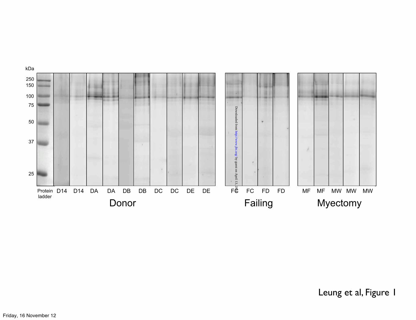
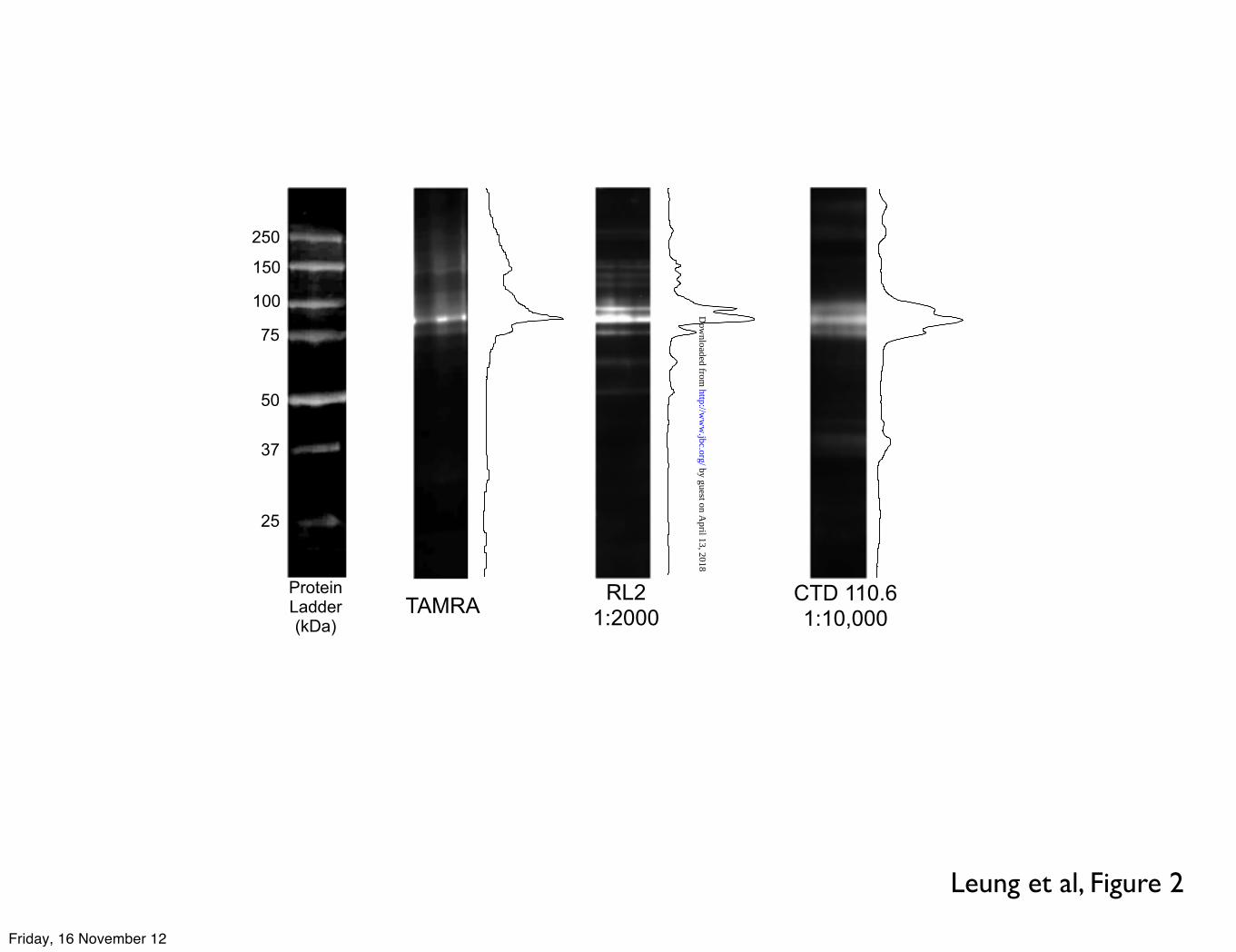
Results using the highly specific O-GlcNAc enzymatic labelling of myofibrils from donor, failing and myectomy human heart muscle are presented in Figure 1. In contrast to previous studies on rodent heart myofibrils, all the samples showed a predominant O-GlcNAc-modified protein at 90 kDa. Two O-GlcNAc specific antibodies, CTD110.6 and RL2 were also used to detect O-GlcNAcylation in human myofibrils. Both antibodies labelled a predominant group of bands in the 90 kDa region and the 90 kDa band was the only labelled band common to all three detection methods, indicating that it is highly likely to be O-GlcNAc-modified rather than non-specifically labelled (Figure 2).
Identification of the 90 kDa band as ZASP
O-GlcNAcylated protein in human heart myofibrillar fractions was enzymatically labelled with TAMRA and separated by SDS-PAGE. The TAMRA-labelled 90 kDa protein band was excised
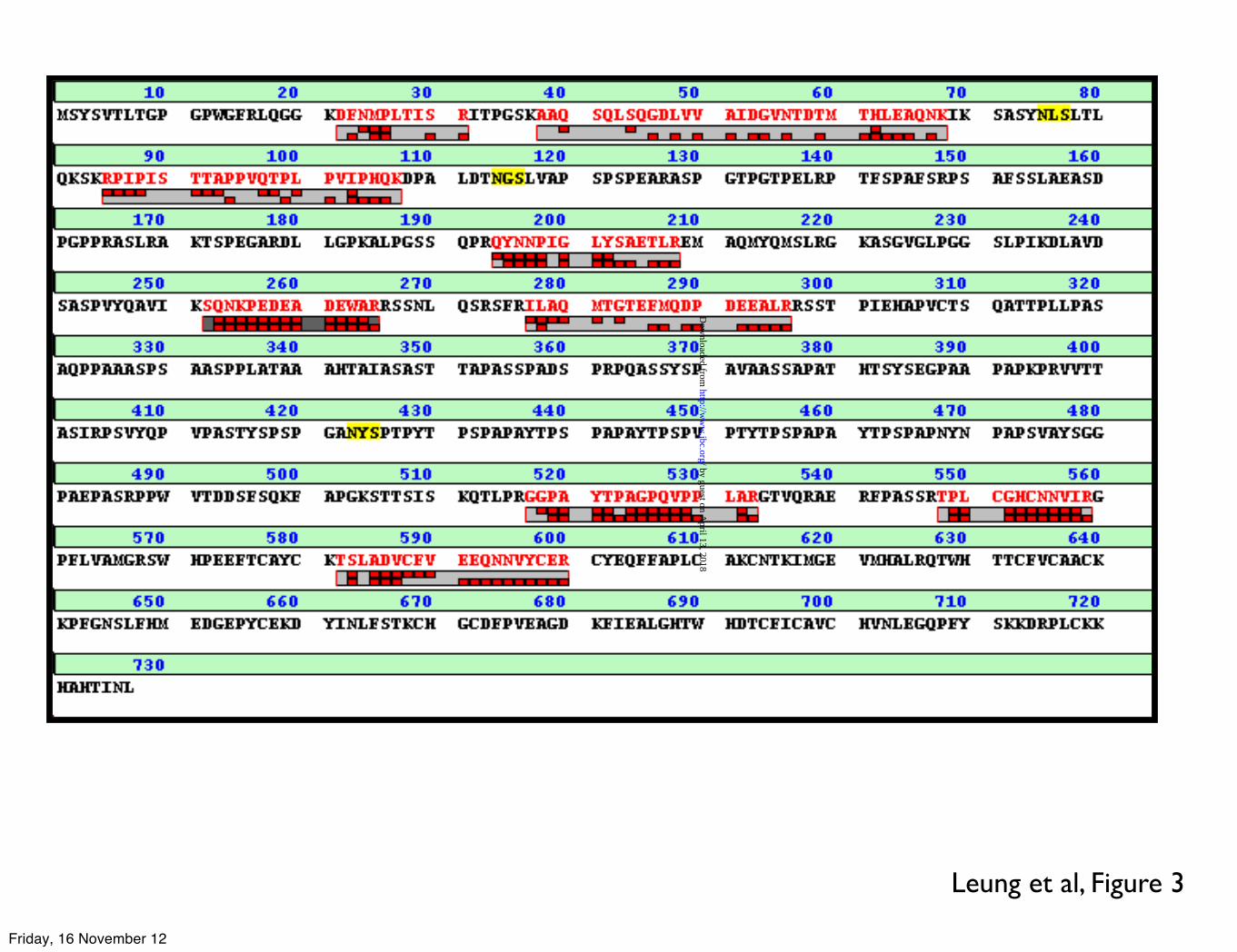
for identification by MALDI-TOF/TOF mass spectrometry. Two separate analyses were performed using different donor heart samples. The possible matches with proteins in the Swiss-Prot human protein database are shown in the data supplement, table 2. The list includes most of the components of the sarcomere, possibly due to smearing in the gel because of their abundance. Both analyses indicated the presence of the LIM domain-binding protein 3 (LDB3 gene; commonly known as Z-band alternatively spliced PDZ-motif protein: ZASP) in the gel band with 2 and 9 unique peptides respectively (Figure 3). Only ZASP, the similar Z-disc protein, enigma homologue (ENH), and α-actinin have molecular weights close to 90 kDa in the lists of proteins identified.
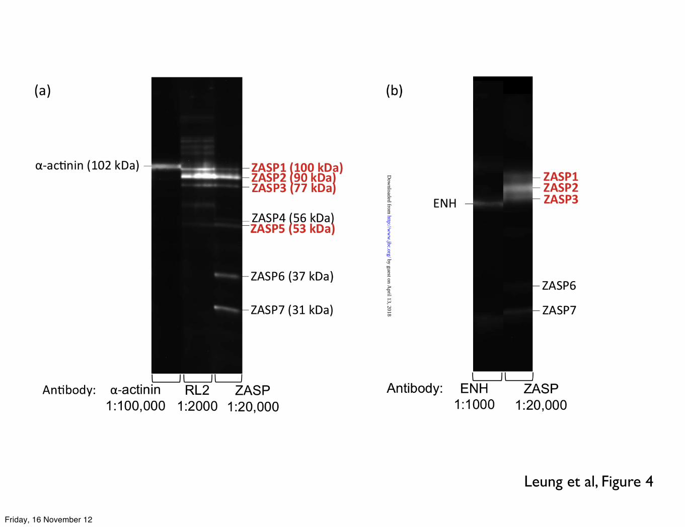
We confirmed that the 90 kDa protein band is ZASP by Western blotting with specific antibodies (Figure 4). The ZASP antibody detected 7 bands, of which the ZASP1, 2, 3 and 5 bands were indicated to be O-GlcNAcylated by the RL2 antibody (Figure 4a). α-actinin was excluded since the α-actinin antibody consistently detected a band with higher molecular mass than any O-GlcNAc or ZASP bands. The possibility of the bands assigned to ZASP being ENH, was eliminated by Western blotting with antibody to ENH (Figure 4b).
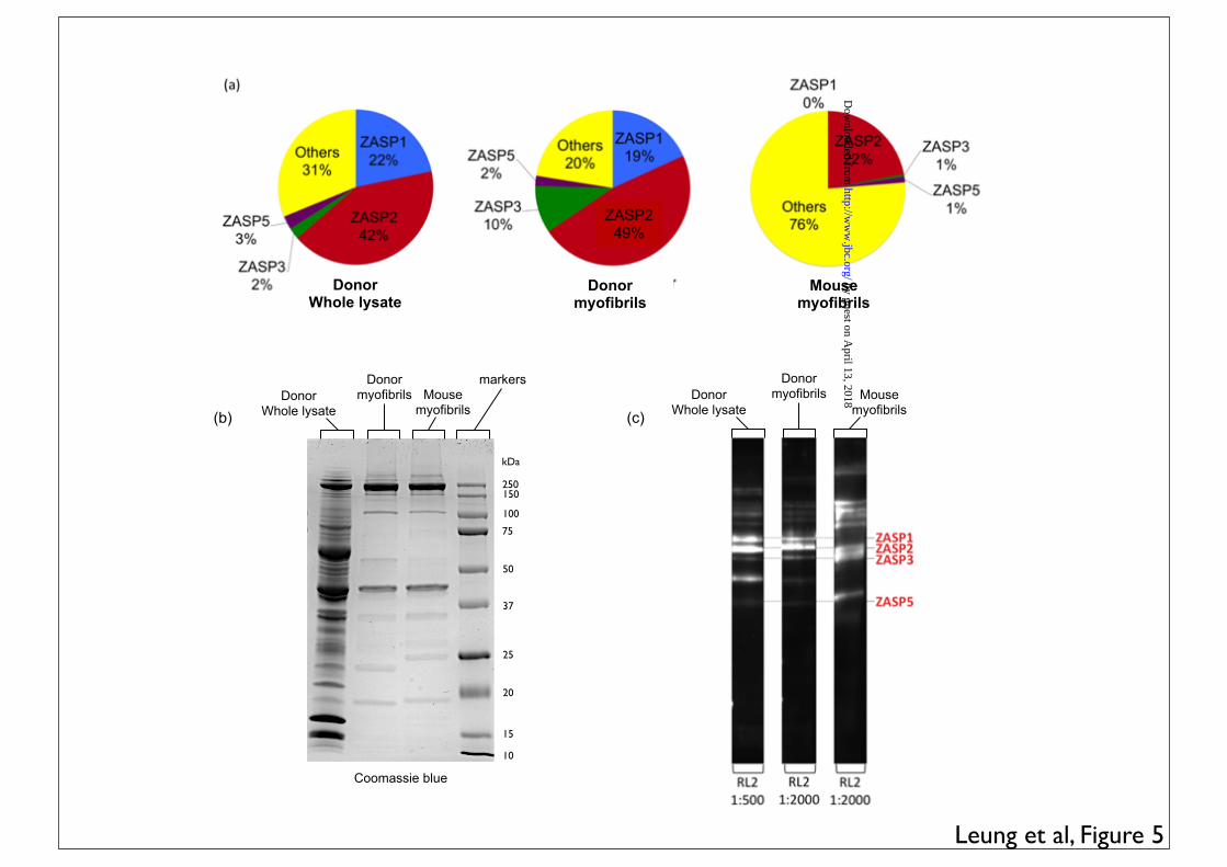
Human cardiac tissue whole lysate gave a similar profile to the human myofibrils when Western blotted with the RL2 anti-O-GlcNAc antibody with 69% of labelling in ZASP bands 1, 2, 3 and 5. However, mouse myofibrils were different from human myofibrils with only 24% of O-GlcNAc detected by RL2 in ZASP bands (Figure 5).
Co-localisation of O-GlcNAcylation with ZASP at the Z-disc
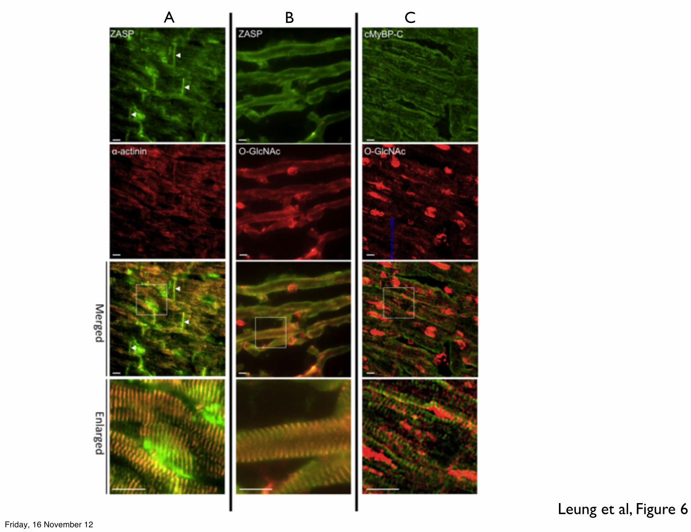
Immunofluorescence microscopy showed that ZASP is present in the Z-disc, co-localising with α-actinin, and also in the intercalated disc (Figure 6, column A). When O-GlcNAc antibody RL2 was used, it showed the presence of O-GlcNAc-modified proteins in the Z-disc, co-localised with ZASP, and also present in the nucleus (column B). Double immunofluorescence probing with O-GlcNAc antibody and cMyBP-C (cardiac myosin binding protein-C) antibody showed that O-GlcNAc modification is not located in the C-zone, where the cMyBP-C is located (Figure 6, column C).
Differences in ZASP and O-GlcNAc Levels Between Health and Diseased Samples
Both the enzymatic labelling and the RL2 antibody showed that the fraction of O-GlcNAc labelling of the ZASP bands was greater in failing heart and
by guest on April 13, 2018
http://ww
w.jbc.org/
Dow
nloaded from
4
myectomy muscle samples (Table 1). This could be due to increased O-GlcNAc labelling of the ZASP or an increased content of ZASP in the Z-disc.
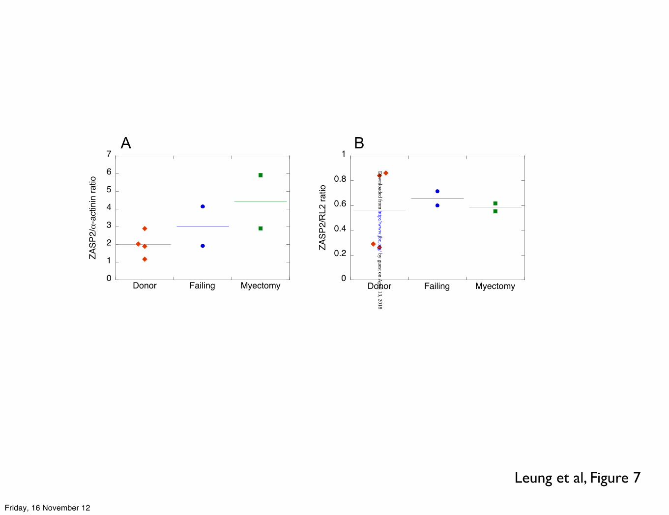
The amount of ZASP2 (probed with antibody to ZASP) relative to α-actinin (probed with EA-53 antibody to α-actinin) present in human myofibrils was measured and compared between samples. From the ZASP2 band/α-actinin band ratio we estimated that the amount of ZASP increased in failing and myectomy samples by approximately two-fold whilst there is no trend for the level of ZASP O-GlcNAcylation (Figure 7).
The natural abundance of ZASP in myofibrils is low and not detectable with Coomassie Blue stain when ~20 µg of myofibril protein was separated on a mini SDS-PAGE gel. Its estimated abundance is less than 1/400th of that of α-actinin (data not shown), thus it is likely that ZASP is highly substituted by O-GlcNAc.
DISCUSSION
Double immunofluorescence of frozen human cardiac tissues demonstrated that the Z-disc is the most O-GlcNAcylated compartment of the sarcomere. Specific enzymatic labelling and probing with two different antibodies to O-GlcNAc showed a 90 kDa protein that we identified as ZASP (Z-band alternatively spliced PDZ-motif protein, LDB3 gene) as the most O-GlcNAcylated human myofibril protein. Significantly, O-GlcNAc transferase, the enzyme that attaches the O-GlcNAc molecule to protein, was also found to localize at the Z-disc (19). Since the quantity of ZASP in the Z-line is estimated to be in the region of 1 per 400 α-actinin molecules, we predict that ZASP is highly modified with O-GlcNAc.
It is remarkable that published studies of O-GlcNAc modifications in heart muscle, based on measurements with rat or mouse heart generally show a large number of bands modified by O-GlcNAc with only a small proportion of O-GlcNAc in bands with apparent Mr in the region of 90 kDa (3,7,8,15,20) and that ZASP has not previously been identified as a highly O-GlcNAc-modifed protein (4,21,22). There are at least three reasons for this: firstly, the widely used CTD110.6 antibody is not wholly specific to O-GlcNAc substituted proteins. CTD110.6 was described to also label N-GlcNAc-modified proteins (23), however, the RL2 antibody and enzymatic labelling methods used here are more specific and in our study the 90 kDa band was identified by all three methods. Secondly, due to its low abundance ZASP may have been excluded in
some types of assays and thirdly, we have shown that the 90 kDa O-GlcNAc band is much less prominent in mouse heart compared with human heart (Figure 5).
The identity of the 90 kDa band as ZASP (also known as cypher in mouse, and LIM domain binding protein, LDB3) was established unambiguously by two independent MALDI-MS experiments and by means of ZASP-specific antibodies. In addition, α-actinin and ENH (enigma homologue, PDLIM5), both Z-disc associated proteins of similar mass to ZASP, were excluded using specific antibodies. α-actinin is larger than ZASP and ENH is smaller (Figure 4). When probed with its specific antibody, seven ZASP bands were observed in human myofibrils including three at around 90 kDa that were detected as the main O-GlcNAcylated bands by RL2 antibody. With RL2, the 90 kDa band (ZASP2) was the most O-GlcNAcylated in all human myofibril and whole lysate samples and ZASP2 was also the only band consistently detected by enzymatic labelling with TAMRA. The ZASP homologue in mouse, cypher, is expressed as 6 isoforms: four long isoforms (723, 679, 661, 622 amino acids) and two short forms (327 and 288 amino acids) that lack the C-terminal LIM domains (24,25); the observed multiple ZASP bands may correspond to these isoforms but could also be due to protein cleavage.
ZASP is classified as a member of the enigma family, with a PDZ domain near its N-terminus and 3 LIM domains near the C-terminus (24,26): see Supp Table 3. It is one of the proteins found in the Z-disc of the sarcomere in both skeletal and cardiac muscles, and binds to other Z-disc proteins such as α-actinin (27), calsarcin and myotilin (28).
ZASP plays an important, but undefined role in development and maintenance of the myofibril. Cypher knockout mice have a lethal phenotype (29) and mutations in the LDB3 gene are associated with cardiomyopathies including dilated cardiomyopathy and left ventricular non-compaction (30-33). ZASP binds to α-actinin via its N-terminal PDZ domain and to other Z-disc proteins to maintain Z-disc structure. It possibly plays a signalling role through its C-terminal LIM domains binding to PKC (10) and is a potential mechanotransducer, in concert with other Z-disc proteins, which respond to mechanosensation (34,35). The LIM domains are only present in the long ZASP isoforms and may be the site of O-GlcNAcylation. Although we do not know yet if ZASP is phosphorylated by PKC in physiological conditions, it is interesting to note, for further investigation, the often-observed ying-yang
by guest on April 13, 2018
http://ww
w.jbc.org/
Dow
nloaded from
5
relationship between phosphorylation and O-GlcNAcylation (2).
Changes in protein O-GlcNAcylation have been associated with pathological conditions in the heart, with increases in global O-GlcNAcylation in human aortic stenosis and in diabetes, myocardial infarction and hypertension in rats (3,19,20,36,37). We observed a corresponding increase in the proportion of total O-GlcNAcylation incorporated into ZASP in end stage failing heart and in myectomy samples from patients with hypertrophic obstructive cardiomyopathy (Table 1). However, this may be a consequence of recruitment of ZASP to the Z-disc rather than an increased level of O-GlcNAcylation.
Since ZASP has both a structural and mechanotransducing role in the Z-disc, our finding that it is modified by the potential signalling adduct O-GlcNAc raises many interesting and important questions to be answered in future studies.
by guest on April 13, 2018
http://ww
w.jbc.org/
Dow
nloaded from
6
REFERENCES
1.# Torres,#C.#R.,#and#Hart,#G.#W.#(1984)#J"Biol"Chem#259,#3308:3317#2.# Hart,#G.#W.,#Slawson,#C.,#Ramirez:Correa,#G.,#and#Lagerlof,#O.#(2011)#Ann"Rev"Biochem#80,#825:
858#3.# Lunde,#I.#G.,#Aronsen,#J.#M.,#Kvaløy,#H.,#Qvigstad,#E.,#Sjaastad,#I.,#Tønnessen,#T.,#Christensen,#G.,#
Grønning:Wang,#L.#M.,#and#Carlson,#C.#R.#(2012)#Physiol"Genomics#44,#162:172#4.# Belke,#D.#D.#(2011)#J"Appl"Physiol#111,#157:162#5.# Hedou,#J.,#Cieniewski:Bernard,#C.,#Leroy,#Y.,#Michalski,#J.:C.,#Mounier,#Y.,#and#Bastide,#B.#(2007)#J"
Biol"Chem#282,#10360:10369#6.# Jones,#S.#P.,#Zachara,#N.#E.,#Ngoh,#G.#A.,#Hill,#B.#G.,#Teshima,#Y.,#Bhatnagar,#A.,#Hart,#G.#W.,#and#
Marban,#E.#(2008)#Circulation#117,#1172:1182#7.# Ramirez:Correa,#G.#A.,#Jin,#W.,#Wang,#Z.,#Zhong,#X.,#Gao,#W.#D.,#Dias,#W.#B.,#Vecoli,#C.,#Hart,#G.#W.,#
and#Murphy,#A.#M.#(2008)#Circ."Res.#103,#1354:1358#8.# Hedou,#J.,#Bastide,#B.,#Page,#A.,#Michalski,#J.:C.,#and#Morelle,#W.#(2009)#Proteomics#9,#2139:2148#9.# Cieniewski:Bernard,#C.,#Bastide,#B.,#Lefebvre,#T.,#Lemoine,#J.,#Mounier,#Y.,#and#Michalski,#J.#C.#
(2004)#Mol"Cell"Proteom#3,#577:585#10.# Sheikh,#F.,#Bang,#M.#L.,#Lange,#S.,#and#Chen,#J.#(2007)#Trends"Cardiovasc"Med#17,#258:262#11.# Jacques,#A.,#Copeland,#O.,#Messer,#A.,#Gallon,#C.,#King,#C.,#McKenna,#W.,#Tsang,#V.,#and#Marston,#S.#
(2008)#J"Mol"Cell"Cardiol#45,#209:216#12.# Jacques,#A.,#Briceno,#N.,#Messer,#A.,#Gallon,#C.,#Jalizadeh,#S.,#Garcia,#E.,#Kikonda:Kanda,#G.,#
Goddard,#J.,#Harding,#S.,#Watkins,#H.,#Tsang,#V.,#McKenna,#W.,#and#Marston,#S.#(2008)#Cardiovasc"Res#79,#481:491#
13.# Messer,#A.,#Gallon,#C.,#McKenna,#W.,#Elliott,#P.,#Dos#Remedios,#C.,#and#Marston,#S.#(2009)#Proteomics"Clin"Appl#3,#1371:1382#
14.# Messer,#A.#E.,#Jacques,#A.#M.,#and#Marston,#S.#B.#(2007)#J"Mol"Cell"Cardiol#42,#247:259#15.# Fulop,#N.,#Mason,#M.#M.,#Dutta,#K.,#Wang,#P.,#Davidoff,#A.#J.,#Marchase,#R.#B.,#and#Chatham,#J.#C.#
(2007)#Am"J"Physiol"292,#C1370:C1378#16.# Khidekel,#N.,#Arndt,#S.,#Lamarre:Vincent,#N.,#Lippert,#A.,#Poulin:Kerstien,#K.#G.,#Ramakrishnan,#B.,#
Qasba,#P.#K.,#and#Hsieh:Wilson,#L.#C.#(2003)#J"Am"Chem"Soc#125,#16162:16163#17.# Khidekel,#N.,#Ficarro,#S.#B.,#Clark,#P.#M.,#Bryan,#M.#C.,#Swaney,#D.#L.,#Rexach,#J.#E.,#Sun,#Y.#E.,#Coon,#J.#
J.,#Peters,#E.#C.,#and#Hsieh:Wilson,#L.#C.#(2007)#Nature"Chemical"Biology#3,#339:348#18.# Bardswell,#S.#C.,#Cuello,#F.,#Rowland,#A.#J.,#Sadayappan,#S.,#Robbins,#J.,#Gautel,#M.,#Walker,#J.#W.,#
Kentish,#J.#C.,#and#Avkiran,#M.#(2009)#J"Biol"Chem#285,#5674:5682#19.# Ramirez,#G.,#Slawson,#C.,#Zeidan,#Q.,#Ding,#W.,#Shen,#X.,#Gao,#W.#D.,#Caceres,#V.,#Paolocci,#N.,#Hart,#
G.#W.,#and#Murphy,#A.#M.#(2012)#Circulation#124,#A16695#20.# Watson,#L.#J.,#Facundo,#H.#T.,#Ngoh,#G.#A.,#Ameen,#M.,#Brainard,#R.#E.,#Lemma,#K.#M.,#Long,#B.#W.,#
Prabhu,#S.#D.,#Xuan,#Y.:T.,#and#Jones,#S.#P.#(2010)#Proc"Natl"Acad"Sci"USA#107,#17797:17802#21.# Wang,#Z.,#Pandey,#A.,#and#Hart,#G.#W.#(2007)#Mol"Cell"Proteomics#6,#1365:1379#22.# Wang,#Z.,#Udeshi,#N.,#O'Malley,#M.,#Shabanowitz,#J.,#Hunt,#D.,#and#Hart,#G.#(2009)#Mol"Cell"
Proteomics##9,,153:160#23.# Isono,#T.#(2011)#PloS"one#6,#e18959#24.# Zheng,#M.,#Cheng,#H.,#Banerjee,#I.,#and#Chen,#J.#(2010)#J"Mol"Cell"Biol#2,#96:102#25.# Huang,#C.,#Zhou,#Q.,#Liang,#P.,#Hollander,#M.#S.,#Sheikh,#F.,#Li,#X.,#Greaser,#M.,#Shelton,#G.#D.,#Evans,#
S.,#and#Chen,#J.#(2003)#J"Biol"Chem#278,#7360:7365#26.# Au,#Y.#H.,#Atkinson,#R.#A.,#Guerrini,#R.,#Kelly,#G.,#Joseph,#C.,#Martin,#S.#R.,#Muskett,#F.#W.,#Pallavicini,#
A.,#Faulkner,#G.,#and#Pastore,#A.#(2004)#Structure#12,#611:622#27.# Faulkner,#G.,#Pallavicini,#A.,#Formentin,#E.,#Comelli,#A.,#Ievolella,#C.,#Trevisan,#S.,#Bortoletto,#G.,#
Scannapieco,#P.,#Salamon,#M.,#Mouly,#V.,#Valle,#G.,#and#Lanfranchi,#G.#(1999)#J"Cell"Biol#146,#465:475#
28.# von#Nandelstadh,#P.,#Ismail,#M.,#Gardin,#C.,#Suila,#H.,#Zara,#I.,#Belgrano,#A.,#Valle,#G.,#Carpen,#O.,#and#Faulkner,#G.#(2009)#Mol"Cell"Biol#29,#822:834#
29.# Zhou,#Q.,#Chu,#P.#H.,#Huang,#C.,#Cheng,#C.#F.,#Martone,#M.#E.,#Knoll,#G.,#Shelton,#G.#D.,#Evans,#S.,#and#Chen,#J.#(2001)#J"Cell"Biol#155,#605:612#
by guest on April 13, 2018
http://ww
w.jbc.org/
Dow
nloaded from

7
30.# Xing,#Y.,#Ichida,#F.,#Matsuoka,#T.,#Isobe,#T.,#Ikemoto,#Y.,#Higaki,#T.,#Tsuji,#T.,#Haneda,#N.,#Kuwabara,#A.,#Chen,#R.,#Futatani,#T.,#Tsubata,#S.,#Watanabe,#S.,#Watanabe,#K.,#Hirono,#K.,#Uese,#K.,#Miyawaki,#T.,#Bowles,#K.#R.,#Bowles,#N.#E.,#and#Towbin,#J.#A.#(2006)#Mol"Genet"Metab#88,#71:77#
31.# Arimura,#T.,#Inagaki,#N.,#Hayashi,#T.,#Shichi,#D.,#Sato,#A.,#Hinohara,#K.,#Vatta,#M.,#Towbin,#J.#A.,#Chikamori,#T.,#Yamashina,#A.,#and#Kimura,#A.#(2009)#Cardiovasc"Res#83,#80:88#
32.# Theis,#J.#L.,#Bos,#J.#M.,#Bartleson,#V.#B.,#Will,#M.#L.,#Binder,#J.,#Vatta,#M.,#Towbin,#J.#A.,#Gersh,#B.#J.,#Ommen,#S.#R.,#and#Ackerman,#M.#J.#(2006)#Biochem"Biophys"Res"Commun#351,#896:902#
33.# Vatta,#M.,#Mohapatra,#B.,#Jimenez,#S.,#Sanchez,#X.,#Faulkner,#G.,#Perles,#Z.,#Sinagra,#G.,#Lin,#J.:H.,#Vu,#T.#M.,#Zhou,#Q.,#Bowles,#K.#R.,#Di#Lenarda,#A.,#Schimmenti,#L.,#Fox,#M.,#Chrisco,#M.#A.,#Murphy,#R.#T.,#McKenna,#W.,#Elliott,#P.,#Bowles,#N.#E.,#Chen,#J.,#Valle,#G.,#and#Towbin,#J.#A.#(2003)#J"Am"Coll"Cardiol#42,#2014:2027#
34.# Knoell,#R.,#Hoshijima,#M.,#and#Chien,#K.#(2003)#J"Mol"Med#81,#750:756#35.# Buyandelger,#B.,#Ng,#K.:E.,#Miocis,#S.,#Gunkel,#S.,#Piotrowska,#I.,#Ku,#C.:H.,#and#Knoell,#R.#(2011)#J"
Cardiovasc"Trans"Res#4,#238:244#36.# Brainard,#R.,#and#Jones,#S.#P.#(2011)#Circulation#124,#A17366#37.# Ramirez:Correa,#G.,#Slawson,#C.,#Wei,#D.,#Hart,#G.,#and#Murphy,#A.#(2011)#Biophys"J#100,#451a#38.# Marston,#S.,#Copeland,#O.,#Jacques,#A.,#Livesey,#K.,#Tsang,#V.,#McKenna,#W.#J.,#Jalilzadeh,#S.,#
Carballo,#S.,#Redwood,#C.,#and#Watkins,#H.#(2009)#Circ"Res#105,#219:222#39.# Copeland,#O.,#Sadayappan,#S.,#Messer,#A.#E.,#Stienen,#G.#J.,#Velden,#J.,#and#Marston,#S.#B.#(2010)#J"
Mol"Cell"Cardiol#49,#1003:1011#40.# Bayliss,#C.#R.,#Jacques,#A.#M.,#Leung,#M.:C.,#Ward,#D.#G.,#Redwood,#C.#S.,#Gallon,#C.#E.,#Copeland,#O.#
a.#n.,#Mckenna,#W.#J.,#Dos#Remedios,#C.,#Marston,#S.#B.,#and#Messer,#A.#E.#(2012)#Cardiovasc"Res#DOI#10.1093/cvr/cvs322#
by guest on April 13, 2018
http://ww
w.jbc.org/
Dow
nloaded from
8
Acknowledgments–#We are grateful to Prof. Cristobal dos Remedios, University of Sydney for human heart muscle samples and to Prof William J. McKenna at the Heart Hospital, London for the HOCM septal myectomy tissue. We thank Prof. Gerald W. Hart, Johns Hopkins University, Baltimore, Maryland for the gift of CTD110.6 antibody. We also wish to thank Dr. Sakthivel Sadayappan, Loyola University Chicago, Illinois for the gift of the cMyBP-C antibody. We thank Mr. O’Neal Copeland for technical assistance. FOOTNOTES *This work was supported by grants from the British Heart Foundation and the European Community's Seventh Framework Programme (2007-2013) Grant Agreement N. 241577 (BIG-Heart). To whom correspondence may be addressed: Steven Marston, Myocardial Function, Imperial Centre for Translational and Experimental Medicine, Du Cane Road, London W12 0NN, UK, Tel.: (+44) 207 594 2732; E-mail: [email protected] The abbreviations used are: O-GlcNAc, O-linked-β-N-acetylglucosamine; ZASP, Z-band alternatively spliced PDZ-motif protein, LDB3 gene; ENH, enigma homolology protein, PDLIM5 gene; MHC, myosin heavy chain; TnI, troponin I; MLC, myosin light chain, HOCM, hypertrophic obstructive cardiomyopathy; TAMRA, Tetramethylrhodamine; cMyBP-C, cardiac myosin binding protein-C;
by guest on April 13, 2018
http://ww
w.jbc.org/
Dow
nloaded from
9
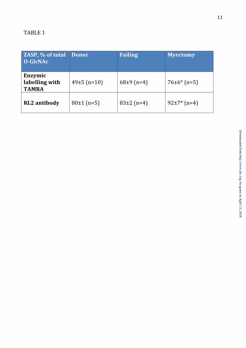
LEGENDS: Table 1. ZASP O-GlcNAcylation (sum of ZASP 1, 2, 3, 4 and 5 as identified in figure 4) expressed as a percentage of total O-GlcNAcylation in donor, failing and myectomy samples. O-GlcNAcylated band volume was measured by densitometry of SDS-PAGE gels probed by enzymatic labelling with TAMRA as shown in Figure 1 and 2 or Western blotting with RL2 antibody, as shown in Figures 2 and 4. The figures are the values of the volume of the ZASP bands as a percentage of the volume of all bands in the same lane averaged for 4-10 lanes. * indicates significant difference (p<0.05, students t test) between O-GlcNAc percentage in donor and myectomy samples. Figure 1. SDS-PAGE gels of myofibrillar proteins (4µg) following specific enzymatic labelling of O-GlcNAc visualised by TAMRA fluorescence. Inverted images are shown here. Duplicate assays of myofibrils from 5 donor hearts (D), 2 failing hearts (F) and 2 myectomy samples (M). Numbers and letters identify the samples that have been described in previous studies (12-14,38-40) Figure 2. Comparison of detection of O-GlcNAc in donor heart myofibrils (4µg) separated by SDS-PAGE and visualised by enzymatic labelling (TAMRA fluorescence) and Western blots using RL2 and CTD110.6 primary antibodies (detected by chemiluminescence). Original gel and corresponding densitometer traces are shown. Bands in the 90 kDa region predominated with all three detection methods. Figure 3. Location of peptides in the ZASP 1 amino acid sequence (77.1 kDa) identified in excised “90 kDa” band from TAMRA- labelled myofibrils separated on SDS-PAGE, as in Figure 1, determined by MALDI-TOF/TOF analyses of trypsin digests (Bruker Ultraflextreme instrument). Peptide identification was performed using the Mascot search engine, specifying human taxonomy for search in the Swiss-Prot database (versions 83.1). Results of the second analysis are shown. Coverage was 22.1% and 9 unique peptides were identified. The first analysis (AB Scitex 4800 instrument) identified the two unique peptides 252K.SQNKPEDEADEWAR.R265 and 194R.QYNNPIGLYSAETLR.E208 Figure 4 Western blots of donor heart myofibrils (4µg) separated by SDS-PAGE. The same gel samples was probed (a) with antibodies specific to O-GlcNAc (RL2), ZASP and α � actinin: ZASP identified as the major O-
GlcNAcylated protein and α � actinin excluded. (b) with antibodies specific to ENH and ZASP showing specificity of the ZASP antibody and excluding
ENH as a major O-GlcNAcylated protein. Figure 5. Patterns of O-GlcNAcylation in whole human heart lysate, myofibrillar fraction and mouse myofibrils (a) Pie charts showing the distribution of O-GlcNAcylated proteins in donor whole lysate and the myofibrillar fraction of donor heart and mouse myofibrils. Individual O-GlcNAcylated proteins were detected in western blots with RL2 antibody (see Figure 2) and expressed as a fraction of total O-GlcNAcylated protein. (b) Total protein in donor whole lysate, donor myofibrils and mouse myofibrils separated by SDS-PAGE, Coomassie Blue staining. (c) O-GlcNAcylated protein in donor whole lysate, donor myofibrils and mouse myofibrils separated by SDS-PAGE. Western blots probed with anti-O-GlcNAc antibody RL2. Figure 6. Indirect double immunofluorescence microscopy study showing localization of A ZASP and �-actinin
by guest on April 13, 2018
http://ww
w.jbc.org/
Dow
nloaded from
10
B ZASP and O-GlcNAc C MyBP-C and O-GlcNAc Cryosections thickness=5 µm. Scale bars=10 µm. White arrowheads indicate intercalated discs Figure 7 Dot-plots showing ratios of ECL signals from Western blots of SDS-PAGE separation of donor myofibrils probed with A anti-ZASP (ZASP2 band) and �-actinin antibody. Increased ratio indicates increased ZASP in the myofibrils. B anti-ZASP (ZASP2 band ) and anti-O-GlcNAc (RL2 antibody). Increased ratio indicates decreased O-GlcNAc content of ZASP.
by guest on April 13, 2018
http://ww
w.jbc.org/
Dow
nloaded from
11
TABLE#1#
#
ZASP,,%,of,total,O8GlcNAc,
Donor, Failing, Myectomy,
Enzymic,labelling,with,TAMRA,
#49±5#(n=10)#
#68±9#(n=4)#
#76±6*#(n=5)#
,RL2,antibody,,
#80±1#(n=5)#
#83±2#(n=4)#
#92±7*#(n=4)#
#
by guest on April 13, 2018
http://ww
w.jbc.org/
Dow
nloaded from
Leung et al, Figure 1
Donor Failing Myectomy
250150
100
75
50
37
25
kDa
Proteinladder
D14 D14 DA DA DB DB DC DC DE DE FC FC FD FD MF MF MW MW MW
Friday, 16 November 12
by guest on April 13, 2018
http://ww
w.jbc.org/
Dow
nloaded from
Rf d
ista
nce
d
ow
n tra
ck
0.0
0.1
0.2
0.3
0.4
0.5
0.6
0.7
0.8
0.9
0
14
0
P
ro
file
h
eig
ht
T
ra
ck 1
Rf d
ista
nce
d
ow
n tra
ck
0.0
0.1
0.2
0.3
0.4
0.5
0.6
0.7
0.8
0.9
0
10
0
50
15
0
20
0
P
ro
file
h
eig
ht
T
ra
ck 1
Rf distance down track
0.0
0.1
0.2
0.3
0.4
0.5
0.6
0.7
0.8
0.9
0
100
50
150
200
Profile height
Track 1
TAMRA RL21:2000
CTD 110.61:10,000
ProteinLadder(kDa)
250
150
100
75
50
37
25
Leung et al, Figure 2
Friday, 16 November 12
by guest on April 13, 2018
http://ww
w.jbc.org/
Dow
nloaded from
Supp. Table 3
The peptides detected by the second analysis are shown in the full human LBD3 amino acid sequence.
Supp Table 4 Characterization of Enigma protein family in vertebrates, modified from (2)
Leung et al, Figure 3
Friday, 16 November 12
by guest on April 13, 2018
http://ww
w.jbc.org/
Dow
nloaded from
Leung et al, Figure 4
Friday, 16 November 12
by guest on April 13, 2018
http://ww
w.jbc.org/
Dow
nloaded from
Leung et al, Figure 5
250150
100
75
50
37
25
20
15
10
DonorWhole lysate
Donormyofibrils
Mousemyofibrils
Mousemyofibrils
DonorWhole lysate
Donormyofibrils
markersMouse
myofibrilsDonor
Whole lysate
Donormyofibrils
Coomassie blue
(b) (c)
kDa
by guest on April 13, 2018
http://ww
w.jbc.org/
Dow
nloaded from
Leung et al, Figure 5Leung et al, Figure 6
A B C
Friday, 16 November 12
by guest on April 13, 2018
http://ww
w.jbc.org/
Dow
nloaded from
0
1
2
3
4
5
6
7
Donor Failing Myectomy
ZASP
2/α
-act
inin
ratio
0
0.2
0.4
0.6
0.8
1
Donor Failing Myectomy
ZASP
2/R
L2 ra
tio
Leung et al, Figure 7
A B
Friday, 16 November 12
by guest on April 13, 2018
http://ww
w.jbc.org/
Dow
nloaded from
MarstonMan Ching Leung, Paul G. Hitchen, Douglas G. Ward, Andrew E. Messer and Steve B.
-N-acetylglucosamine-substituted protein in human heart myofibrilsβZ-band alternatively spliced PDZ-motif protein (ZASP) is the major O-linked-
published online December 27, 2012J. Biol. Chem.
10.1074/jbc.M112.410316Access the most updated version of this article at doi:
Alerts:
When a correction for this article is posted•
When this article is cited•
to choose from all of JBC's e-mail alertsClick here
Supplemental material:
http://www.jbc.org/content/suppl/2012/12/27/M112.410316.DC1
by guest on April 13, 2018
http://ww
w.jbc.org/
Dow
nloaded from