Genetics and Plant Breeding Characterization and molecular ...
β-Thalassemia: New Therapeutic Modalities, Genetics, Complications, and Quality of Life
Anemia
Guest Editors: Mehran Karimi, Sezaneh Haghpanah, Ali T. Taher, and Maria Domenica Cappellini
β-Thalassemia: New Therapeutic Modalities,Genetics, Complications, and Quality of Life
Anemia
β-Thalassemia: New Therapeutic Modalities,Genetics, Complications, and Quality of Life
Guest Editors: Mehran Karimi, Sezaneh Haghpanah,Ali T. Taher, and Maria Domenica Cappellini

Copyright © 2012 Hindawi Publishing Corporation. All rights reserved.
This is a special issue published in “Anemia.” All articles are open access articles distributed under the Creative Commons AttributionLicense, which permits unrestricted use, distribution, and reproduction in any medium, provided the original work is properly cited.
Editorial Board
Bruno Annibale, ItalyEdward J. Benz, USADuran Canatan, TurkeyFernando Ferreira Costa, BrazilEitan Fibach, IsraelMaria Stella Figueiredo, BrazilAjit C. Gorakshakar, IndiaS. Ha, Hong KongH. Heimpel, Germany
Maureen E. Hoatlin, USAHans Joenje, The NetherlandsGeorge J. Kontoghiorghes, CyprusVichai Laosombat, ThailandJohnson M. Liu, USAMaurizio Longinotti, ItalyDimitris Loukopoulos, GreeceIain C. Macdougall, UK
Aurelio Maggio, ItalyJohn Meletis, GreeceA. Piga, ItalyKanokwan Sanchaisuriya, ThailandDonald S. Silverberg, IsraelMaria Tsironi, GreeceGerard R. Vreugdenhil, The NetherlandsJohn S. Waye, Canada

Contents
β-Thalassemia: New Therapeutic Modalities, Genetics, Complications, and Quality of Life,Mehran Karimi, Sezaneh Haghpanah, Ali T. Taher, and Maria Domenica CappelliniVolume 2012, Article ID 902067, 1 page
Health-Related Quality of Life, Treatment Satisfaction, Adherence and Persistence in β-Thalassemia andMyelodysplastic Syndrome Patients with Iron Overload Receiving Deferasirox: Results from the EPICClinical Trial, John Porter, Donald K. Bowden, Marina Economou, Jacques Troncy, Arnold Ganser,Dany Habr, Nicolas Martin, Adam Gater, Diana Rofail, Linda Abetz-Webb, Helen Lau,and Maria Domenica CappelliniVolume 2012, Article ID 297641, 10 pages
Physiopathology of Bone Modifications in β-Thalassemia, Carlo Perisano, Emanuele Marzetti,Maria Silvia Spinelli, Cinzia Anna Maria Calla, Calogero Graci, and Giulio MaccauroVolume 2012, Article ID 320737, 5 pages
Correlation of Oxidative Stress with Serum Trace Element Levels and Antioxidant Enzyme Status in BetaThalassemia Major Patients: A Review of the Literature, Q. Shazia, Z. H. Mohammad, Taibur Rahman,and Hossain Uddin ShekharVolume 2012, Article ID 270923, 7 pages
Thalassemic DNA-Containing Red Blood Cells Are under Oxidative Stress, Mutaz Dana, Eugenia Prus,and Eitan FibachVolume 2012, Article ID 943974, 5 pages
Intracranial Blood Flow Velocity in Patients with β-Thalassemia Intermedia Using Transcranial DopplerSonography: A Case-Control Study, Nahid Ashjazadeh, Sajad Emami, Peyman Petramfar, Ehsan Yaghoubi,and Mehran KarimiVolume 2012, Article ID 798296, 4 pages
Hindawi Publishing CorporationAnemiaVolume 2012, Article ID 902067, 1 pagedoi:10.1155/2012/902067
Editorial
β-Thalassemia: New Therapeutic Modalities, Genetics,Complications, and Quality of Life
Mehran Karimi,1 Sezaneh Haghpanah,1 Ali T. Taher,2 and Maria Domenica Cappellini3
1 Hematology Research Center, Shiraz University of Medical Sciences, Shiraz, Iran2 American University of Beirut Medical Center, Beirut, Lebanon3 IRCCS Ca’ Granda Foundation Maggiore Policlinico Hospital, University of Milan, Milan, Italy
Correspondence should be addressed to Mehran Karimi, [email protected]
Received 19 June 2012; Accepted 19 June 2012
Copyright © 2012 Mehran Karimi et al. This is an open access article distributed under the Creative Commons AttributionLicense, which permits unrestricted use, distribution, and reproduction in any medium, provided the original work is properlycited.
Beta-thalassemia is considered one of the most commongenetic disorders which mass migration is introducing tocountries worldwide and challenging them with its man-agement. Advanced and improved medical care has allowedus to prolong the lives of thalassemia patients, simultane-ously uncovering novel complications and outlining newchallenges. We herein present in this special issue of Anemiasome of the most cutting-edge research outcomes pertainingto the latest complications and the novel treatment strategiesin iron-chelation therapy. We also study the effects of theseinterventions on the quality of life of patients. It is studies likethese that serve as the basis of evidence-based medicine andguide clinicians through their process of decision making.
One article of this special issue addresses the new methodof using transactional Doppler ultrasonography for measur-ing intracranial blood flow velocity in patients with beta-thalassemia intermedia. This study shows higher blood flowvelocity in these patients compared to the control group,which may point to a higher risk of ischemic events in thefuture.
Another paper discusses that thalassemic DNA-con-taining red blood cells are under oxidative stress, whichinduces externalization of phosphatidylserine. This mech-anism is involved in the removal of these cells from thecirculation by the spleen, similar to that of the removal ofsenescent red blood cells.
This special issue also includes a paper explaining thepathophysiology of bone modifications in beta-thalassemia.Imbalance in mineral turnover resulting in abnormal regula-tion of bone metabolism may be related to hormonal and
genetic factors, iron overload, and iron chelation therapy.These factors and their contribution are addressed.
Moreover, a review article that addresses the mechanismof tissue damage arising from iron overload in patients withβ-thalassemia major is presented. It is the result of oxidativestress from free radical production, altered antioxidantenzymes, and its interaction with other essential trace ele-ment levels.
The last paper presented is a study comparing the qualityof life in patients with β-thalassemia major and myelo-dysplastic syndrome with iron-overload treated either withdeferasirox or deferoxamine injections. The results showthat deferasirox can improve health-related quality of lifetreatment satisfaction and adherence compared to subcuta-neous deferoxamine injection. This issue is crucial and oftenneglected in the long-term treatment of patients with ironoverload.
Mehran KarimiSezaneh Haghpanah
Ali T. TaherMaria Domenica Cappellini
Hindawi Publishing CorporationAnemiaVolume 2012, Article ID 297641, 10 pagesdoi:10.1155/2012/297641
Research Article
Health-Related Quality of Life, Treatment Satisfaction,Adherence and Persistence in β-Thalassemia and MyelodysplasticSyndrome Patients with Iron Overload Receiving Deferasirox:Results from the EPIC Clinical Trial
John Porter,1 Donald K. Bowden,2 Marina Economou,3 Jacques Troncy,4 Arnold Ganser,5
Dany Habr,6 Nicolas Martin,7 Adam Gater,8 Diana Rofail,8 Linda Abetz-Webb,8
Helen Lau,6 and Maria Domenica Cappellini9
1 Department of Haematology, UCL Cancer Institute, University College London, Paul O’Gorman Building, 72 Huntley Street,London WC1E 6BT, UK
2 Monash Medical Centre, Melbourne, VIC 3168, Australia3 Thalassemia Clinical Care Services Unit, Hippokration General Hospital Thessaloniki, Egnatia Street 106, 54622 Thessaloniki, Greece4 Hematology, Hopital Edouard Herriot, 6 Rue Antoine Lumiere, 69008 Lyon, France5 Medizinische Hochschule Hannover (MHH), Department of Hematology, Hemostasis, Oncology and Stem Cell Transplantation,Carl-Neuberg Strasse 1, 30625 Hannover, Germany
6 Novartis Pharmaceutical Corporation, 180 Park Avenue, 105-3E065, Florham Park, NJ 07932-1080, USA7 Novartis Pharma AG Postfach, 4002 Basel, Switzerland8 Adelphi Values, Adelphi Mill, Grimshaw Lane, Bollington, Cheshire SK10 5JB, UK9 Universita di Milano, Can Granda Foundation IRCCS, Via F. Sforza 35, 20122 Milan, Italy
Correspondence should be addressed to John Porter, [email protected]
Received 20 January 2012; Revised 1 May 2012; Accepted 19 May 2012
Academic Editor: Sezaneh Haghpanah
Copyright © 2012 John Porter et al. This is an open access article distributed under the Creative Commons Attribution License,which permits unrestricted use, distribution, and reproduction in any medium, provided the original work is properly cited.
Treatment of iron overload using deferoxamine (DFO) is associated with significant deficits in patients’ health-related quality oflife (HRQOL) and low treatment satisfaction. The current article presents patient-reported HRQOL, satisfaction, adherence, andpersistence data from β-thalassemia (n = 274) and myelodysplastic syndrome (MDS) patients (n = 168) patients participatingin the Evaluation of Patients’ Iron Chelation with Exjade (EPIC) study (NCT00171821); a large-scale 1-year, phase IIIb studyinvestigating the efficacy and safety of the once-daily oral iron chelator, deferasirox. HRQOL and satisfaction, adherence, andpersistence to iron chelation therapy (ICT) data were collected at baseline and end of study using the Medical Outcomes Short-Form 36-item Health Survey (SF-36v2) and the Satisfaction with ICT Questionnaire (SICT). Compared to age-matched norms,β-thalassemia and MDS patients reported lower SF-36 domain scores at baseline. Low levels of treatment satisfaction, adherence,and persistence were also observed. HRQOL improved following treatment with deferasirox, particularly among β-thalassemiapatients. Furthermore, patients reported high levels of satisfaction with deferasirox at end of study and greater ICT adherence,and persistence. Findings suggest deferasirox improves HRQOL, treatment satisfaction, adherence, and persistence with ICT inβ-thalassemia and MDS patients. Improving such outcomes is an important long-term goal for patients with iron overload.
1. Introduction
Regular blood transfusions are essential for the manage-ment of haematological conditions such as β-thalassemiamajor and myelodysplastic syndromes (MDS). As a result,
however, patients with these conditions are susceptible tothe development of transfusion-dependent iron overload(hemosiderosis or secondary iron overload). In the absenceof a naturally occurring physiological mechanism for theremoval of excess iron in the body, life-long treatment and
2 Anemia
adherence to iron chelation therapy (ICT) are necessary toprevent the morbidity and mortality that may result if excessiron is allowed to accumulate [1, 2].
Deferoxamine (DFO), most commonly delivered by con-tinuous subcutaneous infusion over 8 to 12 hours a day,is the oldest available form of ICT used by patients withtransfusion-dependent disorders. Prior research, albeit insmall sample sizes, has indicated significant deficits in health-related quality of life (HRQOL) among patients receivingDFO for the treatment of transfusion-dependent iron over-load, compared to values from age-matched normative pop-ulations [3, 4]. In particular, the time-consuming nature ofDFO regimens and side effects associated with this formof ICT (including local site reactions) [5–7] can have adetrimental impact on numerous facets of patients’ lives,including work; social activities; sex life; sleep; emotionalwell-being [8]. As a result, patient satisfaction with DFOtreatment regimens is low and suboptimal adherence iscommon among patients [3, 4]. Improvements in ICTadministration convenience and tolerability are expected toimprove patient’s satisfaction with ICT and HRQOL, thuspromoting adherence to ICT regimens and potentially reduc-ing iron overload-related morbidity/mortality and associatedhealthcare costs [1, 9, 10].
Deferasirox (Exjade) is an oral ICT first approved in2005 and is the most widely prescribed ICT today [11].Deferasirox has been shown to be an efficacious and generallywell-tolerated therapy for the treatment of iron overload inβ-thalassemia and MDS patients [12, 13]. Findings fromrandomised control trials comparing outcomes in patientswith iron overload treated using either deferasirox or DFOhave also suggested the superiority of deferasirox in termsof treatment satisfaction and adherence [14, 15]. How-ever, additional research using validated patient-reportedoutcome (PRO) measures is needed in order to betterunderstand the added benefits of deferasirox over DFO interms of reducing HRQOL burden and improving treatmentsatisfaction, adherence, and persistence among patients withtransfusion-dependent iron overload. The current workseeks to address these needs by presenting and discussingPRO findings from the Evaluation of Patients’ Iron Chelationwith Exjade (EPIC) study (NCT00171821), a large-scaleprospective study designed to investigate the efficacy andsafety of deferasirox in patients diagnosed with transfusion-dependent iron overload [12, 13].
2. Methods
2.1. Study Design. The EPIC study was a prospective, 1-year,multicentre, open-label phase IIIb trial conducted by 136investigators across 23 countries [12, 13]. A PRO sub-study within the EPIC trial was conducted to assess self-reported HRQOL and treatment satisfaction, adherence,and persistence in patients with transfusion-dependent ironoverload. Based on the availability of validated questionnairetranslations, the PRO substudy included participating studysites in Australia, Belgium, France, Germany, Greece, Italy,the Netherlands, and the UK. Study findings reported herefocus specifically on data from adult patients (≥16 years of
age) with β-thalassemia and MDS. Based on the inherentdifferences in the underlying disease and patient profiles,study findings for patients with β-thalassemia and MDS willbe reported separately. Of note, however, PRO data were alsocollected from patients with a variety of other transfusion-dependent disorders (including sickle cell disease, aplasticanemia, and other rare anemias) but sample sizes wereconsidered too small (n < 30) for evaluable analysis as sep-arate subgroups.
In accordance with EPIC trial selection criteria, all pa-tients enrolled in this study were required to have trans-fusion-related iron overload, evident by a serum ferritin levelof ≥1000 ng/mL or with LIC > 2 mg Fe/g dw, as determinedby R2-Magnetic Resonance Imaging (MRI) [12, 13]. Patientsunsuitable for participation in a clinical study from a clinicalperspective (e.g., presence of systemic diseases which wouldprevent the patient from undergoing treatment) or a practi-cal perspective (e.g., history of non-compliance to medicalregimens) were excluded from the study. Assessments ofHRQOL and satisfaction, adherence, and persistence to ICTwere collected at baseline and at the end of the study (EOS;week 52 or at time of early study discontinuation), whereappropriate, using the Medical Outcomes Short-Form 36-item Health Survey (SF-36v2) and the Satisfaction withICT Questionnaire (SICT). Questionnaires were providedto patients in the native language of the respective countryin which the patient was enrolled. Both questionnaireshave been linguistically validated for use in the respectivecountries, ensuring the cross-cultural equivalence of thequestionnaires and enabling data collected from differentcountries to be considered in a single-pooled dataset.
2.2. Ethics. The EPIC study was conducted in accordancewith the Declaration of Helsinki; the International Confer-ence on Harmonization (ICH) Tripartite Guidelines forGood Clinical Practice 1996; the Rules Governing Medic-inal Products in the European Community (Directive91/507/EEC); the US 21 Code of Federal Regulations dealingwith clinical studies. Written informed consent was obtainedfrom all patients prior to participation in the PRO substudy.
2.3. PRO Measures
2.3.1. The Medical Outcomes Short-Form 36-Item Health Sur-vey (SF-36v2). The SF-36v2 is a self-administered question-naire comprising 36-items measuring eight dimensions ofgeneral HRQOL: physical functioning (10 items), role limi-tation due to physical health problems (4 items), bodily pain(2 items), general health perceptions (5 items), vitality (4items), social functioning (2 items), role limitations due toemotional problems (3 items), and general mental health (5items). In addition to scores for individual dimensions, twosummary scores assessing physical and mental dimensions ofhealth and well-being can also be calculated: Physical Com-ponent Summary (PCS) score and the Mental ComponentSummary (MCS) score, respectively.
Although specific “tools” for the assessment of HRQOLhave been developed for thalassemia [16], the SF-36 has theadvantage of having been used extensively within clinical
Anemia 3
trials and academic studies, across a wide range of diseaseareas, including β-thalassemia and MDS [3, 17–20]. Thepsychometric validity and reliability of the instrument asa generic measure of health-related functional status andwell-being is well established [21–23]. In this study, the SF-36v2 was collected from patients at both baseline and EOS.All data were handled and scored in accordance with thedeveloper’s instructions: item scores for each dimension werecoded, summed, and transformed to a scale from 0 (worstpossible health state) to 100 (best possible health state),whereby higher values indicate better HRQOL. Domainscores were only calculated if at least half of all itemscomprising a domain were completed by the patient; missingdata was not imputed [22].
2.3.2. Satisfaction with ICT Questionnaire (SICT). The SICTis a questionnaire designed specifically to assess patientsatisfaction with ICT regimens [24]. It comprises 19 itemsassessing four domains of patient satisfaction: perceivedeffectiveness of ICT (PE), burden of ICT (BD), acceptanceof ICT (AC), and side effects of ICT (SE). Patients rate allitems on a response scale from 1 “very dissatisfied” to 5“very satisfied”. Domain scores are calculated as the meanscore across constituent items and a higher score indicatesgreater satisfaction with respect to the questionnaire domain.As with the SF-36v2, domain scores were calculated if at leasthalf of all items comprising a domain were completed by thepatient; no missing data was imputed [24].
In addition, the SICT also includes three individual itemsdesigned to assess adherence to ICT “How often did you fol-low the chelation therapy regimen exactly as recommendedby your doctor?”, ICT persistence “How often did you thinkabout stopping your chelation therapy?”, and difficultiesremembering to take ICT “How often did you have troubleremembering to take your chelation therapy?”. All three itemsare assessed on a 5-point Likert scale from 1 “Always” to5 “Never” and are designed to be interpreted as standaloneitems of the respective concepts.
Previous studies in patients with a variety of transfusion-dependent haematological disorders have provided evidencethat the SICT is a reliable and valid measure of iron overloadpatients’ satisfaction, adherence, and persistence to ICTregimens [24, 25]. All patients participating in the PROsubstudy completed the SICT at EOS. Only patients withprior history of ICT were required to complete the SICT atbaseline; the SICT was not relevant at this timepoint for thosepatients with no prior history of ICT.
2.3.3. Statistical Analyses and Data Interpretation. Descrip-tive statistics for subscale domains and summary componentscores of the SF-36v2 were computed at baseline and EOS. Tohighlight the HRQOL burden associated with iron overload,mean SF-36 domain, and summary scores at baseline, andEOS were compared to published data of patients with β-thalassemia or MDS and age-matched norms derived fromthe UK general population [3, 17, 22, 26]. Confidenceinterval estimates were used to evaluate the significance ofdifferences in observed study means relative to other refer-ence groups. SICT domain scores and responses to questions
regarding patient-reported adherence and persistence withICT therapy utilization were also summarized at baseline andEOS.
Relevant differences in group means between study andother reference populations for SF-36 domain scores (e.g.,disease-specific and UK general population) were evaluatedusing a distribution-based approach for establishing clini-cally meaningful difference. In this regard, differences thatare 0.5 standard deviation (SD) units of a baseline score werecharacterized as clinically meaningful [27–29].
Analysis of questionnaires at baseline and EOS (e.g., week52 or at time of early study discontinuation) was undertakenonly in cases where sample sizes were large enough (n >30) for statistical analyses. Data were presented separatelyfor patients with underlying β-thalassemia and MDS dueto inherent differences in disease populations and patientprofiles.
3. Results
3.1. Demographic and Clinical Characteristics. The demo-graphic and clinical characteristics of β-thalassemia (n =274) and MDS (n = 168) patients evaluated in this PRO sub-study are displayed in Table 1 and are generally similar tothose of the overall β-thalassemia and MDS populationsenrolled in the EPIC trial [12, 13]. As expected, the meanage of patients with MDS was considerably higher thanthe mean age of patients with β-thalassemia. Almost all β-thalassemia patients (n = 270; 98.5%) had a history of priorICT, with 66.4% (n = 184) having previously received DFOmonotherapy and 30.3% (n = 84) having previously receivedDFO and deferiprone. Only 51.8% (n = 87) of MDS patientshad a history of prior ICT, however, with 37.5% (n = 63)of the MDS sample having previously received DFO mono-therapy and 8.3% (n = 14) having previously received DFOand deferiprone. As such, where relevant, differences be-tween MDS patients with a history of ICT and ICT-naıvepatients are highlighted. Note, however, that sample sizes ofevaluable data do not allow for statistical comparison of dif-ferences between these two groups.
3.2. Changes in HRQOL following Treatment with Deferasirox
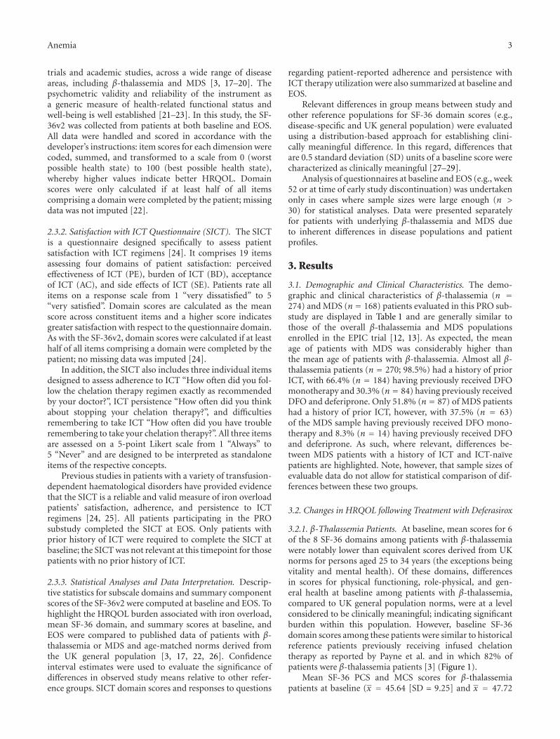
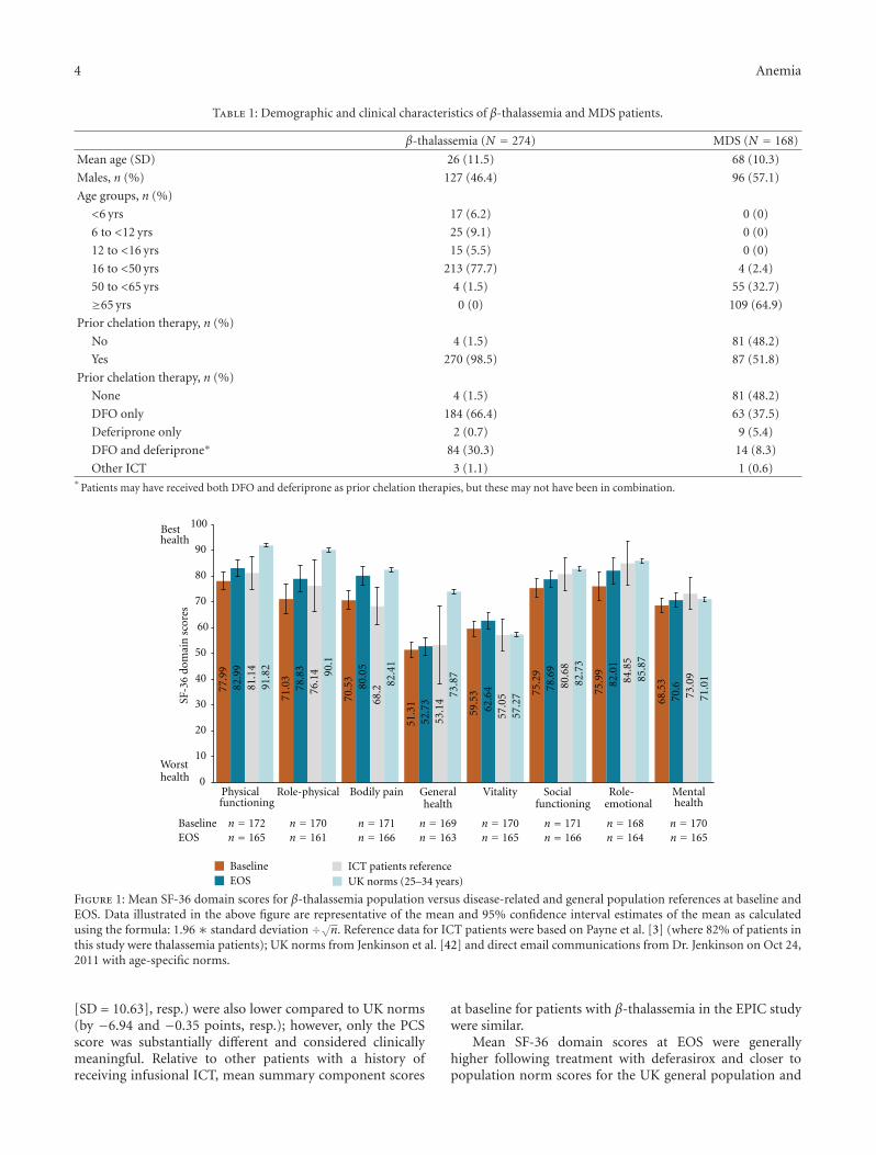
3.2.1. β-Thalassemia Patients. At baseline, mean scores for 6of the 8 SF-36 domains among patients with β-thalassemiawere notably lower than equivalent scores derived from UKnorms for persons aged 25 to 34 years (the exceptions beingvitality and mental health). Of these domains, differencesin scores for physical functioning, role-physical, and gen-eral health at baseline among patients with β-thalassemia,compared to UK general population norms, were at a levelconsidered to be clinically meaningful; indicating significantburden within this population. However, baseline SF-36domain scores among these patients were similar to historicalreference patients previously receiving infused chelationtherapy as reported by Payne et al. and in which 82% ofpatients were β-thalassemia patients [3] (Figure 1).
Mean SF-36 PCS and MCS scores for β-thalassemiapatients at baseline (x = 45.64 [SD = 9.25] and x = 47.72
4 Anemia
Table 1: Demographic and clinical characteristics of β-thalassemia and MDS patients.
β-thalassemia (N = 274) MDS (N = 168)
Mean age (SD) 26 (11.5) 68 (10.3)
Males, n (%) 127 (46.4) 96 (57.1)
Age groups, n (%)
<6 yrs 17 (6.2) 0 (0)
6 to <12 yrs 25 (9.1) 0 (0)
12 to <16 yrs 15 (5.5) 0 (0)
16 to <50 yrs 213 (77.7) 4 (2.4)
50 to <65 yrs 4 (1.5) 55 (32.7)
≥65 yrs 0 (0) 109 (64.9)
Prior chelation therapy, n (%)
No 4 (1.5) 81 (48.2)
Yes 270 (98.5) 87 (51.8)
Prior chelation therapy, n (%)
None 4 (1.5) 81 (48.2)
DFO only 184 (66.4) 63 (37.5)
Deferiprone only 2 (0.7) 9 (5.4)
DFO and deferiprone∗ 84 (30.3) 14 (8.3)
Other ICT 3 (1.1) 1 (0.6)∗
Patients may have received both DFO and deferiprone as prior chelation therapies, but these may not have been in combination.
77.9
9
71.0
3
70.5
3
51.3
1
59.5
3 75.2
9
75.9
9
68.5
382.9
9
78.8
3
80.0
5
52.7
3
62.6
4 78.6
9
82.0
1
70.681
.14
76.1
4
68.2
53.1
4
57.0
5
80.6
8
84.8
5
73.0
9
91.8
2 90.1
82.4
1
73.8
7
57.2
7
82.7
3
85.8
7
71.0
1
0
10
20
30
40
50
60
70
80
90
100
Role-physical Bodily pain Vitality
BaselineEOS
BaselineEOS
ICT patients referenceUK norms (25–34 years)
Besthealth
Worsthealth
SF-3
6 do
mai
n s
core
s
Physicalfunctioning
Socialfunctioning
Mentalhealth
Generalhealth
n = 170n = 165
n = 168n = 164
n = 171n = 166
n = 170n = 165
n = 169n = 163
n = 171n = 166
n = 170n = 161
n = 172n = 165
Role-emotional
Figure 1: Mean SF-36 domain scores for β-thalassemia population versus disease-related and general population references at baseline andEOS. Data illustrated in the above figure are representative of the mean and 95% confidence interval estimates of the mean as calculatedusing the formula: 1.96 ∗ standard deviation ÷√n. Reference data for ICT patients were based on Payne et al. [3] (where 82% of patients inthis study were thalassemia patients); UK norms from Jenkinson et al. [42] and direct email communications from Dr. Jenkinson on Oct 24,2011 with age-specific norms.
[SD = 10.63], resp.) were also lower compared to UK norms(by −6.94 and −0.35 points, resp.); however, only the PCSscore was substantially different and considered clinicallymeaningful. Relative to other patients with a history ofreceiving infusional ICT, mean summary component scores
at baseline for patients with β-thalassemia in the EPIC studywere similar.
Mean SF-36 domain scores at EOS were generallyhigher following treatment with deferasirox and closer topopulation norm scores for the UK general population and
Anemia 5
50.3
4
32.7
2
65.2
8
41.9
9
41.0
9
69.4
6
58.9
6
67.6
1
48.0
7
29.9
5
61.5
9
39.4
8
41.6
2
62.6
7
46.3
0 62.1
5
48.1
39.2
66.6
41.5 50
.6
67
59
72.377
.42
78.1
6
71.5
2
66.2
4
57.7
9
81.0
8
84.1
1
73.8
4
0
10
20
30
40
50
60
70
80
90
100Besthealth
Worsthealth
SF-3
6 do
mai
n s
core
s
Role-physical Bodily pain Vitality Role-emotionalPhysicalfunctioning
Socialfunctioning
Mentalhealth
Generalhealth
BaselineEOS
n = 97n = 75
n = 95n = 74
n = 97n = 74
n = 95n = 71
n = 93n = 72
n = 97n = 74
n = 93n = 72
n = 93n = 72
BaselineEOS
MDS patient referenceUK norms (55–64 years)
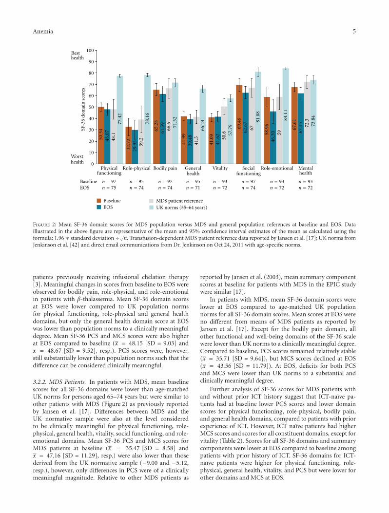
Figure 2: Mean SF-36 domain scores for MDS population versus MDS and general population references at baseline and EOS. Dataillustrated in the above figure are representative of the mean and 95% confidence interval estimates of the mean as calculated using theformula: 1.96∗ standard deviation ÷√n. Transfusion-dependent MDS patient reference data reported by Jansen et al. [17]; UK norms fromJenkinson et al. [42] and direct email communications from Dr. Jenkinson on Oct 24, 2011 with age-specific norms.
patients previously receiving infusional chelation therapy[3]. Meaningful changes in scores from baseline to EOS wereobserved for bodily pain, role-physical, and role-emotionalin patients with β-thalassemia. Mean SF-36 domain scoresat EOS were lower compared to UK population normsfor physical functioning, role-physical and general healthdomains, but only the general health domain score at EOSwas lower than population norms to a clinically meaningfuldegree. Mean SF-36 PCS and MCS scores were also higherat EOS compared to baseline (x = 48.15 [SD = 9.03] andx = 48.67 [SD = 9.52], resp.). PCS scores were, however,still substantially lower than population norms such that thedifference can be considered clinically meaningful.
3.2.2. MDS Patients. In patients with MDS, mean baselinescores for all SF-36 domains were lower than age-matchedUK norms for persons aged 65–74 years but were similar toother patients with MDS (Figure 2) as previously reportedby Jansen et al. [17]. Differences between MDS and theUK normative sample were also at the level consideredto be clinically meaningful for physical functioning, role-physical, general health, vitality, social functioning, and role-emotional domains. Mean SF-36 PCS and MCS scores forMDS patients at baseline (x = 35.47 [SD = 8.58] andx = 47.16 [SD = 11.29], resp.) were also lower than thosederived from the UK normative sample (−9.00 and −5.12,resp.), however, only differences in PCS were of a clinicallymeaningful magnitude. Relative to other MDS patients as
reported by Jansen et al. (2003), mean summary componentscores at baseline for patients with MDS in the EPIC studywere similar [17].
In patients with MDS, mean SF-36 domain scores werelower at EOS compared to age-matched UK populationnorms for all SF-36 domain scores. Mean scores at EOS wereno different from means of MDS patients as reported byJansen et al. [17]. Except for the bodily pain domain, allother functional and well-being domains of the SF-36 scalewere lower than UK norms to a clinically meaningful degree.Compared to baseline, PCS scores remained relatively stable(x = 35.71 [SD = 9.64]), but MCS scores declined at EOS(x = 43.56 [SD = 11.79]). At EOS, deficits for both PCSand MCS were lower than UK norms to a substantial andclinically meaningful degree.
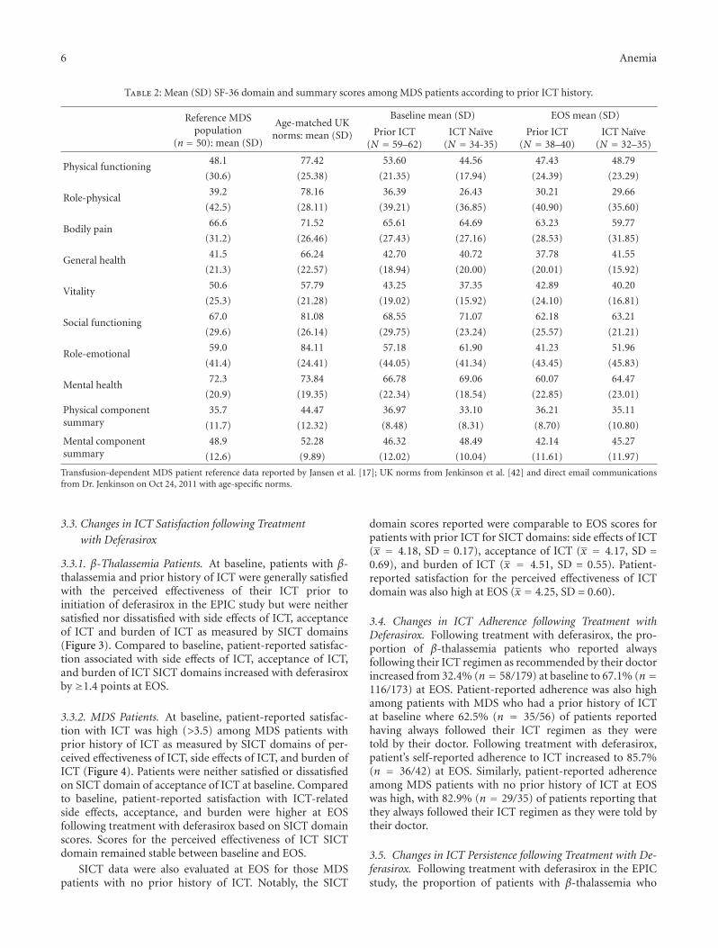
Further analysis of SF-36 scores for MDS patients withand without prior ICT history suggest that ICT-naıve pa-tients had at baseline lower PCS scores and lower domainscores for physical functioning, role-physical, bodily pain,and general health domains, compared to patients with priorexperience of ICT. However, ICT naıve patients had higherMCS scores and scores for all constituent domains, except forvitality (Table 2). Scores for all SF-36 domains and summarycomponents were lower at EOS compared to baseline amongpatients with prior history of ICT. SF-36 domains for ICT-naıve patients were higher for physical functioning, role-physical, general health, vitality, and PCS but were lower forother domains and MCS at EOS.
6 Anemia
Table 2: Mean (SD) SF-36 domain and summary scores among MDS patients according to prior ICT history.
Baseline mean (SD) EOS mean (SD)Reference MDSpopulation
(n = 50): mean (SD)
Age-matched UKnorms: mean (SD) Prior ICT
(N = 59–62)ICT Naıve
(N = 34-35)Prior ICT
(N = 38–40)ICT Naıve
(N = 32–35)
Physical functioning48.1 77.42 53.60 44.56 47.43 48.79
(30.6) (25.38) (21.35) (17.94) (24.39) (23.29)
Role-physical39.2 78.16 36.39 26.43 30.21 29.66
(42.5) (28.11) (39.21) (36.85) (40.90) (35.60)
Bodily pain66.6 71.52 65.61 64.69 63.23 59.77
(31.2) (26.46) (27.43) (27.16) (28.53) (31.85)
General health41.5 66.24 42.70 40.72 37.78 41.55
(21.3) (22.57) (18.94) (20.00) (20.01) (15.92)
Vitality50.6 57.79 43.25 37.35 42.89 40.20
(25.3) (21.28) (19.02) (15.92) (24.10) (16.81)
Social functioning67.0 81.08 68.55 71.07 62.18 63.21
(29.6) (26.14) (29.75) (23.24) (25.57) (21.21)
Role-emotional59.0 84.11 57.18 61.90 41.23 51.96
(41.4) (24.41) (44.05) (41.34) (43.45) (45.83)
Mental health72.3 73.84 66.78 69.06 60.07 64.47
(20.9) (19.35) (22.34) (18.54) (22.85) (23.01)
Physical componentsummary
35.7 44.47 36.97 33.10 36.21 35.11
(11.7) (12.32) (8.48) (8.31) (8.70) (10.80)
Mental componentsummary
48.9 52.28 46.32 48.49 42.14 45.27
(12.6) (9.89) (12.02) (10.04) (11.61) (11.97)
Transfusion-dependent MDS patient reference data reported by Jansen et al. [17]; UK norms from Jenkinson et al. [42] and direct email communicationsfrom Dr. Jenkinson on Oct 24, 2011 with age-specific norms.
3.3. Changes in ICT Satisfaction following Treatment
with Deferasirox
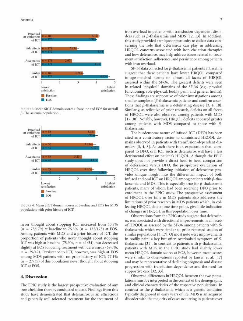
3.3.1. β-Thalassemia Patients. At baseline, patients with β-thalassemia and prior history of ICT were generally satisfiedwith the perceived effectiveness of their ICT prior toinitiation of deferasirox in the EPIC study but were neithersatisfied nor dissatisfied with side effects of ICT, acceptanceof ICT and burden of ICT as measured by SICT domains(Figure 3). Compared to baseline, patient-reported satisfac-tion associated with side effects of ICT, acceptance of ICT,and burden of ICT SICT domains increased with deferasiroxby ≥1.4 points at EOS.
3.3.2. MDS Patients. At baseline, patient-reported satisfac-tion with ICT was high (>3.5) among MDS patients withprior history of ICT as measured by SICT domains of per-ceived effectiveness of ICT, side effects of ICT, and burden ofICT (Figure 4). Patients were neither satisfied or dissatisfiedon SICT domain of acceptance of ICT at baseline. Comparedto baseline, patient-reported satisfaction with ICT-relatedside effects, acceptance, and burden were higher at EOSfollowing treatment with deferasirox based on SICT domainscores. Scores for the perceived effectiveness of ICT SICTdomain remained stable between baseline and EOS.
SICT data were also evaluated at EOS for those MDSpatients with no prior history of ICT. Notably, the SICT
domain scores reported were comparable to EOS scores forpatients with prior ICT for SICT domains: side effects of ICT(x = 4.18, SD = 0.17), acceptance of ICT (x = 4.17, SD =0.69), and burden of ICT (x = 4.51, SD = 0.55). Patient-reported satisfaction for the perceived effectiveness of ICTdomain was also high at EOS (x = 4.25, SD = 0.60).
3.4. Changes in ICT Adherence following Treatment withDeferasirox. Following treatment with deferasirox, the pro-portion of β-thalassemia patients who reported alwaysfollowing their ICT regimen as recommended by their doctorincreased from 32.4% (n = 58/179) at baseline to 67.1% (n =116/173) at EOS. Patient-reported adherence was also highamong patients with MDS who had a prior history of ICTat baseline where 62.5% (n = 35/56) of patients reportedhaving always followed their ICT regimen as they weretold by their doctor. Following treatment with deferasirox,patient’s self-reported adherence to ICT increased to 85.7%(n = 36/42) at EOS. Similarly, patient-reported adherenceamong MDS patients with no prior history of ICT at EOSwas high, with 82.9% (n = 29/35) of patients reporting thatthey always followed their ICT regimen as they were told bytheir doctor.
3.5. Changes in ICT Persistence following Treatment with De-ferasirox. Following treatment with deferasirox in the EPICstudy, the proportion of patients with β-thalassemia who
Anemia 7
4.63
4.27
4.41
3.98
3.26
2.67
2.93
4.12
1 2 3 4 5
Lowest Highest
BaselineEOS
satisfactionsatisfaction
n = 178n = 172
n = 179n = 171
n = 180n = 172
n = 180n = 173
Acceptanceof ICT
Burdenof ICT
Side effectsof ICT
Perceivedeff ectiveness
of ICT
Figure 3: Mean SICT domain scores at baseline and EOS for overallβ-Thalassemia population.
4.56
4.11
4.17
3.95
3.77
3.23
3.83
3.95
1 2 3 4 5
Lowest
Highest
BaselineEOS
satisfactionsatisfaction
n= 55
n = 56
n = 56
n = 56
n = 41
n = 41
n = 42
n = 40Acceptance
of ICT
Burdenof ICT
Side effectsof ICT
Perceivedeff ectiveness
of ICT
Figure 4: Mean SICT domain scores at baseline and EOS for MDSpopulation with prior history of ICT.
never thought about stopping ICT increased from 40.8%(n = 73/179) at baseline to 76.3% (n = 132/173) at EOS.Among patients with MDS and a prior history of ICT, theproportion of patients who never thought about stoppingICT was high at baseline (75.9%, n = 41/54), but decreasedslightly at EOS following treatment with deferasirox (69.0%,n = 29/42). Persistence to ICT, however, was high at EOSamong MDS patients with no prior history of ICT; 77.1%(n = 27/35) of this population never thought about stoppingICT at EOS.
4. Discussion
The EPIC study is the largest prospective evaluation of anyiron chelation therapy conducted to date. Findings from thisstudy have demonstrated that deferasirox is an efficaciousand generally well-tolerated treatment for the treatment of
iron overload in patients with transfusion-dependent disor-ders such as β-thalassemia and MDS [12, 13]. In addition,this study provided a unique opportunity to collect data con-cerning the role that deferasirox can play in addressingHRQOL concerns associated with iron chelation therapiesand how deferasirox may help address issues related to treat-ment satisfaction, adherence, and persistence among patientswith iron overload.
SF-36 data collected for β-thalassemia patients at baselinesuggest that these patients have lower HRQOL comparedto age-matched norms on almost all facets of HRQOLassessed within the SF-36. The greatest deficits were seenin related “physical” domains of the SF-36 (e.g., physicalfunctioning, role-physical, bodily pain, and general health).These findings are supportive of prior investigations amongsmaller samples of β-thalassemia patients and confirm asser-tions that β-thalassemia is a debilitating disease [3, 4, 18].Similarly, as reflective of prior research, deficits on all facetsof HRQOL were also observed among patients with MDS[17, 30]. Notably, however, HRQOL deficits appeared greateramong patients with MDS compared to those with β-thalassemia.
The burdensome nature of infused ICT (DFO) has beencited as a contributory factor to diminished HRQOL do-mains observed in patients with transfusion-dependent dis-orders [3, 4, 8]. As such there is an expectation that, com-pared to DFO, oral ICT such as deferasirox will have a lessdetrimental effect on patient’s HRQOL. Although the EPICstudy does not provide a direct head-to-head comparisonof deferasirox versus DFO, the prospective evaluation ofHRQOL over time following initiation of deferasirox pro-vides unique insight into the differential impact of bothinfused and oral ICT on HRQOL among patients with β-tha-lassemia and MDS. This is especially true for β-thalassemiapatients, many of whom had been receiving DFO prior toenrolment in the EPIC study. The prospective assessmentof HRQOL over time in MDS patients also addresses thelimitations of prior research in MDS patients which, in col-lecting HRQOL data at one time point, give little indicationof changes in HRQOL in this population over time.
Observations from the EPIC study suggest that deferasir-ox was associated with directional improvements in all facetsof HRQOL as assessed by the SF-36 among patients with β-thalassemia which were similar to prior reported studies ofsimilar populations [3, 17]. Of most note were improvementsin bodily pain; a key but often overlooked symptom of β-thalassemia [31]. In contrast to patients with β-thalassemia,patients with MDS in the EPIC study had slightly lowermean HRQOL domain scores at EOS, however, mean scoreswere similar to observations reported by Jansen et al. [17]and may be representative of declining prognosis and diseaseprogression with transfusion dependence and the need forsupportive care [32, 33].
Observed differences in HRQOL between the two popu-lations must be interpreted in the context of the demographicand clinical characteristics of the respective populations. Incontrast to the β-thalassemia which is a genetic conditiontypically diagnosed in early years of life, MDS is an acquireddisorder with the majority of cases occurring in patients over
8 Anemia
the age of 60 [34]; a difference reflected in the substudysamples for the EPIC trial (mean age of 26 versus 68 years,resp.). In addition, modern-day therapy has increased lifeexpectancy for β-thalassemia patients, such that patients cannow live for decades [35, 36]. Past research has also indi-cated that HRQOL in patients with β-thalassemia remainsrelatively stable over time; adding confidence that changesobserved in this study are a result of the study treatment(e.g., switch to deferasirox) as opposed to statistical artifact[37]. By contrast, the prognosis of patients with MDS isgenerally poor due to disease progression, deterioration, andincreasing transfusion dependence [38]. The presence ofcomorbid conditions (e.g., diabetes, coronary heart disease,or chronic pulmonary obstructive disease) which are increas-ingly prevalent among elderly populations also complicatethe management of MDS and contribute to poor risk amongthese patients [39]. As such, HRQOL may be expected todeteriorate among MDS populations over time, independentof the form of ICT that is received by the patient.
The EPIC study provides unique insight into the impactthat deferasirox may have on patients HRQOL over a one-year period. However, the key to minimising long-term mor-bidity and mortality in patients with transfusion-dependentdisorders is ensuring patient’s adherence to recommendedICT regimens. The burdensome nature of infusional ICTregimens or iterative ICT combination regimens may com-plicate compliance and result in suboptimal adherence, andpersistence [3, 4]. This was also evident in the presentstudy, particularly among patients with β-thalassemia whereapproximately 40% of patients at baseline self-reported thatthey always followed their treatment regimen as recom-mended by their doctor (e.g., adherence) and never thoughtabout stopping ICT treatment (e.g., persistence). However,self-reported adherence and persistence among patients withβ-thalassemia increased at EOS following treatment withdeferasirox (67.1% and 76.3%, resp.). Likewise, the propor-tion of MDS patients with prior history of ICT who reportedalways following their ICT regimen as recommended by theirdoctor increased at EOS compared to baseline. Self-reportedpersistence with ICT was slightly lower at EOS compared tobaseline (75.9% and 69.0% never thought about stoppingtheir ICT at baseline and EOS, resp.) among MDS patientswith prior history of ICT which may be associated withunderlying disease progression.
Patients who are less satisfied with prescribed treatmentare expected to be less likely to adhere to recommendedtreatment protocols. Consistent with this, patients with β-thalassemia in the EPIC study demonstrated a higher levelof satisfaction with ICT at EOS following treatment withdeferasirox, particularly in relation to practical aspects of ICTsuch as side-effects, burden, and acceptability of treatmentregimen which coincided with higher self-reported adher-ence and persistence with deferasirox at EOS. In MDS pa-tients with prior history of ICT, notable improvements insatisfaction with ICT were recorded between baseline andEOS and higher self-reported adherence. This observationsuggests that the directional changes in HRQOL in patientswith MDS may be associated with other factors such asprogressive underlying disease, complications of older age,
among others, and the may not be related to study treatment.Furthermore, levels of satisfaction among patients with MDSand no prior history of ICT were also high at EOS, offeringfurther support that the directional changes in aspects ofHRQOL between baseline and EOS may be attributable toother factors.
In reflecting on potential limitations of the present study,the number of MDS patients for whom there was evaluabledata should be considered. The high rate of study discontin-uations among MDS patients participating in the EPIC studyis acknowledged as source of potential bias in these data andis a key factor limiting the availability of evaluable data inthis substudy. As detailed by Gattermann et al. (2010), of341 patients with MDS enrolled, 175 patients completed thestudy (median duration of treatment: 50.6 weeks) whereas166 discontinued (rate: 48.7%) with the primary reason forstudy discontinuation being due to adverse events (n =78) [13]. Nonetheless, when interpreting findings from thecurrent study, it is necessary to consider that HRQOL,treatment satisfaction, adherence, and persistence may benegatively affected in those patients discontinuing treatmentdue to adverse events.
In interpreting the findings of this study, it is also impor-tant to appreciate that the defining features of controlledclinical studies (e.g., study selection criteria, predeterminedassessment schedules, study treatment) may introduce bias tothe evaluation of health outcomes (e.g., HRQOL, treatmentsatisfaction, adherence, persistence, etc.). In particular, theexclusion of participants with a history of poor complianceto medical regimens may have affected ratings of adherence,and persistence at baseline and EOS. As such, further con-sideration of naturalistic studies would help to establish thereal-world validity of observed changes in HRQOL, treat-ment satisfaction, adherence and persistence associated withdeferasirox observed in the EPIC study. A recent retrospec-tive assessment of iron chelation adherence from the Tha-lassemia Clinical Research Network (TRCN) also documentshigh patient-reported adherence with deferasirox [40, 41].With this end in mind, the present study provides furtherevidence of the validity of the SF-36 and SICT as measures ofHRQOL and treatment satisfaction/adherence/persistence,respectively, and supports their use in future studies oftransfusion-dependent conditions and ICTs. Standardisationof PRO assessments in future studies (using the SF-36 andSICT, e.g.) is particularly important for enabling meaningfulcomparisons to be made between varying ICT regimens.
5. Conclusions
This substudy represents the largest prospective evaluation ofpatient-reported outcomes with deferasirox to date. Findingsindicate improvements in patient-reported HRQOL, ICTsatisfaction, adherence, and persistence following treatmentwith deferasirox particularly among β-thalassemia patients,the majority of whom had been using infused ICT prior toenrolment in the study. Patient satisfaction with deferasiroxis high and patients receiving deferasirox report being morelikely to adhere and persist with ICT. Such evaluations are
Anemia 9
vital for improving both the long-term health outcomesand survival of patients with transfusion-dependent ironoverload and minimising future health resource use.
Funding
Novartis commissioned Adelphi Values to provide adviceon patient reported outcome strategies for the clinical trial:NCT00171821.
Acknowledgments
The authors would like to thank all the patients and inves-tigators who took part in the EPIC study.
References
[1] T. E. Delea, J. Edelsberg, O. Sofrygin et al., “Consequences andcosts of noncompliance with iron chelation therapy in patientswith transfusion-dependent thalassemia: a literature review,”Transfusion, vol. 47, no. 10, pp. 1919–1929, 2007.
[2] V. Gabutti and A. Piga, “Results of long-term iron-chelatingtherapy,” Acta Haematologica, vol. 95, no. 1, pp. 26–36, 1996.
[3] K. A. Payne, M. P. Desrosiers, J. J. Caro et al., “Clinical andeconomic burden of infused iron chelation therapy in theUnited States,” Transfusion, vol. 47, no. 10, pp. 1820–1829,2007.
[4] K. A. Payne, D. Rofail, J. F. Baladi et al., “Iron chelation ther-apy: clinical effectiveness, economic burden and quality of lifein patients with iron overload,” Advances in Therapy, vol. 25,no. 8, pp. 725–742, 2008.
[5] V. Alymara, D. Bourantas, A. Chaidos et al., “Effectiveness andsafety of combined iron-chelation therapy with deferoxamineand deferiprone,” Hematology Journal, vol. 5, no. 6, pp. 475–479, 2004.
[6] P. J. Giardina and R. W. Grady, “Chelation therapy in β-tha-lassemia: an optimistic update,” Seminars in Hematology, vol.38, no. 4, pp. 360–366, 2001.
[7] P. Rebulla and The Cooleycare Cooperative Group, “Transfu-sion reactions in thalassemia. A survey from the CooleycareProgramme,” Haematologica, vol. 75, no. 5, pp. 122–127, 1990.
[8] L. Abetz, J. F. Baladi, P. Jones, and D. Rofail, “The impact ofiron overload and its treatment on quality of life: results froma literature review,” Health and Quality of Life Outcomes, vol.4, article 73, 2006.
[9] T. E. Delea, M. Hagiwara, S. K. Thomas, J. F. Baladi, P. D.Phatak, and T. D. Coates, “Outcomes, utilization, and costsamong thalassemia and sickle cell disease patients receivingdeferoxamine therapy in the United States,” American Journalof Hematology, vol. 83, no. 4, pp. 263–270, 2008.
[10] M. Evangeli, K. Mughal, and J. B. Porter, “Which psychosocialfactors are related to chelation adherence in thalassemia asystematic review,” Hemoglobin, vol. 34, no. 3, pp. 305–321,2010.
[11] J. L. Kwiatkowski, “Real-world use of iron chelators,” ASHEducation Book, vol. 2011, pp. 451–458, 2011.
[12] M. D. Cappellini, J. Porter, A. El-Beshlawy et al., “Tailoringiron chelation by iron intake and serum ferritin: the pro-spective EPIC study of deferasirox in 1744 patients with trans-fusion-dependent anemias,” Haematologica, vol. 95, no. 4, pp.557–566, 2010.
[13] N. Gattermann, C. Finelli, M. D. Porta et al., “Deferasiroxin iron-overloaded patients with transfusion-dependent
myelodysplastic syndromes: results from the large 1-year EPICstudy,” Leukemia Research, vol. 34, no. 9, pp. 1143–1150, 2010.
[14] M. D. Cappellini, M. Bejaoui, L. Agaoglu et al., “Prospectiveevaluation of patient-reported outcomes during treatmentwith deferasirox or deferoxamine for iron overload in patientswith β-thalassemia,” Clinical Therapeutics, vol. 29, no. 5, pp.909–917, 2007.
[15] E. Vichinsky, Z. Pakbaz, O. Onyekwere et al., “Patient-reportedoutcomes of deferasirox (Exjade, ICL670) versus deferox-amine in sickle cell disease patients with transfusional hemo-siderosis: substudy of a randomized open-label phase II trial,”Acta Haematologica, vol. 119, no. 3, pp. 133–141, 2008.
[16] S. Ratip, D. Skuse, J. Porter, B. Wonke, A. Yardumian, andB. Modell, “Psychosocial and clinical burden of thalassaemiaintermedia and its implications for prenatal diagnosis,” Ar-chives of Disease in Childhood, vol. 72, no. 5, pp. 408–412, 1995.
[17] A. J. G. Jansen, M. L. Essink-Bot, E. A. M. Beckers, W. C.J. Hop, M. R. Schipperus, and D. J. Van Rhenen, “Qualityof life measurement in patients with transfusion-dependentmyelodysplastic syndromes,” British Journal of Haematology,vol. 121, no. 2, pp. 270–274, 2003.
[18] A. Sobota, R. Yamashita, Y. Xu et al., “Quality of life in tha-lassemia: a comparison of SF-36 results from the thalassemialongitudinal cohort to reported literature and the US norms,”American Journal of Hematology, vol. 86, no. 1, pp. 92–95,2011.
[19] L. Mednick, S. Yu, F. Trachtenberg et al., “Symptoms of depres-sion and anxiety in patients with thalassemia: prevalence andcorrelates in the thalassemia longitudinal cohort,” AmericanJournal of Hematology, vol. 85, no. 10, pp. 802–805, 2010.
[20] L. Scalone, L. G. Mantovani, M. Krol et al., “Costs, qualityof life, treatment satisfaction and compliance in patients withβ-thalassemia major undergoing iron chelation therapy: theITHACA study,” Current Medical Research and Opinion, vol.24, no. 7, pp. 1905–1917, 2008.
[21] J. E. Ware and C. D. Sherbourne, “The MOS 36-item short-form health survey (SF-36). I. Conceptual framework anditem selection,” Medical Care, vol. 30, no. 6, pp. 473–483, 1992.
[22] J. E. Ware, M. Kosinski, and J. E. Dewey, How to Score versiontwo of the SF-36 Health Survey, QualityMetric, Lincoln, RI,USA, 2000.
[23] J. E. Ware, M. Kosinski, and J. B. Bjorner, User’s Manual for theSF-36v2 Health Survey, QualityMetric, Lincoln, RI, USA, 2ndedition, 2007.
[24] D. Rofail, L. Abetz, M. Viala, C. Gait, J. F. Baladi, and K. Payne,“Satisfaction and adherence in patients with iron overloadreceiving iron chelation therapy as assessed by a newly devel-oped patient instrument,” Value in Health, vol. 12, no. 1, pp.109–117, 2009.
[25] D. Rofail, M. Viala, A. Gater, L. Abetz-Webb, J. F. Baladi, andM. D. Cappellini, “An instrument assessing satisfaction withiron chelation therapy: psychometric testing from an open-label clinical trial,” Advances in Therapy, vol. 27, no. 8, pp. 533–546, 2010.
[26] C. Jenkinson, S. Stewart-Brown, and S. Petersen, “Assess-ment and evaluation of the SF36 Version II,” Health Ser-vices Research Unit, University of Oxford, 2006, http://www.hsru.ox.ac.uk/sf36v2.htm.
[27] G. H. Guyatt, D. Osoba, A. W. Wu et al., “Methods to explainthe clinical significance of health status measures,” Mayo ClinicProceedings, vol. 77, no. 4, pp. 371–383, 2002.
[28] J. A. Sloan, D. Cella, and R. D. Hays, “Clinical significanceof patient-reported questionnaire data: another step toward
10 Anemia
consensus,” Journal of Clinical Epidemiology, vol. 58, no. 12,pp. 1217–1219, 2005.
[29] K. W. Wyrwich, W. M. Tierney, and F. D. Wolinsky, “Furtherevidence supporting an SEM-based criterion for identifyingmeaningful intra-individual changes in health-related qualityof life,” Journal of Clinical Epidemiology, vol. 52, no. 9, pp. 861–873, 1999.
[30] D. J. Pinchon, S. J. Stanworth, C. Doree, S. Brunskill, and D.R. Norfolk, “Quality of life and use of red cell transfusionin patients with myelodysplastic syndromes. A systematicreview,” American Journal of Hematology, vol. 84, no. 10, pp.671–677, 2009.
[31] D. Haines, S. Carson, S. Green, M. Martin, T. D. Coates, O. O.Vega et al., “Phenomenon of pain in thalassemia: a prospectiveanalysis by the thalassemia clinical research network (TCRN),”Blood (ASH Annual Meeting Abstracts), vol. 116, article 253,2010.
[32] L. Malcovati, M. G. Della Porta, C. Pascutto et al., “Prognosticfactors and life expectancy in myelodysplastic syndromesclassified according to WHO criteria: a basis for clinicaldecision making,” Journal of Clinical Oncology, vol. 23, no. 30,pp. 7594–7603, 2005.
[33] L. Malcovati, U. Germing, A. Kuendgen et al., “Time-de-pendent prognostic scoring system for predicting survival andleukemic evolution in myelodysplastic syndromes,” Journal ofClinical Oncology, vol. 25, no. 23, pp. 3503–3510, 2007.
[34] X. Ma, M. Does, A. Raza, and S. T. Mayne, “Myelodysplasticsyndromes: incidence and survival in the United States,”Cancer, vol. 109, no. 8, pp. 1536–1542, 2007.
[35] H. A. Pearson, A. R. Cohen, P. J. V. Giardina, and H. H.Kazazian, “The changing profile of homozygous β-thalas-semia: demography, ethnicity, and age distribution of currentNorth American patients and changes in two decades,”Pediatrics, vol. 97, no. 3, pp. 352–356, 1996.
[36] P. Telfer, “Update on survival in thalassemia major,” Hemo-globin, vol. 33, no. 1, pp. S76–S80, 2009.
[37] R. Yamashita, Y. Xu, F. Trachtenberg, P. Kohlbry, D. A. Kleinert,P. Giardina et al., “Changes in health status and quality of lifein adults with thalassemia: year 1 report of the thalassemialongitudinal cohort study,” Blood (ASH Annual Meeting Abs-tracts), vol. 116, article 1533, 2010.
[38] L. Malcovati, M. G. Della Porta, and M. Cazzola, “Predictingsurvival and leukemic evolution in patients with myelodys-plastic syndrome,” Haematologica, vol. 91, no. 12, pp. 1588–1590, 2006.
[39] M. F. Fey and M. Dreyling, “Acute myeloblastic leukaemiasand myelodysplastic syndromes in adult patients: ESMO clin-ical practice guidelines for diagnosis, treatment and follow-up,” Annals of Oncology, vol. 21, supplement 5, pp. v158–v161,2010.
[40] J. L. Kwiatkowski, H. Y. Kim, A. A. Thompson, C. T. Quinn,B. U. Mueller, I. Odame et al., “Chelation use and iron burdenin North American and British thalassemia patients: a reportfrom the thalassemia longitudinal cohort,” Blood, vol. 119, no.12, pp. 2746–2753, 2012.
[41] F. Trachtenberg, E. Vichinsky, D. Haines et al., “Iron chelationadherence to deferoxamine and deferasirox in thalassemia,”American Journal of Hematology, vol. 86, no. 5, pp. 433–436,2011.
[42] C. Jenkinson, S. Stewart-Brown S Petersen, and C. Paice,“Assessment of the SF-36 version 2 in the United Kingdom,”Journal of Epidemiology and Community Health, vol. 53, no. 1,pp. 46–50, 1999.
Hindawi Publishing CorporationAnemiaVolume 2012, Article ID 320737, 5 pagesdoi:10.1155/2012/320737
Review Article
Physiopathology of Bone Modifications in β-Thalassemia
Carlo Perisano,1 Emanuele Marzetti,1 Maria Silvia Spinelli,1 Cinzia Anna Maria Calla,2
Calogero Graci,1 and Giulio Maccauro1
1 Department of Orthopaedics and Traumatology, University Hospital Agostino Gemelli,Catholic University of the Sacred Heart School of Medicine, Largo A. Gemelli 1, 00168 Rome, Italy
2 Department of Biochemistry and Clinical Biochemistry, University Hospital Agostino Gemelli,Catholic University of the Sacred Heart School of Medicine, Largo A. Gemelli 1, 00168 Rome, Italy
Correspondence should be addressed to Carlo Perisano, [email protected]
Received 22 December 2011; Accepted 7 April 2012
Academic Editor: Sezaneh Haghpanah
Copyright © 2012 Carlo Perisano et al. This is an open access article distributed under the Creative Commons Attribution License,which permits unrestricted use, distribution, and reproduction in any medium, provided the original work is properly cited.
β-thalassemia major (βTM) or Cooley anemia is characterized by significantly reduced or absent synthesis of β-globin chains,which induces important pathologic consequences including hemolytic anemia, altered erythropoiesis, and bone marrowoverstimulation. The pathogenesis of bone changes in patients with βTM is not yet completely understood. However, an unbalancein bone mineral turnover resulting from increased resorption and suppression of osteoblast activity has been detected in βTMpatients. The abnormal regulation of bone metabolism may be related to hormonal and genetic factors, iron overload and ironchelation therapy, nutritional deficits, and decreased levels of physical activity. Here, we review the most recent findings on thephysiopathology of bone abnormalities in βTM. Clinical presentation and radiological features of βTM-related bone changes arealso discussed.
1. Introduction
β-thalassemia, firstly described by Cooley and Lee [1],comprises a group of inherited, autosomal, recessive, andhematologic disorders characterized by decreased or absentsynthesis of β-globin chains. The mature hemoglobin (Hb)molecule is a tetramer composed of two α-globin and twoβ-globin chains, along with a heme prosthetic group. β-globin synthesis is controlled by one gene located on eachchromosome 11 [2]. Defects are usually secondary to pointmutations and rarely occur as a consequence of deletions[2]. In β-thalassemia, β-globin chain production can rangefrom near to normal to completely absent, leading to varyingdegrees of excess α-globin chains and disease severity [2]. β-thalassemia trait (minor), resulting from heterozygosity forβ-thalassemia, is clinically asymptomatic and manifests withmicrocytosis and mild anemia. β-thalassemia intermediacomprises a clinically and genotypically heterogeneous groupof disorders, ranging in severity from the asymptomaticcarrier state to severe, transfusion-dependent disease. β-thalassemia major (βTM) or Cooley anemia is characterizedby severely reduced or absent synthesis of β-globin chainsfrom both genes, with symptoms and signs beginning at
about six months of age (abdominal swelling, growth retar-dation, irritability, jaundice, pallor, skeletal abnormalities,and splenomegaly) [2].
In βTM, the defective synthesis of β chains, togetherwith excess α chains, leads to hemolytic anemia, altered ery-thropoiesis, reduced erythrocyte survival, and bone marrowoverstimulation [1–4]. Patients need blood transfusions tocorrect anemia and iron-chelating therapy to control ironoverload [1–4]. Anemia, excess body iron, and iron-chelationtherapy can result in endocrine disorders (e.g., diabetes mel-litus, hypogonadism, hypothyroidism, hypoparathyroidism,hypopituitarism, and Addison’s disease), growth retardation,liver and cardiac failure, and splenomegaly. The latter canworsen anemia and occasionally causes thrombocytopeniaand neutropenia, thereby increasing the risk of infections andhemostatic disorders [2–5]. Heart failure is the leading causeof death in patients with βTM [3, 6].
2. Epidemiology
The worldwide prevalence of α- and β-thalassemia traitis 1.7% [4]. Males and females are equally affected. The
2 Anemia
incidence of thalassemia trait is 4.4 per 10,000 live births [4].β-thalassemia in its various presentations is more commonin the Mediterranean area, Africa, and Southeastern Asia.
3. Pathogenesis of Bone Changesin β-Thalassemia
The pathogenesis of bone changes in βTM patients is notyet completely understood [7]. In spite of the improvedtreatment of the hematologic disorder and its complications,β-thalassemia patients exhibit an unbalance in bone mineralturnover with increased resorptive rates and suppressionof osteoblast activity, resulting in diminished bone mineraldensity (BMD) more evident in the lumbar spine [8,9]. Putative mechanisms involved in the pathogenesis ofbone abnormalities in βTM are discussed in the followingsubsections.
3.1. Impairments in Osteoblast Activity. Mahachoklertwat-tana et al. [7, 10] reported growth retardation and delayedbone age, reduced BMD (especially of the lumbar spine),and low serum IGF-I levels in children and adolescentswith βTM. In these patients, bone histomorphometryrevealed increased osteoid thickness and delayed osteoidmaturation and mineralization, indicating impaired bonematrix maturation and defective mineralization [7]. Inaddition, iron depots were detected along mineralizationfronts and osteoid surfaces, while focal-thickened osteoidseams were found together with iron deposits. Dynamic boneformation studies revealed reduced bone formation rates.These findings indicate that delayed bone maturation andfocal osteomalacia contribute to the pathogenesis of bonedisease in suboptimally blood-transfused βTM patients withiron overload. Iron depots within bones and low circulatingIGF-I levels may partly contribute to skeletal abnormalities[7].
Morabito et al. [11] showed that βTM patients displayedan unbalanced bone turnover, characterized by enhancedresorption rates (indicated by high levels of pyridiniumcross-links) and a decreased neoformation phase (evidencedby low levels of osteocalcin, an osteoblast-derived protein)[11]. Voskaridou and colleagues [12] found increased serumlevels of Dickkopf-1 (Dkk1), a soluble inhibitor of winglesstype (Wnt) signaling, and sclerostin [13], a Wnt inhibitor,specifically expressed by osteocytes, in βTM patients. Highercirculating levels of Dkk1 and sclerostin correlated withreduced bone mineral density of lumbar spine and distalradius as well as with increased bone resorption andreduced bone formation markers. These findings indicatethat disruption of Wnt signaling in patients with thalassemiaand osteoporosis leads to osteoblast deregulation. Therefore,sclerostin and Dkk-1 have been proposed as potentialtargets for treatment in patients with thalassemia-inducedosteoporosis [13].
3.2. Abnormal Osteoclast Activity. Besides impairments inosteoblast activity, which are thought to be a major causeof osteopenia/osteoporosis in βTM, an enhanced activation
of osteoclasts is also invoked as a contributing factor [14].This provides the rationale for the use of bisphosphonates,which are potent inhibitors of osteoclast function, for themanagement of βTM-induced osteoporosis [15].
An association between increased circulating levels ofproresorptive cytokines and altered bone turnover hasbeen detected in βTM patients [16]. The receptor acti-vator of nuclear factor-kappa B (RANK)/RANK ligand(RANKL)/osteoprotegerin (OPG) pathway has recently beenrecognized as the final, dominant mediator of osteoclastproliferation and activation [9]. The OPG/RANKL systemacts as an important paracrine mediator of bone metabolismalso in thalassemic patients. Indeed, these patients showedno differences in plasma levels of OPG in the presenceof higher circulating levels of RANKL, with consequentlower OPG/RANKL ratio and increased osteoclastic activity[16]. Urinary levels of pyridinium cross-links, a markerof bone resorption, were higher in βTM patients thancontrols and were positively correlated with plasma levelsof RANKL, pointing to a central role of the OPG/RANKLsystem in development of bone abnormalities in βTM.It is suggested that the OPG/RANKL pathway may beinvolved in mediating the skeletal actions of sex steroidsin βTM patients, as indicated by the negative correlationexisting between serum levels of RANKL and sex hormones[16]. Therefore, the improvement of patient compliance tohormonal replacement therapy could correct the alterationsof the OPG/RANKL system, potentially normalizing boneturnover. Furthermore, since the degree of bone resorptiondepends on Hb levels and the severity of hypogonadism, ade-quate hormonal replacement therapy and annual monitoringof bone conditions may be of benefit in young adult βTMpatients [17].
The negative relationship between Hb and RANKL levelsas well as between erythropoietin and OPG/RANKL ratioalso suggests that medullary expansion may act throughenhanced RANKL levels in increasing bone resorption.Indeed, anemia, by continuously stimulating erythropoietinsynthesis and hence determining bone marrow hyperplasia,may increase bone resorption through enhanced RNAKLlevels [11]. In addition, the expansion of bone marrow cancause mechanical interruption of bone, cortical thinning,bone distortion, and increased fragility [14].
3.3. Hormonal Factors. Hormonal abnormalities, includ-ing diabetes, thyroid/parathyroid dysfunction, and hypogo-nadism, are believed to underlie the altered bone turnoverobserved in βTM [14]. In female βTM patients, low estrogenand progesterone levels enhance osteoclast activity andreduce bone formation, while in males, low testosteronelevels result in a decrease in its stimulatory effects onosteoblast proliferation and differentiation [14]. In addi-tion, insufficiency of the GH-IGF-1 axis leads to impairedosteoblast proliferation and bone matrix formation, whileincreasing osteoclast activation [14].
3.4. Genetic Factors. Genetic factors have been shown to playa role in the pathogenesis of osteopenia/osteoporosis in βTM
Anemia 3
patients [14]. For instance, a polymorphism G→T or TTin the regulatory region of COLIA1 at the recognition sitefor transcription factor Sp1 is associated with the presenceof osteoporosis [18]. Sp1 polymorphism occurs more fre-quently in females but is not specific to any ethnic group.In βTM male patients, the presence of the Sp1 mutation isassociated with more severe osteoporosis of the spine andthe hip compared with female patients [18]. In addition,male βTM patients who are heterozygous or homozygousat the polymorphic Sp1 site have lower BMD than femalesand no improvements in spinal osteoporosis in response totreatment with bisphosphonates [18]. Another study showeda consistent association between Sp1 polymorphism andvertebral osteoporosis in a sample of Italian βTM patients,suggesting the possibility that genotyping of the Sp1 sitecould be of clinical value for the identification of thalassemicpatients at risk for osteoporosis and fractures [19].
Vitamin D receptor (VDR) polymorphisms at exon 2(FokI) and intron 8 (BsmI) may be involved in determiningthe stature and BMD at femoral neck (FBMD) and lumbarspine (LBMD) in βTM patients [20]. Indeed, significantlyshorter stature and lower LBMD and FBMD were observedin patients harboring the CC VDR genotype, while signifi-cant shorter height and lower LBMD have been reported inprepubertal and pubertal female patients with the BB VDRgenotype [20].
3.5. Iron Overload and Iron-Chelation Therapy. Iron over-load impairs osteoid maturation and inhibits local mineral-ization, resulting in focal osteomalacia [14]. In addition, theincorporation of iron in calcium hydroxyapatite affects thegrowth of crystals, leading to defective mineralization [14].
Deferoxamine, the most commonly used iron chelator,inhibits DNA synthesis, osteoblast and fibroblast prolif-eration, osteoblast precursor differentiation, and collagenformation, while enhancing osteoblast apoptosis [14].
3.6. Miscellanea. Nutritional deficits are commonly observedin βTM patients and may contribute to bone abnormalities.In particular, vitamin C deficiency can lead to impairedosteoblast activation and reduced collagen synthesis. Lowvitamin D levels are associated with alterations in cal-cium/phosphate homeostasis, reduced osteoblast activity,and increased bone resorption rates [14]. Finally, decreasedlevels of physical activity, due to disease complications and/oroverprotection, negatively influence bone turnover, leadingto reduced bone formation and enhanced resorption [14].
4. Clinical Features
Bone marrow expansion and extramedullary hemopoiesiscan result in the classical enlargement of cranial and facialbones with mongoloid appearance, as originally describedby Cooley [1, 3]. Novel transfusion regimens and earlyiron-chelating therapy have improved the survival of βTMpatients [21] and have substituted the marked bone abnor-malities previously described [1] with less severe skeletallesions. Yet, sequelae of osteopenia and severe osteoporosis
represent the leading cause of morbidity in βTM patients[14, 22]. Indeed, the prevalence of osteoporosis in thesepatients is as high as 50%, with higher rates in males [23, 24].
In βTM patients, bone fractures range incidence between38 and 41% and occur as a consequence of falls in over 50%of cases [14, 25]. Fractures more frequently involve the upperlimb, while spine, hips, and pelvis are affected in 10% of cases[14, 25]. Due to the high bone fragility of βTM patients,fractures of long bones, especially those involving the femur,should be treated as pathological fractures and require thestabilization of the entire bone with intramedullary nailing[26].
βTM patients may also develop the so-called thalassemicosteoarthropathy, a nonerosive seronegative osteoarthropa-thy of varying severity, characterized by soft tissue swellingand pain, usually localized at the ankle joints [27]. Otherskeletal abnormalities relatively common in βTM patientsinclude lower and upper limb length discrepancy due topremature fusion of the epiphyseal line [28], axial deviationof the limbs, osteochondrosis, and short stature [14, 29, 30].Involvement of the spine is frequent and can manifest asspinal deformities (e.g., scoliosis, kyphosis), vertebral col-lapse, cord compression, or intervertebral disc degeneration[9, 31–35].
5. Radiological Features
In βTM patients, the most evident radiological changesare those caused by intense marrow hyperplasia [36]. Suchabnormalities include bone cortex thinning and wideningof intratrabecular spaces, usually seen in hands, but alsoin the pelvis and ribs [36]. Extramedullary hemopoietictissue sometimes grows beneath the periosteum, producinga scalloped cortex edge in hands, feet, tibiae, fibulae, knees,radii, and ulnae. In other cases, extramedullary hemopoietictissues can appear as large intrathoracic masses, simulatingparavertebral tumors. In the skull, significant thickening ofthe cranium can take place, and overgrowth of the facialbones can impede pneumatization of sinuses [36].
6. Conclusions
Bone changes are frequent in βTM patients and occuras a consequence of the hematological disorder and itscomplications as well as iron overload, iron-chelation ther-apy, nutritional deficits, and sedentarism. The sequalae ofosteoporosis, especially vertebral and long bone fractures,represent a major cause of morbidity in these patients.A better understanding of the pathogenetic mechanismsunderlying bone abnormalities in βTM is needed to developtargeted treatments. As of now, the early detection ofosteoporosis and the eventual institution of bisphosphonatetreatment are the most effective strategies to reduce theincidence and severity of skeletal complications. The useof new-generation iron chelators may avoid the negativeeffects of deferoxamine on bone metabolism. Finally, theidentification and correction of nutritional and hormonaldeficits and the engagement in physical training programs
4 Anemia
should be pursued in βTM patients to reduce the incidenceof osteoporosis and increase overall bone strength.
References
[1] T. B. Cooley and P. Lee, “A series of cases of splenomegaly inchildren with anemia and peculiar bone changes,” Transactionsof the American Pediatric Society, vol. 37, pp. 29–30, 1925.
[2] H. L. Muncie Jr. and J. S. Campbell, “Alpha and β thalassemia,”American Family Physician, vol. 80, no. 4, pp. 339–344, 2009.
[3] R. Di Matteo, F. Liuzza, F. Pezzillo, L. Gerardino, and G.Maccauro, “Subtrocanteric femoral fracture in a 26 year oldwoman affected by β-talassemia major due to minor trauma:analysis of bone modification causing the complication,”Clinica Terapeutica, vol. 158, no. 5, pp. 425–429, 2007.
[4] D. Rund and E. Rachmilewitz, “β-thalassemia,” The NewEngland Journal of Medicine, vol. 353, no. 11, pp. 1135–1146,2005.
[5] C. Borgna-Pignatti, M. D. Cappellini, P. De Stefano et al.,“Survival and complications in thalassemia,” Annals of the NewYork Academy of Sciences, vol. 1054, pp. 40–47, 2005.
[6] C. Borgna-Pignatti, S. Rugolotto, P. De Stefano et al., “Survivaland complications in patients with thalassemia major treatedwith transfusion and deferoxamine,” Haematologica, vol. 89,no. 10, pp. 1187–1193, 2004.
[7] P. Mahachoklertwattana, V. Sirikulchayanonta, A. Chuansum-rit et al., “Bone histomorphometry in children and adolescentswith β-thalassemia disease: iron-associated focal osteomala-cia,” Journal of Clinical Endocrinology and Metabolism, vol. 88,no. 8, pp. 3966–3972, 2003.
[8] E. Carmina, G. Di Fede, N. Napoli et al., “Hypogonadism andhormone replacement therapy on bone mass of adult womenwith thalassemia major,” Calcified Tissue International, vol. 74,no. 1, pp. 68–71, 2004.
[9] E. Voskaridou and E. Terpos, “New insights into the patho-physiology and management of osteoporosis in patients withβ thalassaemia,” British Journal of Haematology, vol. 127, no.2, pp. 127–139, 2004.
[10] P. Mahachoklertwattana, A. Chuansumrit, R. Sirisriro, L.Choubtum, A. Sriphrapradang, and R. Rajatanavin, “Bonemineral density, biochemical and hormonal profiles in subop-timally treated children and adolescents with β-thalassaemiadisease,” Clinical Endocrinology, vol. 58, no. 3, pp. 273–279,2003.
[11] N. Morabito, A. Gaudio, A. Lasco et al., “Osteoprotegerinand RANKL in the pathogenesis of thalassemia-inducedosteoporosis: new pieces of the puzzle,” Journal of Bone andMineral Research, vol. 19, no. 5, pp. 722–727, 2004.
[12] E. Voskaridou, D. Christoulas, C. Xirakia et al., “SerumDickkopf-1 is increased and correlates with reduced bonemineral density in patients with thalassemia-induced osteo-porosis. Reduction post-zoledronic acid administration,”Haematologica, vol. 94, no. 8, article 1182, 2009.
[13] E. Voskaridou, D. Christoulas, A. Papatheodorou et al., “Highcirculating levels of sclerostin correlate with bone mineraldensity in patients with thalassemia and osteoporosis: therole of the Wnt signaling in the pathogenesis of bone loss inthalassemia,” Blood (ASH Annual Meeting Abstracts), vol. 116,article 1010, 2010.
[14] R. Haidar, K. M. Musallam, and A. T. Taher, “Bone disease andskeletal complications in patients with β thalassemia major,”Bone, vol. 48, no. 3, pp. 425–432, 2011.
[15] E. Voskaridou, A. Anagnostopoulos, K. Konstantopoulos et al.,“Zoledronic acid for the treatment of osteoporosis in patientswith β-thalassemia: results from a single-center, randomized,placebo-controlled trial,” Haematologica, vol. 91, no. 9, pp.1193–1202, 2006.
[16] N. Morabito, G. T. Russo, A. Gaudio et al., “The “lively”cytokines network in β-thalassemia major-related osteoporo-sis,” Bone, vol. 40, no. 6, pp. 1588–1594, 2007.
[17] E. Voskaridou, M. C. Kyrtsonis, E. Terpos et al., “Boneresorption is increased in young adults with thalassaemiamajor,” British Journal of Haematology, vol. 112, no. 1, pp. 36–41, 2001.
[18] B. Wonke, C. Jensen, J. J. Hanslip et al., “Genetic andacquired predisposing factors and treatment of osteoporosisin thalassaemia major,” Journal of Pediatric Endocrinology andMetabolism, vol. 11, supplement 3, pp. 795–801, 1998.
[19] S. Perrotta, M. D. Cappellini, F. Bertoldo et al., “Prospectivescreening by a panfungal polymerase chain reaction assayin patients at risk for fungal infections: Implications forthe management of febrile neutropenia,” British Journal ofHaematology, vol. 111, no. 2, pp. 461–466, 2000.
[20] M. Ferrara, S. M. R. Matarese, M. Francese et al., “Effect ofVDR polymorphisms on growth and bone mineral density inhomozygous β thalassaemia,” British Journal of Haematology,vol. 117, no. 2, pp. 436–440, 2002.
[21] N. F. Olivieri, “The β-thalassemias,” The New England Journalof Medicine, vol. 341, no. 2, pp. 99–109, 1999.
[22] E. P. Vichinsky, “The morbidity of bone disease in tha-lassemia,” Annals of the New York Academy of Sciences, vol. 850,pp. 344–348, 1998.
[23] C. E. Jensen, S. M. Tuck, J. E. Agnew et al., “High prevalenceof low bone mass in thalassaemia major,” British Journal ofHaematology, vol. 103, no. 4, pp. 911–915, 1998.
[24] M. G. Vogiatzi, K. A. Autio, J. E. Mait, R. Schneider, M. Lesser,and P. J. Giardina, “Low bone mineral density in adolescentswith β-thalassemia,” Annals of the New York Academy ofSciences, vol. 1054, pp. 462–466, 2005.
[25] E. B. Fung, P. R. Harmatz, P. D. K. Lee et al., “Increasedprevalence of iron-overload associated endocrinopathy inthalassaemia versus sickle-cell disease,” British Journal ofHaematology, vol. 135, no. 4, pp. 574–582, 2006.
[26] R. Di Matteo, F. Liuzza, P. F. Manicone et al., “Bone andmaxillofacial abnormalities in thalassemia: a review of theliterature,” Journal of Biological Regulators and HomeostaticAgents, vol. 22, no. 4, pp. 211–216, 2008.
[27] G. M. Gratwick, P. G. Bullough, W. H. O. Bohne, A. L.Markenson, and C. M. Peterson, “Thalassemic osteoarthropa-thy,” Annals of Internal Medicine, vol. 88, no. 4, pp. 494–501,1978.
[28] G. Currarino and M. E. Erlandson, “Premature fusion ofepiphyses in cooley’s anemia,” Radiology, vol. 83, pp. 656–664,1964.
[29] L. N. Grinberg, E. A. Rachmilewitz, N. Kitrossky, and M.Chevion, “Hydroxyl radical generation in β-thalassemic redblood cells,” Free Radical Biology and Medicine, vol. 18, no. 3,pp. 611–615, 1995.
[30] O. Onur, A. Sivri, F. Gumruk, and C. Altay, “β thalassaemia:a report of 20 children,” Clinical Rheumatology, vol. 18, no. 1,pp. 42–44, 1999.
[31] B. Wonke, “Bone disease in β-thalassaemia major,” BritishJournal of Haematology, vol. 103, no. 4, pp. 897–901, 1998.
[32] R. Haidar, H. Mhaidli, K. Musallam, and A. T. Taher, “Thespine in β thalassemia syndromes,” Spine, vol. 37, no. 4, pp.334–339, 2012.
Anemia 5
[33] S. Desigan, M. A. Hall-Craggs, C. P. Ho, J. Eliahoo, and J. B.Porter, “Degenerative disc disease as a cause of back pain inthe thalassaemic population: a case-control study using MRIand plain radiographs,” Skeletal Radiology, vol. 35, no. 2, pp.95–102, 2006.
[34] P. Korovessis, D. Papanastasiou, M. Tiniakou, and N. G.Beratis, “Incidence of scoliosis in β-thalassemia and follow-upevaluation,” Spine, vol. 21, no. 15, pp. 1798–1801, 1996.
[35] R. Haidar, H. Mhaidli, and A. T. Taher, “Paraspinalextramedullary hematopoiesis in patients with thalassemiaintermedia,” European Spine Journal, vol. 19, no. 6, pp. 871–878, 2010.
[36] J. H. Middlemiss and A. B. Raper, “Skeletal changes in thehaemoglobinopathies,” Journal of Bone and Joint Surgery B,vol. 48, no. 4, pp. 693–702, 1966.

Hindawi Publishing CorporationAnemiaVolume 2012, Article ID 270923, 7 pagesdoi:10.1155/2012/270923
Review Article
Correlation of Oxidative Stress with Serum Trace ElementLevels and Antioxidant Enzyme Status in Beta ThalassemiaMajor Patients: A Review of the Literature
Q. Shazia,1 Z. H. Mohammad,1 Taibur Rahman,2 and Hossain Uddin Shekhar2
1 School of Medicine, Universiti Malaysia Sabah (UMS), Locked Bag 2073, 88999 Kota Kinabalu, Sabah, Malaysia2 Department of Biochemistry and Molecular Biology, University of Dhaka, Dhaka 1000, Bangladesh
Correspondence should be addressed to Hossain Uddin Shekhar, [email protected]
Received 4 January 2012; Accepted 25 February 2012
Academic Editor: Mehran Karimi
Copyright © 2012 Q. Shazia et al. This is an open access article distributed under the Creative Commons Attribution License,which permits unrestricted use, distribution, and reproduction in any medium, provided the original work is properly cited.
Beta thalassemia major is an inherited disease resulting from reduction or total lack of beta globin chains. Patients with thisdisease need repeated blood transfusion for survival. This may cause oxidative stress and tissue injury due to iron overload, alteredantioxidant enzymes, and other essential trace element levels. The aim of this review is to scrutinize the relationship betweenoxidative stress and serum trace elements, degree of damage caused by oxidative stress, and the role of antioxidant enzymes inbeta thalassemia major patients. The findings indicate that oxidative stress in patients with beta thalassemia major is mainlycaused by tissue injury due to over production of free radical by secondary iron overload, alteration in serum trace elementsand antioxidant enzymes level. The role of trace elements like selenium, copper, iron, and zinc in beta thalassemia major patientsreveals a significant change of these trace elements. Studies published on the status of antioxidant enzymes like catalase, superoxidedismutase, glutathione, and glutathione S-transferase in beta thalassemia patients also showed variable results. The administrationof selective antioxidants along with essential trace elements and minerals to reduce the extent of oxidative damage and relatedcomplications in beta thalassemia major still need further evaluation.
1. Introduction
Beta thalassemia is one of the most common inherited singlegene disorder caused by about 200 mutations in the beta glo-bin genes. In beta thalassemia where there is no or reducedproduction of beta globin chains, the alpha chain produc-tion will continue to occur. This increased synthesis of alphachains makes the developing erythrocytes more fragile lead-ing to early damage, ineffective erythropoeisis and anemia.Beta thalassemia exists in different forms depending uponthe beta globin chains deficit. The most severe form amongthem is beta thalassemia major which occurs as a result ofinheritance of two beta globin chain mutations either inhomozygous or compound heterozygous states. Patients withbeta thalassemia major need repeated blood transfusions forsurvival due to severe anemia. The beta globin chain deficitfor beta-thalassemia trait (minor) is 50%, while that forbeta-thalassemia major is 100% and between 50–80% for
beta-thalassemia intermediate [1]. Malaysia has a multiracialpopulation of 27.7 million, consisting of 50.8% Malays,23.0% Chinese, and 6.9% Indians, indigenous people ofSabah and Sarawak (11.0%), and other minority groups(8.3%) [2]. According to the report of Malaysian Ministryof Health, one (1) out of twenty (20) Malaysians is acarrier of thalassemia. In Malaysia, there are about 600,000–1(one) million thalassemia carriers. They are common amongMalays, Chinese, and Sabahans but rarely among Indiansand native Sarawakians. Thalassemia major is considered tobe one of the life-threatening genetic disorders in Malaysiawith the gene frequency of 3.4–4.5% [3]. Recurrent bloodtransfusions in beta thalassemia major lead to accumulationof excess iron in the body tissues. This secondary ironoverload is responsible for peroxidative damage by increasedproduction of reactive oxygen species within the erythrocytesleading to oxidative stress. This oxidative stress will causegrowth failure as well as liver, cardiovascular, endocrine,
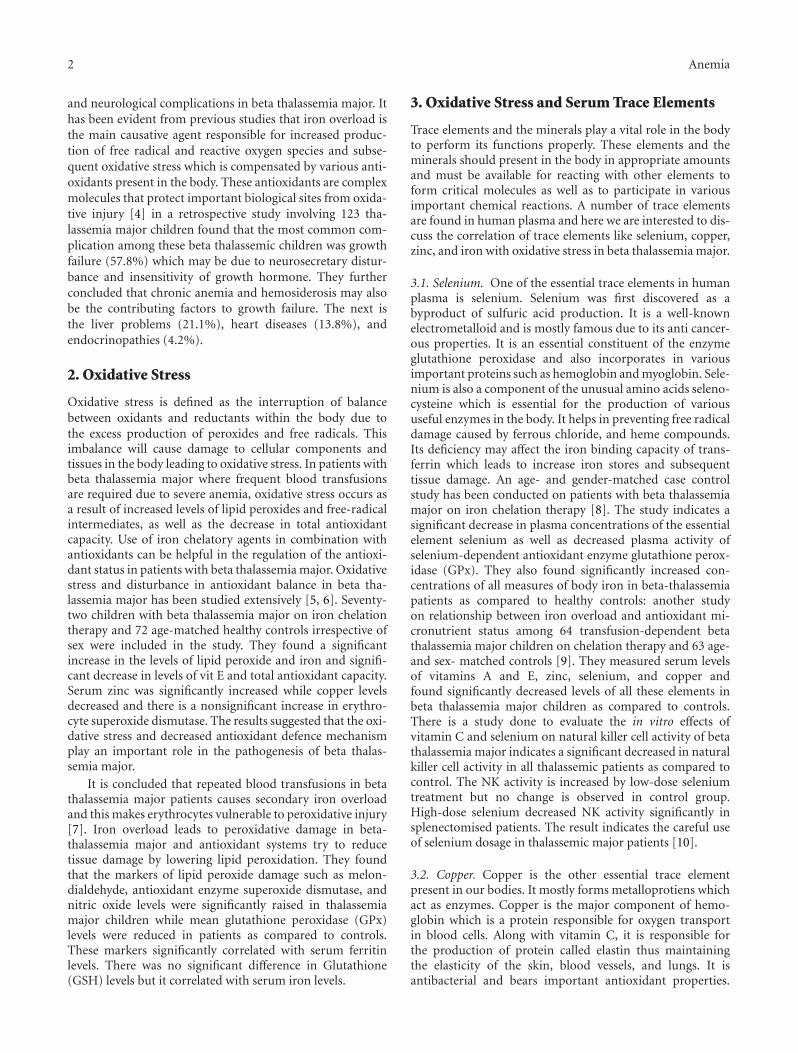
2 Anemia
and neurological complications in beta thalassemia major. Ithas been evident from previous studies that iron overload isthe main causative agent responsible for increased produc-tion of free radical and reactive oxygen species and subse-quent oxidative stress which is compensated by various anti-oxidants present in the body. These antioxidants are complexmolecules that protect important biological sites from oxida-tive injury [4] in a retrospective study involving 123 tha-lassemia major children found that the most common com-plication among these beta thalassemic children was growthfailure (57.8%) which may be due to neurosecretary distur-bance and insensitivity of growth hormone. They furtherconcluded that chronic anemia and hemosiderosis may alsobe the contributing factors to growth failure. The next isthe liver problems (21.1%), heart diseases (13.8%), andendocrinopathies (4.2%).
2. Oxidative Stress
Oxidative stress is defined as the interruption of balancebetween oxidants and reductants within the body due tothe excess production of peroxides and free radicals. Thisimbalance will cause damage to cellular components andtissues in the body leading to oxidative stress. In patients withbeta thalassemia major where frequent blood transfusionsare required due to severe anemia, oxidative stress occurs asa result of increased levels of lipid peroxides and free-radicalintermediates, as well as the decrease in total antioxidantcapacity. Use of iron chelatory agents in combination withantioxidants can be helpful in the regulation of the antioxi-dant status in patients with beta thalassemia major. Oxidativestress and disturbance in antioxidant balance in beta tha-lassemia major has been studied extensively [5, 6]. Seventy-two children with beta thalassemia major on iron chelationtherapy and 72 age-matched healthy controls irrespective ofsex were included in the study. They found a significantincrease in the levels of lipid peroxide and iron and signifi-cant decrease in levels of vit E and total antioxidant capacity.Serum zinc was significantly increased while copper levelsdecreased and there is a nonsignificant increase in erythro-cyte superoxide dismutase. The results suggested that the oxi-dative stress and decreased antioxidant defence mechanismplay an important role in the pathogenesis of beta thalas-semia major.
It is concluded that repeated blood transfusions in betathalassemia major patients causes secondary iron overloadand this makes erythrocytes vulnerable to peroxidative injury[7]. Iron overload leads to peroxidative damage in beta-thalassemia major and antioxidant systems try to reducetissue damage by lowering lipid peroxidation. They foundthat the markers of lipid peroxide damage such as melon-dialdehyde, antioxidant enzyme superoxide dismutase, andnitric oxide levels were significantly raised in thalassemiamajor children while mean glutathione peroxidase (GPx)levels were reduced in patients as compared to controls.These markers significantly correlated with serum ferritinlevels. There was no significant difference in Glutathione(GSH) levels but it correlated with serum iron levels.
3. Oxidative Stress and Serum Trace Elements
Trace elements and the minerals play a vital role in the bodyto perform its functions properly. These elements and theminerals should present in the body in appropriate amountsand must be available for reacting with other elements toform critical molecules as well as to participate in variousimportant chemical reactions. A number of trace elementsare found in human plasma and here we are interested to dis-cuss the correlation of trace elements like selenium, copper,zinc, and iron with oxidative stress in beta thalassemia major.
3.1. Selenium. One of the essential trace elements in humanplasma is selenium. Selenium was first discovered as abyproduct of sulfuric acid production. It is a well-knownelectrometalloid and is mostly famous due to its anti cancer-ous properties. It is an essential constituent of the enzymeglutathione peroxidase and also incorporates in variousimportant proteins such as hemoglobin and myoglobin. Sele-nium is also a component of the unusual amino acids seleno-cysteine which is essential for the production of varioususeful enzymes in the body. It helps in preventing free radicaldamage caused by ferrous chloride, and heme compounds.Its deficiency may affect the iron binding capacity of trans-ferrin which leads to increase iron stores and subsequenttissue damage. An age- and gender-matched case controlstudy has been conducted on patients with beta thalassemiamajor on iron chelation therapy [8]. The study indicates asignificant decrease in plasma concentrations of the essentialelement selenium as well as decreased plasma activity ofselenium-dependent antioxidant enzyme glutathione perox-idase (GPx). They also found significantly increased con-centrations of all measures of body iron in beta-thalassemiapatients as compared to healthy controls: another studyon relationship between iron overload and antioxidant mi-cronutrient status among 64 transfusion-dependent betathalassemia major children on chelation therapy and 63 age-and sex- matched controls [9]. They measured serum levelsof vitamins A and E, zinc, selenium, and copper andfound significantly decreased levels of all these elements inbeta thalassemia major children as compared to controls.There is a study done to evaluate the in vitro effects ofvitamin C and selenium on natural killer cell activity of betathalassemia major indicates a significant decreased in naturalkiller cell activity in all thalassemic patients as compared tocontrol. The NK activity is increased by low-dose seleniumtreatment but no change is observed in control group.High-dose selenium decreased NK activity significantly insplenectomised patients. The result indicates the careful useof selenium dosage in thalassemic major patients [10].
3.2. Copper. Copper is the other essential trace elementpresent in our bodies. It mostly forms metalloprotiens whichact as enzymes. Copper is the major component of hemo-globin which is a protein responsible for oxygen transportin blood cells. Along with vitamin C, it is responsible forthe production of protein called elastin thus maintainingthe elasticity of the skin, blood vessels, and lungs. It isantibacterial and bears important antioxidant properties.
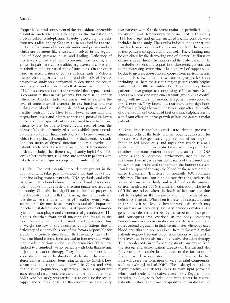
Anemia 3
Copper is a central component of the antioxidant superoxidedismutase molecule and also helps in the formation ofprotein called ceruloplasmin thereby protecting the cellsfrom free-radical injury. Copper is also required for the pro-duction of hormones like nor adrenaline and prostaglandinswhich are hormone-like chemicals involved in the regula-tion of blood pressure, pulse, and healing. Deficiency ofthis trace element will lead to anemia, neutropenia, andgrowth impairment, abnormalities in glucose and cholesterolmetabolism, and increased rate of infections. On the otherhand, an accumulation of copper in body leads to Wilson’sdisease with copper accumulation and cirrhosis of liver. Aprospective study was performed to determine the serumlevels of zinc and copper in beta thalassemia major children[11]. This cross-sectional study revealed that hypozincemiais common in thalassemic patients, but there is no copperdeficiency. Another study was carried out to evaluate thelevel of some essential elements in one hundred and fivethalassemic blood-transfusion-dependent patients and 54healthy controls [12]. They found lower serum zinc andmagnesium levels and higher copper and potassium levelsin thalassemic major patients as compared to controls. Zincdeficiency may be due to hyperzincuria resulted from therelease of zinc from hemolyzed red cells while hypercupremiaoccurs in acute and chronic infections and hemochromatosiswhich is the principal complication of thalassemia. A studydone on status of thyroid function and iron overload inpatients with beta thalassemia major on Deferoxamine inJordan concluded that there is significantly high (P < 0.05)levels of serum ferritin, FT3, zinc, and copper in patients withbeta thalassemia major as compared to controls [13].
3.3. Zinc. The next essential trace element present in thebody is zinc. It takes part in various important body func-tions including protein synthesis, DNA synthesis, and cellu-lar growth. It is found almost in every cell and plays a vitalrole in body’s immune system affecting innate and acquiredimmunity. Zinc also has significant antioxidant propertiesthereby protecting the cells from damage due to free radicals.It is the active site for a number of metalloenzymes whichare required for nucleic acid synthesis and also importantfor other host defense mechanisms like production of mono-cytes and macrophages and chemotaxis of granulocytes [14].Zinc is absorbed from small intestine and found in theblood bound to albumin. Impaired growths, alopecia, lossof weight are few of the associated complications due todeficiency of zinc which is one of the factors responsible forgrowth and puberty disorders in thalassemic patients [15].Frequent blood transfusions can lead to iron overload whichmay result in various endocrine abnormalities. They havestudied two hundred twenty patients with beta thalassemiamajor on chelation therapy. They found that there is anassociation between the duration of chelation therapy andabnormalities in lumbar bone mineral density (BMD). Lowserum zinc and copper was observed in 79.6% and 68%of the study population, respectively. There is significantassociation of serum zinc levels with lumbar but not femoralBMD. Another study was carried out to evaluate the serumcopper and zinc in Jordanian thalassaemic patients. Forty
two patients with β-thalassemia major on periodical bloodtransfusion and Deferoxamine were included in this study[16]. Forty age- and gender-matched healthy controls wereincluded in the study. The results indicate that copper andzinc levels were significantly increased in beta thalassemiamajor patients compared with controls. These finding maybe explained by the decreasing rate of glomerular filtrationof zinc seen in chronic hemolysis and the disturbance in themetabolism of zinc and copper in thalassaemic patients dueto the increasing serum zinc. The high level of copper couldbe due to increase absorption of copper from gastrointestinaltract. It is shown that a case control prospective studyincluding 100 beta thalassemia major patients with heightswithin 3rd to 10th percentile [17]. They randomly dividepatients in two groups ach comprising of 50 patients. Group1 was given oral zinc supplements while group 2 is a controlgroup with no zinc supplements. The patients were observedfor 18 months. They found out that there is no significantdifference in height between the two groups after 18 monthsof observation and concluded that oral zinc sulphate has nosignificant effect on linear growth of beta thalassemia majorpatient.
3.4. Iron. Iron is another essential trace element present inalmost all cells of the body. Human body requires iron forthe synthesis of oxygen carrying protein called haemoglobinfound in red blood cells, and myoglobin which is also aprotein found in muscles. It also takes part in the productionof other important proteins in the body such as for DNAsynthesis and cell division. Furthermore, iron is used inthe connective tissues in our body, some of the neurotrans-mitters in our brain, and to maintain the immune system.Iron is transported through the blood by the serum protein,called transferrin. Transferrin is normally 30% saturatedwith iron. The total iron-binding capacity (tibc) reflects thestatus of iron in the body and is defined as the amountof iron needed for 100% transferrin saturation. The levelsof TIBC are raised when the levels of iron are low thuswill be helpful in the diagnosis and monitoring of irondeficiency anaemia. When iron is present in excess amountsin the body it will lead to hemochromatosis, which maybe primary or secondary. Primary hemochromatosis is agenetic disorder characterized by increased iron absorptionand consequent iron overload in the body. Secondaryhemochromatosis occurs in diseases like thalassemia due toiron overload especially in thalassemia major where repeatedblood transfusions are required. Beta thalassemia majorpatients require frequent blood transfusions which lead toiron overload in the absence of effective chelation therapy.This iron deposits in thalassemic patients can exceed fromthe storage and detoxification capacity of ferritin and alsofully saturates transferrin and leads to the formation offree iron which accumulates in blood and tissues. This freeiron will cause the formation of very harmful compounds,such as hydroxyl radical (OH). The hydroxyl radicals arehighly reactive and attacks lipids to form lipid peroxideswhich contribute to oxidative stress [18]. Regular bloodtransfusions along with chelation therapy in beta thalassemiapatients drastically improve the quality and duration of life

4 Anemia
to third and fourth decades. Iron overload is serious compli-cation of long-term blood transfusion. It requires adequatetreatment in thalassemics so that the early deaths especiallyfrom iron-induced cardiomyopathies will be prevented. Ithas been shown that the cardiovascular involvement in betathalassemia major patients without cardiac iron overload[19]. They involved twenty six patients with beta thalassemiamajor on chelation therapy without cardiac iron overloadand thirty age- and gender-matched healthy controls in thestudy. The results indicated aortic stiffening associated withincreased left ventricular mass and left atrial enlargementin the beta thalassemia patients as compared to controls.These changes may represent the signs of early cardiovascularinvolvement in beta thalassemia patients without cardiaciron overload. A retrospective chart view study revealedthat three hundred and sixty transfusion-dependent betathalassemic patients treated with Deferoxamine [20]. Allpatients were followed and treated from 1990–2004, dis-ease complications were assessed by the measuring ironoverload and mean serum ferritin concentrations yearly forall patients. The result showed that cardiac complicationsbeing the first most important cause of death followedby infections. Complications and deaths among these betathalassemic major patients is iron-related organ dysfunctionand age related. Furthermore, serum ferritin levels werefound significantly higher in patients who died as comparedto those who survived. It was also found that majority of thecomplicated patients were on nonoptimal chelation therapyand noncompliance. Early detection of iron overload onthe heart is crucial in the management of beta thalassemiamajor. Serum ferritin is the poor indicator of myocardial irondeposition during early iron overload stage [21].
3.5. Magnesium. Magnesium is another trace element whichis essential for maintaining proper body functions. It isvital for body’s immune system, cardiovascular, and mus-culoskeletal systems. Deficiency of this element will leadto hypertension, diabetes, and cardiovascular diseases. Astudy was carried out to evaluate the level of some essen-tial elements in one hundred and five thalassemic blood-transfusion-dependent patients and 54 healthy controls [12].They found lower serum zinc and magnesium levels andhigher copper and potassium levels in thalassemic majorpatients as compared to controls. Zinc deficiency may bedue to hyperzincuria resulted from the release of zinc fromhemolyzed red cells while hypercupremia occurs in acuteand chronic infections and hemochromatosis which is theprincipal complication of thalassemia.
3.6. Iodine. The other vital trace element present in the bodyis iodine and is one of the powerful antioxidants presentin the body. Bernard Courtois, a French chemist, was firstdiscovered iodine in 1811. Iodine is present in almost everybody tissue but found in greater quantities in thyroid,breast, stomach, liver, lungs, heart, adrenals, and ovaries.Iodine is important for mental and physical developmentand maintaining healthy immune system. It takes part in theproduction of thyroid hormones including thyroxin and tri-iodothyronine. These hormones are of primary importance
in maintaining the body metabolism and brain development.Deficiency of this trace element may lead to cancer, diabetes,heart diseases, and multiple sclerosis. A study revealedincreased sensitivity to the inhibitory effect of excess iodideon thyroid functions in 25 beta thalassemia major patientswith normal thyroid functions [22]. The patients weregiven 20 mg of iodine three times daily for three weeks.They found significant decrease in concentration of thyroidhormones and significant increase in TSH concentrationswith 56% of the patients reached to hypothyroid levels.They concluded that beta thalassemia major patients shouldnot be given excess iodide due to increased sensitivity toinhibitory effects on thyroid functions as it may lead topermanent hypothyroidism. A study was carried out onlong-term intensive combined chelation therapy on thyroidfunction in 51 beta thalassemia major patients after theyachieved negative iron balance [23]. While on Deferoxaminemonotherapy, eighteen patients required thyroxin but aftercombined therapy with deferrioxamine and deferipronethere is significant (P < 0.0001) decrease in iron overload anda significant increase in mean FT4 and FT3 concentrationswith mean decrease in TSH. They concluded that negativeiron balance can be achieved rapidly with combinationchelation therapy than with monotherapy as well as there isreversal of hypothyroidism with this regime.
3.7. Calcium. Calcium is one of the most abundant traceelements present in the body. It is important for regulatingcardiovascular, musculoskeletal, and nervous systems of thebody. Deficiency of calcium may lead to rickets, osteomala-cia, and osteoporosis. Excess of this trace element may lead tokidney stones, impaired renal functions, and prostate cancer.Studies have shown that calcium may activate the enzymesinvolved in the production of reactive oxygen species and freeradicals by the mitochondria. It has been shown that seventy-five percent of patients had a low calcium level and 72.5% ofpatients had hypothyroidism [24]. The low calcium level wasprobably caused by a combination of hypoparathyroidismand osteomalacia resulting from deficient calcium intake.A case control study done on the effects of intramuscularinjection of a megadose of cholecalciferol involving 40 betathalassemia major patients and 40 nonthalassemic controls[25]. They found that among thalassemia major patients,two had hypoparathyroidism and low 25-OH D, and twohad hypocalcaemia with hypophosphatemia, high alkalinephosphatase (ALP), high PTH, and serum 25-OH D belowng/mL. The remaining patients had low 25-OH D concen-trations with normal serum Ca and PO4 concentrations.Vitamin D deficiency is present in 100% of thalassemia majorpatients and treatment with megadose injection of cholecal-ciferol is effective for hypovitaminosis D for 3 months. A casestudy done on 14-year-old girl with beta thalassemia majordiagnosed since the age of 9 months came to their centerwith generalized tonic clonic seizure [26]. The investigationsrevealed diffuse intracranial calcifications in deep whitematter, posterior fossa, basal ganglia, and both thalami.The laboratory and neuroimaging also indicate hypoparathy-roidism. They recommend periodic assessment and controlof serum calcium in all patients with thalassemia major
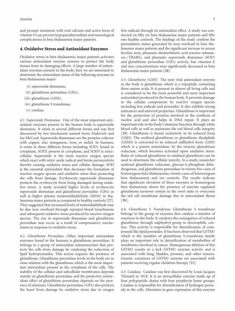
Anemia 5
and prompt treatment with oral calcium and active form ofvitamin D can prevent hypoparathyroidism and neurologicalcomplications in beta thalassemia major patients.
4. Oxidative Stress and Antioxidant Enzymes
Oxidative stress in beta thalassemia major patients activatesvarious antioxidant enzyme systems to protect the bodytissues from its damaging effects. A large number of antiox-idant enzymes present in the body, here we are interested todetermine the antioxidant status of the following enzymes inbeta thalassemia major:
(i) superoxide dismutase,
(ii) glutathione peroxidase (GPx),
(iii) glutathione (GSH),
(iv) glutathione S transferase,
(v) catalase.
4.1. Superoxide Dismutase. One of the most important anti-oxidant enzymes present in the human body is superoxidedismutase. It exists in several different forms and was firstdiscovered by two biochemist named Irwin Fridovich andJoe McCord. Superoxide dismutases are the proteins cofactorwith copper, zinc manganese, iron, or nickel. In humans,it exists in three different forms including SOD1 found incytoplasm, SOD2 present in cytoplasm, and SOD3 is extra-cellular. Superoxide is the main reactive oxygen specieswhich react with nitric oxide radical and forms peroxynitritethereby causing oxidative stress and cellular damage. SODis the essential antioxidant that decreases the formation ofreactive oxygen species and oxidative stress thus protectingthe cells from damage. Erythrocyte superoxide dismutaseprotects the erythrocyte from being damaged during oxida-tive stress. A study revealed higher levels of erythrocytesuperoxide dismutase and glutathione peroxidise (GPx) aswell as higher plasma melanoyldialdehyde (MDA) in tha-lassemia major patients as compared to healthy controls [27].They suggested that increased levels of malondialdehyde maybe due iron overload through repeated blood transfusionsand subsequent oxidative stress produced by reactive oxygenspecies. The rise in superoxide dismutase and glutathioneperoxidase may occur as a result of compensatory mecha-nisms in response to oxidative stress.
4.2. Glutathione Peroxidase. Other important antioxidantenzymes found in the humans is glutathione peroxidase. Itbelongs to a group of antioxidant selenoenzymes that pro-tects the cells from damage by catalyzing the reduction oflipid hydroperoxides. This action requires the presence ofglutathione. Glutathione peroxidase levels in the body are inclose relation with the glutathione which is the most impor-tant antioxidant present in the cytoplasm of the cells. Thestability of the cellular and subcellular membranes dependsmainly on glutathione peroxidase and the protective antiox-idant effect of glutathione peroxidase depends on the pres-ence of selenium. Glutathione peroxidase (GPx) also protectsthe heart from damage by oxidative stress due to oxygen
free radicals through its antioxidant effect. A study was con-ducted on fifty six beta thalassemia major patients and fiftyone healthy controls. The findings of the study confirm theperoxidative status generated by iron overload in beta tha-lassemia major patients and the significant increase in serumferritin, iron, plasmatic thiobarbituric acid reactive substan-ces (TBARS), and plasmatic superoxide dismutase (SOD)and glutathione peroxidase (GPx) activity, but vitamins Eand zinc concentrations were significantly decreased in betathalassemia major patients [28].
4.3. Glutathione (GSH). The next vital antioxidant enzymein the body is glutathione which is a tripeptide containingthree amino acids. It is present in almost all living cells andis considered to be the most powerful and most importantantioxidant produced in the human body. It prevents damageto the cellular components by reactive oxygen speciesincluding free radicals and peroxides. It also exhibits stronganticancer and antiviral properties. Glutathione is importantfor the protection of proteins involved in the synthesis ofnucleic acid and also helps in DNA repair. It plays animportant role in the body’s immune function through whiteblood cells as well as maintains the red blood cells integrity[29]. Glutathione is found exclusively in its reduced form(GSH). The oxidized glutathione or glutathione disulphide(GSSH) is converted to its reduced sulfhydryl form (GSH)which is a potent antioxidant, by the enzyme glutathionereductase, which becomes activated upon oxidative stress.Ratio of reduced glutathione to oxidized glutathione can beused to determine the cellular toxicity. In a study, researcheranalysed glutathione reductase, glucose-6-phosphate dehy-drogenase, and glutathione peroxidase in twenty five cases ofhomozygous beta thalassaemia, twenty cases of heterozygousbeta thalassaemia and ten controls. The results indicatethat significant elevation of these enzymes in homozygousbeta thalassemia shows the presence of enzyme regulatedglutathione turnover system in the overt state to overcomethe red cell membrane damage due to autooxidant threat[30].
4.4. Glutathione S Transferase. Glutathione S transferasebelongs to the group of enzymes that catalyze a number ofreactions in the body. It catalyzes the conjugation of reducedglutathione through sulphydryl group to electrophilic cen-tres. This activity is responsible for detoxification of com-pounds like lipid peroxides. It has been observed that GSTM1which is the member of glutathione S-transferase familyplays an important role in detoxification of metabolites ofxenobiotics involved in cancer. Homogenous deletion of thisGSTM1 results in a lack GSTM1 enzyme activity and isassociated with lung, bladder, prostate, and other tumors.Genetic variations of GSTM1 enzyme are associated withpatients receiving regular chelation therapy [31].
4.5. Catalase. Catalase was first discovered by Louis JacquesThenard in 1818. It is an intracellular enzyme made up offour polypeptide chains with four porphyrin heme groups.Catalase is responsible for detoxification of hydrogen perox-ide in the cells. Alteration in gene expression of this enzyme
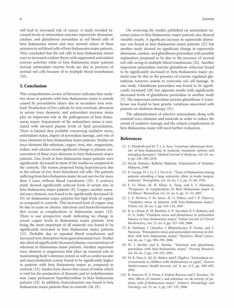
6 Anemia
will lead to increased risk of cancer. A study revealed in-creased levels of antioxidant enzymes superoxide dismutase,catalase, and glutathione peroxidase in red blood cells ofbeta thalassemia minor and near normal values of theseenzymes in red blood cells of beta thalassemia major patients.They concluded that the red cells in beta thalassemia minorreact to increased oxidant threat with augmented antioxidantenzyme activities while in beta thalassemia major patientsnormal antioxidant enzyme levels are due to presence ofnormal red cells because of to multiple blood transfusions[32].
5. Conclusion
This comprehensive review of literature indicates that oxida-tive stress in patients with beta thalassemia major is mainlycaused by peroxidative injury due to secondary iron over-load. Production of free radicals by iron overload, alterationin serum trace elements, and antioxidant enzymes statusplay an important role in the pathogenesis of beta thalas-semia major. Impairment of the antioxidant status is asso-ciated with elevated plasma levels of lipid peroxidation.There is limited data available concerning oxidative stress,antioxidant status, degree of peroxidase damage, and role oftrace elements in beta thalassemia major patients. Studies ontrace elements like selenium, copper, iron, zinc, magnesium,iodine, and calcium reveal significant change in plasma con-centration of these trace elements in beta thalassemia majorpatients. Zinc levels in beta thalassemia major patients weresignificantly decreased in most of the studies as compared tothe controls. The reason proposed being hyperzincuria dueto the release of zinc from hemolysed red cells. The patientssuffering from beta thalasemia major do not survive for morethan 5 years without blood transfusion [33]. A contrarystudy showed significantly reduced levels of serum zinc inbeta thalassemia major patients [9]. Copper, another essen-tial trace element, was found to be significantly decreased [9,15] on thalassemia major patients but high levels of copperas compared to controls. This increased level of copper maybe due to acute or chronic infections and hemochromatosisthat occurs as complications in thalassemia major [12].There is one prospective study indicating no change inserum copper levels in thalassemia major patients. Ironbeing the most important of all minerals was found to besignificantly increased in beta thalassemia major patients[11]. Probably due to repeated blood transfusions andincreased iron absorption from gastrointestinal tract. Studiesalso showed significantly decreased plasma concentrations ofselenium in thalassemia major patients. Another importanttrace element is magnesium that plays an essential role inmaintaining body’s immune system as well as cardiovascularand musculoskeletal system found to be significantly higherin patients with beta thalassemia major as compared tocontrols [12]. Studies have shown that excess of iodine whichis vital for the production of thyroxin and tri-iodothyroninemay cause permanent hypothyroidism in beta thalassemiapatients [22]. In addition, hypocalcaemia was found in betathalassemia major patients than in controls [24, 25].
On reviewing the studies published on antioxidant en-zymes status in beta thalassemia, major patients also showedvariable results. A significant increase in superoxide dismu-tase was found in beta thalassemia major patients [27] butanother study showed no significant change in superoxidedismutase, catalase, and glutathione peroxidase with possibleexplanation proposed to be due to the presence of normalred cells owing to multiple blood transfusions [32]. Anotherimportant antioxidant enzyme glutathione reductase foundto be significantly increased in beta thalassemia major pa-tients may be due to the presence of enzyme regulated glu-tathione turnover system to overcome red cell damage. Inone study, Glutathione peroxidase was found to be signifi-cantly increased [28] but opposite results with significantlydecreased levels of glutathione peroxidase in another study[7]. The important antioxidant enzyme glutathione S-trans-ferase was found to have genetic variations associated withpatients on chelation therapy [31].
The administration of selective antioxidants along withessential trace elements and minerals in order to reduce theextent of oxidative damage and the related complications inbeta thalassemia major still need further evaluation.
References
[1] G. Elizabeth and M. T. J. A. Ann, “Genotype-phenotype diver-sity of beta-thalassemia in malaysia: treatment options andemerging therapies,” Medical Journal of Malaysia, vol. 65, no.4, pp. 256–260, 2010.
[2] Social Statistics Bulletin Malaysia: Department of StatisticsMalaysia, 2008.
[3] E. George, H. J. Li, Y. J. Fei et al., “Types of thalassemia amongpatients attending a large university clinic in kuala lumpur,malaysia,” Hemoglobin, vol. 16, no. 1-2, pp. 51–66, 1992.
[4] F. Ur. Khan, M. H. Khan, A. Tariq, and S. S. Hamayun,“Frequency of complications In Beta thalassemia major inD.I.Khan,” Biomedical, vol. 23, no. 6, pp. 31–33, 2007.
[5] L. E. Pavlova, V. M. Savov, H. G. Petkov, and I. P. Charova,“Oxidative stress in patients with beta-thalassemia major,”Prilozi, vol. 28, no. 1, pp. 145–154, 2007.
[6] R. A. Ghone, K. M. Kumbar, A. N. Suryakar, R. V. Katkam, andN. G. Joshi, “Oxidative stress and disturbance in antioxidantbalance in beta thalassemia major,” Indian Journal of ClinicalBiochemistry, vol. 23, no. 4, pp. 337–340, 2008.
[7] R. Naithanj, J. Chandra, J. Bhattacharjee, P. Verma, and S.Naravan, “Peroxidative stress and antioxidant enzymes in chil-dren with beta thalassemia major,” Paediatric Blood Cancer,vol. 46, no. 7, pp. 780–785, 2006.
[8] W. J. Bartlay and E. Bartfay, “Selenium and glutathioneperoxidase with beta-thalassemia major,” Nursing Research,vol. 50, no. 3, pp. 178–183, 2001.
[9] M. R. Nasr, S. Ali, M. Shaker, and E. Elgabry, “Antioxidant mi-cronutrients in children with thalassaemia in egypt,” EasternMediterranean Health Journal, vol. 8, no. 4-5, pp. 490–495,2002.
[10] B. Atasever, N. Z. Ertan, S. Erdem-Kuruca, and Z. Karakas, “Invitro effects of vitamin c and selenium on nk activity of pa-tients with β-thalassemia major,” Pediatric Hematology andOncology, vol. 23, no. 3, pp. 187–197, 2006.
Anemia 7
[11] A. Mahyar, P. Ayazi, A. A. Pahlevan, H. Mojabi, M. R. Sehhat,and A. Javadi, “Zinc and copper status in children with beta-thalassemia major,” Iranian Journal of Pediatrics, vol. 20, no. 3,pp. 297–302, 2010.
[12] A. H. Al-Samarrai, M. H. Adaay, K. A. Al-Tikriti, and M. M.Al-Anzy, “Evaluation of some essential element levels in tha-lassemia major patients in mosul district, iraq,” Saudi MedicalJournal, vol. 29, no. 1, pp. 94–97, 2008.
[13] F. Irshaid and K. Mansi, “Status of thyroid function and ironoverload in adolescents and young adults with beta- thalas-semia major treated with deferoxamine in jordan,” Proceedingsof World Academy of Science, Engineering and Technology, vol.58, pp. 658–663, 2009.
[14] R. B. William, “Zinc and immune system,” in Encyclopaedia ofImmunology, pp. 2515–2516, Elsevier, Amsterdam, TheNetherlands, 2nd edition, 2004.
[15] A. A. Shamshirsaz, M. R. Bekheirnia, M. Kamgar et al., “Met-abolic and endocrinologic complications in beta-thalassemiamajor: a multicenter study in tehran,” BMC Endocrine Disor-ders, vol. 3, article no. 4, 2003.
[16] M. Kamal, A. Talal, B. Moussa, and N. Hamzeh, “Copper andzinc status in Jordanian patients with β-thalassemia majortreated with Deferoxamine,” Research Journal of BiologicalSciences, vol. 4, no. 5, pp. 566–572, 2009.
[17] M. Faranoush, M. S. Rahiminejad, Z. Karamizadeh, R.Ghorbani, and S. M. Owji, “Zinc supplementation effect onlinear growth in transfusion dependent beta thalassemia,”Iranian Journal of Blood and Cancer, vol. 1, no. 1, pp. 29–32,2008.
[18] P. Raghuveer, P. Vidya, and R. S. Prabhu, “Iron overload in betaThalasemia—a review,” Journal of Bioscience and Technology,vol. 1, no. 1, pp. 20–31, 2009.
[19] D. A. Stakos, D. Margaritis, D. N. Tziakas et al., “Cardiovascu-lar involvement in patients with β-thalassemia major withoutcardiac iron overload,” International Journal of Cardiology, vol.134, no. 2, pp. 207–211, 2009.
[20] S. K. Al Jaouni, “Survival and disease complication ofthalassemia major: experience of 14 years at King AbdulazizUniversity Hospital, Jeddah, KSA,” Medical Science Journal,vol. 17, no. 1, pp. 19–28, 2009.
[21] S. K. Al Jaouni, “Serum Ferritin is a poor indicator ofMyocardial iron Content in Early Stage of Iron Overload inThalasemia Mojor,” The Egyptian Journal of Haematology, vol.32, no. 3, pp. 171–176, 2007.
[22] T. Alexandrides, N. Georgopoulos, S. Yarmenitis, and A.G. Vagenakis, “Increased sensitivity to the inhibitory effectof excess iodide on thyroid function in patients with β-thalassemia major and iron overload and the subsequent de-velopment of hypothyroidism,” European Journal of Endo-crinology, vol. 143, no. 3, pp. 319–325, 2000.
[23] K. Farmaki, I. Tzoumari, and C. Pappa, “Reversal of hypothy-roidism in well chelated βthalassemia major patients,” inProceedings of the 50th Annual Meeting of ASH, San Francisco,Calif, USA, December 2008.
[24] A. A. Tantawy, M. El Kholy, T. Moustafa, and H. H. Elsedfy,“Bone mineral density and calcium metabolism in adolescentswith beta thalassemia major,” Pediatric Endocrinology Reviews,vol. 6, no. 1, pp. 132–135, 2008.
[25] A. Soliman, A. Adel, M. Wagdy, M. Al Ali, and N. ElMulla,“Calcium homeostasis in 40 adolescents with beta-thalassemiamajor: a case-control study of the effects of intramuscularinjection of a megadose of cholecalciferol,” Pediatric Endo-crinology Reviews, vol. 6, no. 1, pp. 149–154, 2008.
[26] M. Mahmoodi, V. De Sanctis, and M. Karimi, “Diffuse intrac-erebral calcification in beta thalassemia major with hypothy-roidism: acese report,” Pediatric Endocrinology Review, supple-ment 2, pp. 331–333, 2011.
[27] S. Filiz, O. Gulyuz, K. Sabri, E. Deniz, and H. Alev, “Oxidantand antioxidant status in beta thalassemia major patients,”Journal of Ankara University Faculty of Medicine, vol. 58, no.1, pp. 34–38, 2005.
[28] A. Kassab-Chekir, S. Laradi, S. Ferchichi et al., “Oxidant,antioxidant status and metabolic data in patients with beta-thalassemia,” Clinica Chimica Acta, vol. 338, no. 1-2, pp. 79–86, 2003.
[29] P. M. Kidd, “Glutathione: systemic protectant against oxida-tive and free radical damage,” Alternative Medicine Review, vol.2, no. 3, pp. 155–176, 1997.
[30] S. Ponnazhagan and R. Sarkar, “Enzymes of the pentose phos-phate pathway in glutathione-regulated membrane protectionin β-thalassaemia,” European Journal of Clinical Chemistry andClinical Biochemistry, vol. 30, no. 8, pp. 481–484, 1992.
[31] R. Origa, S. Satta, G. Matta, and R. Galanello, “Glutathione s-transferase gene polymorphism and cardiac iron overload inthalassaemia major,” British Journal of Haematology, vol. 142,no. 1, pp. 143–145, 2008.
[32] G. C. Gerli, L. Beretta, M. Bianchi, P. ellegatta, and A. G.Agostoni, “Erythrocytesuperoxide dismutase, catalase, andglutathione peroxidise activities in beta thalassemia(majorand minor),” Scandavian Journal of Haematology, vol. 25, no.1, pp. 87–92, 1980.
[33] H. U. Shekhar, Y. Kabir, M. Hossain et al., “Blood trans-fusion-mediated viral infections in thalassemic children inbangladesh,” Journal of Medical Sciences, vol. 7, no. 1, pp. 131–135, 2007.
Hindawi Publishing CorporationAnemiaVolume 2012, Article ID 943974, 5 pagesdoi:10.1155/2012/943974
Research Article
Thalassemic DNA-Containing Red Blood Cells Are underOxidative Stress
Mutaz Dana, Eugenia Prus, and Eitan Fibach
Department of Hematology, Hadassah-Hebrew University Medical Center, Ein-Kerem, P.O. Box 12000, Jerusalem 91120, Israel
Correspondence should be addressed to Eitan Fibach, [email protected]
Received 12 September 2011; Revised 1 December 2011; Accepted 4 December 2011
Academic Editor: Maria Cappellini
Copyright © 2012 Mutaz Dana et al. This is an open access article distributed under the Creative Commons Attribution License,which permits unrestricted use, distribution, and reproduction in any medium, provided the original work is properly cited.
We studied the nature of enucleated RBCs containing DNA remnants, Howell-Jolly (HJ) RBCs and reticulocytes (retics), that arecharacteristically present in the circulation of thalassemic patients, especially after splenectomy. Using flow cytometry methodol-ogy, we measured oxidative status parameters of these cells in patients with β-thalassemia. In each patient studied, these cells hadhigher content of reactive oxygen species and exposed phosphatidylserine compared with their DNA-free counterparts. These re-sults suggest that oxidative stress in thalassemic developing erythroid precursors might, through DNA-breakage, generate HJ-reticsand HJ-RBCs and that oxidative stress-induced externalization of phosphatidylserine is involved in the removal of these cells fromthe circulation by the spleen, a mechanism similar to that of the removal of senescent RBCs.
1. Introduction
The development of red blood cells (RBCs) from their pro-genitors in the bone marrow includes the process of enuclea-tion in which the final stages of nucleated erythroid precur-sors (orthochromatic normoblasts) expel their nuclei to gen-erate enucleated reticulocytes (retics), which leave the mar-row and mature into RBCs [1]. Normally, nucleated RBCs(normoblasts) are undetectable in the circulation, but insome hematological pathologies (e.g., thalassemia and sicklecell disease) they can be found in large numbers [1]. Thesediseases are also characterized by mature RBCs and reticsthat contain DNA remnants, that are called Howell-Jolly (HJ)bodies [1]. The frequency of these cells, which is very low, hasbeen quantified using a flow cytometry technique [2–4].
The spleen is the major site of the reticuloendothelialsystem where senescent RBCs at the end of their life-span areremoved by erythrophagocytosis [5]. It also removes fromthe circulation normoblasts and HJ-cells; thus, in thalassemiaand sickle cell disease, the number of these cells in thepatients’ circulation increases considerably following sple-nectomy [1].
The removal of senescent RBCs has been attributed tovarious mechanisms [5], including exposure (externaliza-tion) of phosphatidylserine (PS) on their surface [6]. The
macrophages of the reticuloendothelial system carry surfacereceptors that specifically bind PS, by which they internalizesenescent RBCs [7]. The mechanism by which normoblastsand HJ-cells are removed from the circulation is unknown.
We have previously shown that in hemolytic anemias,including thalassemia and sickle cell disease, RBCs are underoxidative stress [8], and they generate more reactive oxygenspecies (ROS) and contain less reduced glutathione thannormal RBCs, which results in membrane changes such aslipid peroxidation and externalization of PS.
Using flow cytometry, in the present study we show thatHJ-RBCs and retics are under oxidative stress and carryexposed PS, which may present the trigger for their phago-cytosis by macrophage and removal in the spleen.
2. Materials and Methods
Blood Samples. Peripheral blood (PB) samples were obtainedfrom normal donors and splenectomized and nonsplenec-tomized patients with β-thalassemia intermedia and major.The samples were obtained from the counting vials afterall diagnostic laboratory tests were completed. The researchwas approved by the Hadassah-Hebrew University MedicalCentre Human Experimentation Review Board. The patients’
2 Anemia
mutations and some relevant clinical parameters (e.g., trans-fusion and chelation therapy, splenectomy) were previouslysummarized [9]. In polytransfused patients, blood sampleswere obtained before transfusion, that is, at least 3 weeksfollowing the previous transfusion. Informed consent wasobtained in all cases.
Flow Cytometry Measurements of Oxidative Stress Markers.Cells were stained for transferrin-receptor by incubating with5 μL of APC-conjugated antibodies (Ab) to CD71 at 4◦C for30 minutes. The sample was washed and then divided intotwo aliquots: one aliquot was stained for ROS with 2′-7′-dichlorofluorescin diacetate (DCFH, Sigma, St, Louis, MO),at final concentration of 0.1 mM, at 37◦C for 15 minutes,then washed three times with Ca++- and Mg++-free Dul-becco’s phosphate-buffered-saline (PBS) (Biological Indus-tries, Beit-HaEmek, Israel). A stock solution of 20 mM DCFwas prepared in methanol (Bio Lab, Jerusalem, Israel). Theother aliquot was stained for external phosphatidylserine(PS), by suspending the cells in 100 μL of calcium buffer((10 mM HEPES, 140 mM NaCl and 2.5 mM CaCl2 (pH 7.4))and 2 μL of FITC-conjugated Annexin-V (IQ Products, Gro-ningen, The Netherlands). After 15 minutes at room tem-perature, in the dark, the cells were washed three times withcalcium buffer and resuspended in 0.5 mL of the same buffer.
For every assay, 2 μL of propidium iodide (PI, Mallinck-rodt Chemical Works, St. Louis, MO), dissolved in 0.1%sodium citrate, was added before analysis. Cells stained withanti-CD71 Ab alone, cells stained with anti-CD71 Ab andannexin-V, or cells stained with anti-CD71 Ab and DCF wereused as controls to set the compensation levels. Followingtreatment as indicated above, the cells were analyzed witha Fluorescence Activated Cell Sorter (FACS-calibur, Becton-Dickinson, Immunofluorometry systems, Mountain View,CA). Instrument calibration and settings were performedusing CaliBRITE-3 beads (Becton-Dickinson). The cells werepassed at a rate of ∼1,000 per second, using saline as thesheath fluid. A 488 nm argon laser beam was used for excita-tion. Threshold was set on forward light scatter (FSC) toexclude platelets and cell debris. Gates were set on RBCs,HJ-RBCs, retics, HJ-retics, normoblasts, and WBCs. Cellslabeled with DCF and annexin-V were detected by the FL-1PMT, and cells labeled with APC-conjugated anti-CD-71 Aband PI were detected by the FL-4 and FL-2 PMT, respectively.All PMTs were set on log amplification. The Mean Fluo-rescence Intensities (MFIs) and the percentages of positivecells were calculated using the FACS-equipped CellQuestsoftware (Becton-Dickinson). The results are expressed as theaverage ± standard deviation (SD) and compared using thetwo-sample Student’s t-test for differences in means.
3. Results and Discussion
PB cells were simultaneously stained with an anti-CD71 Aband PI, and either DCF or annexin-V. The anti-CD71 Abmarks the transferrin receptor, and PI the nucleic acid con-tent. To evaluate the contribution of RNA (particularly in
retics which contain small amounts of residual RNA) to thePI staining, PB cells were stained with PI in the presence orabsence of RNase (0.4 mg/mL, Invitrogen, Carlsbad, CA). Nodifference was noted in the pattern of PI staining betweenthese samples. The staining procedure identified cells asRBCs (CD71-PI-), HJ-RBCs (CD71-PI+), WBCs (CD71-PI++), retics (CD71+PI-), HJ-retics (CD71+PI+), and nor-moblasts (CD71+PI++). Figure 1(a) shows a flow cytometrydot-plot (PI versus CD71) analysis of a blood sample derivedfrom a representative splenectomized β-thalassemic patient,indicating the various cell populations. The fluorescencedistribution histograms of each cell population with respectto DCF-fluorescence, indicating generation of ROS, and an-nexin V-fluorescence, indicating exposed PS, with theirMFIs, are shown in Figures 1(b) and 1(c), respectively. Theresults indicate higher ROS and PS in retics than in matureRBCs, and, more critically, in HJ-cells compared with theirnon-HJ counterparts: in the experiment presented in Figure1(b), showing ROS results, the MFI of HJ-RBCs was 2.3-foldhigher than that of RBCs, and the MFI of HJ-retics was 2.4-fold higher than retics. In Figure 1(c), showing PS results, theMFI of HJ-RBCs was 15.3-fold higher than that of RBCs, andthe MFI of HJ-retics was 12.1-fold higher than retics.
Figure 2(a) depicts the frequency of HJ-RBCs in the PB ofnormal donors and in thalassemic patients. The results showno HJ-RBCs in normal donors and much higher frequencyof HJ-RBCs in splenectomized patients compared with non-splenectomized patients. Figures 2(b)-2(c), which summa-rize the average ROS generation and percentage of PS-expos-ing cells, show that both parameters were significantly higherin HJ-RBCs versus RBCs and in HJ-retics versus retics. Theresults also show that both parameters are higher in cellsfrom splenectomized versus nonsplenectomized patients,suggesting that the spleen removes the most damaged cells.
Although the process of nuclear expulsion from develop-ing RBC precursors has been studied extensively [10, 11], thereasons for nuclear remnants (HJ-bodies) leftover in enucle-ated retics and RBCs in certain diseases have not been studiedbefore. We now report that in β-thalassemia the generationof ROS and the externalization of PS, both parameters ofoxidative stress, are elevated in HJ-retics and HJ-RBCs com-pared with their no-HJ-containing counterparts. ROS maybe the cause of HJ formation. They are known to cause DNAbreaks [12] that may generate micronuclei in various celltypes [13], including lymphocytes and neutrophils. The oc-currence of micronuclei has been used as a biomarker forcytogenetic damage [14, 15]. These micronuclei are equiva-lent to the HJ bodies in RBCs. The mechanism of HJ bodies’formation must occur prior to nuclear expulsion. We havepreviously demonstrated that thalassemic erythroid precur-sors, including orthochromatic normoblasts, are at higheroxidative status than their normal counterparts [9]. It mightbe hypothesized that DNA/nuclear breaks induced by oxida-tive stress might result in incomplete expulsion of the nuclearmaterial, resulting in nuclear remnants which remain inretics and mature RBCs.
Several studies [16], including our own [9], indicatedthat ROS stimulate PS externalization on RBCs. Exposed
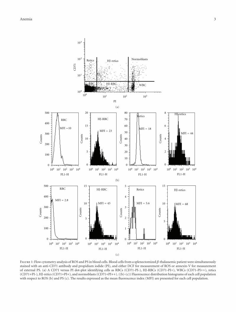
Anemia 3
100
101
102
103
100101 102 103
Retics HJ-retics Normoblasts
RBC HJ-RBC WBC
CD
71
PI
(a)
RBC HJ-RBCRetics
HJ-retics
MFI =10
0
100
200
300
400
500
100 101 102 103 104
FL1-H
0
5
10
15
20
0
10
20
30
40
50
60
70
80
0
2
4
6
8
100 101 102 103 104 100 101 102 103 104 100 101 102 103 104
FL1-H FL1-H FL1-H
Cou
nts
Cou
nts
Cou
nts
Cou
nts
MFI = 23MFI = 18
MFI = 44
(b)
0
100
200
300
400
500
0
5
10
15
0
1
2
3
4
5
0
5
10
15
Cou
nts
100 101 102 103 104
FL1-H
RBC HJ-RBC Retics HJ-retics
100 101 102 103 104
FL1-H
100 101 102 103 104
FL1-H
100 101 102 103 104
FL1-H
Cou
nts
Cou
nts
Cou
nts
MFI = 2.8MFI = 43 MFI = 5.6 MFI = 68
(c)
Figure 1: Flow cytometry analysis of ROS and PS in blood cells. Blood cells from a splenectomized β-thalassemic patient were simultaneouslystained with an anti-CD71 antibody and propidium iodide (PI), and either DCF for measurement of ROS or annexin-V for measurementof external PS. (a) A CD71 versus PI dot-plot identifying cells as RBCs (CD71-PI-), HJ-RBCs (CD71-PI+), WBCs (CD71-PI++), retics(CD71+PI-), HJ-retics (CD71+PI+), and normoblasts (CD71+PI++). ((b)-(c)) Fluorescence distribution histograms of each cell populationwith respect to ROS (b) and PS (c). The results expressed as the mean fluorescence index (MFI) are presented for each cell population.
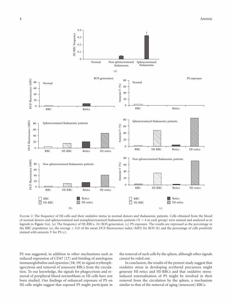
4 Anemia
0
0.1
0.2
0.3
0.4
Normal
HJ-
RB
C fr
equ
ency
Non splenectomized Splenectomizedthalassemia thalassemia
(a)
HJ-RBC
0
20
40
60
80ROS generation
Normal
RBC Retics
Splenectomized thalassemic patients
Non splenectomized thalassemic patients
HJ-retics
0
20
40
60
80
RBC Retics
RBC Retics
HJ-RBC HJ-retics
0
20
40
60
80
RBC ReticsHJ-RBC HJ-retics
DC
F-fl
uor
esce
nce
(M
FI)
DC
F-fl
uor
esce
nce
(M
FI)
DC
F-fl
uor
esce
nce
(M
FI)
(b)
PS exposure
0
20
40
60
80Normal
RBC Retics
Non splenectomized thalassemic patients
Splenectomized thalassemic patients
0
20
40
60
80
RBC ReticsHJ-RBC HJ-retics
0
20
40
60
80
RBC ReticsHJ-RBC HJ-retics
An
nex
in V
(%
)A
nn
exin
V (
%)
An
nex
in V
(%
)
HJ-RBC HJ-reticsRBC Retics
(c)
Figure 2: The frequency of HJ-cells and their oxidative status in normal donors and thalassemic patients. Cells obtained from the bloodof normal donors and splenectomized and nonsplenectomized thalassemic patients (N = 6 in each group) were stained and analyzed as inlegends to Figure 1(a). (a) The frequency of HJ-RBCs. (b) ROS generation. (c) PS exposure. The results are expressed as the percentage inthe RBC population (a), the average ± S.D of the mean DCF-fluorescence index (MFI) for ROS (b) and the percentage of cells positivelystained with annexin-V for PS (c).
PS was suggested, in addition to other mechanisms such asreduced expression of CD47 [17] and binding of autologousimmunoglobulins and opsonins [18, 19] to signal erythroph-agocytosis and removal of senescent RBCs from the circula-tion. To our knowledge, the signals for phagocytosis and re-moval of peripheral blood normoblasts or HJ-cells have notbeen studied. Our findings of enhanced exposure of PS onHJ-cells might suggest that exposed PS might participate in
the removal of such cells by the spleen, although other signalscannot be ruled out.
In conclusion, the results of the present study suggest thatoxidative stress in developing erythroid precursors mightgenerate HJ-retics and HJ-RBCs and that oxidative stress-induced externalization of PS might be involved in theirremoval from the circulation by the spleen, a mechanismsimilar to that of the removal of aging (senescent) RBCs.
Anemia 5
References
[1] J. Jandl, Blood—Textbook of Hematology, Little, Brown andCompany, Boston, Mass, USA, 1996.
[2] S. D. Dertinger, Y. Chen, R. K. Miller et al., “MicronucleatedCD71-positive reticulocytes: a blood-based endpoint of cyto-genetic damage in humans,” Mutation Research, vol. 542, no.1-2, pp. 77–87, 2003.
[3] T. Offer, A. Bhagat, A. Lal et al., “Measuring chromosomebreaks in patients with thalassemia,” Annals of the New YorkAcademy of Sciences, vol. 1054, pp. 439–444, 2005.
[4] V. L. Harrod, T. A. Howard, S. A. Zimmerman, S. D. Dertinger,and R. E. Ware, “Quantitative analysis of Howell-Jolly bodiesin children with sickle cell disease,” Experimental Hematology,vol. 35, no. 2, pp. 179–183, 2007.
[5] D. Bratosin, J. Mazurier, J. P. Tissier et al., “Cellular and mol-ecular mechanisms of senescent erythrocyte phagocytosis bymacrophages. A review,” Biochimie, vol. 80, no. 2, pp. 173–195,1998.
[6] V. A. Fadok, D. L. Bratton, S. C. Frasch, M. L. Warner, and P.M. Henson, “The role of phosphatidylserine in recognition ofapoptotic cells by phagocytes,” Cell Death and Differentiation,vol. 5, no. 7, pp. 551–562, 1998.
[7] Z. Zhou, “New phosphatidylserine receptors: clearance ofapoptotic cells and more,” Developmental Cell, vol. 13, no. 6,pp. 759–760, 2007.
[8] J. Amer, A. Goldfarb, and E. Fibach, “Flow cytometric analysisof the oxidative status of normal and thalassemic red bloodcells,” Cytometry A, vol. 60, no. 1, pp. 73–80, 2004.
[9] I. Freikman, J. Amer, J. S. Cohen, I. Ringel, and E. Fibach,“Oxidative stress causes membrane phospholipid rearrange-ment and shedding from RBC membranes—an NMR study,”Biochimica et Biophysica Acta, vol. 1778, no. 10, pp. 2388–2394,2008.
[10] H. Yoshida, K. Kawane, M. Koike, Y. Mori, Y. Uchiyama, andS. Nagata, “Phosphatidylserine-dependent engulfment by ma-crophages of nuclei from erythroid precursor cells,” Nature,vol. 437, no. 7059, pp. 754–758, 2005.
[11] G. Keerthivasan, A. Wickrema, and J. D. Crispino, “Erythrob-last enucleation,” Stem Cells International, vol. 2011, Article ID139851, 9 pages, 2011.
[12] J. Cadet, T. Douki, and J. L. Ravanat, “Oxidatively generatedbase damage to cellular DNA,” Free Radical Biology and Med-icine, vol. 49, no. 1, pp. 9–21, 2010.
[13] P. Belloni, P. Latini, and F. Palitti, “Radiation-induced bystand-er effect in healthy G0 human lymphocytes: biological andclinical significance,” Mutation Research, vol. 713, no. 1-2, pp.32–38, 2011.
[14] D. F. Smith, J. T. MacGregor, R. A. Hiatt et al., “Micronucleatederythrocytes as an index of cytogenetic damages in humans:demographic and dietary factors associated with micro-nucleated erythrocytes in splenectomized subjects,” CancerResearch, vol. 50, no. 16, pp. 5049–5054, 1990.
[15] A. Vral, M. Fenech, and H. Thierens, “The micronucleus assayas a biological dosimeter of in vivo ionising radiation expo-sure,” Mutagenesis, vol. 26, no. 1, pp. 11–17, 2011.
[16] M. Foller, S. M. Huber, and F. Lang, “Erythrocyte pro-grammed cell death,” IUBMB Life, vol. 60, no. 10, pp. 661–668,2008.
[17] S. Khandelwal, N. van Rooijen, and R. K. Saxena, “Reducedexpression of CD47 during murine red blood cell (RBC)senescence and its role in RBC clearance from the circulation,”Transfusion, vol. 47, no. 9, pp. 1725–1732, 2007.
[18] U. Galili, I. Flechner, and E. A. Rachmilewitz, “A naturallyoccurring anti-alpha-galactosyl IgG recognizing senescenthuman red cells,” Progress in Clinical and Biological Research,vol. 195, pp. 263–278, 1985.
[19] A. Pantaleo, G. Giribaldi, F. Mannu, P. Arese, and F. Turrini,“Naturally occurring anti-band 3 antibodies and red blood cellremoval under physiological and pathological conditions,”Autoimmunity Reviews, vol. 7, no. 6, pp. 457–462, 2008.
Hindawi Publishing CorporationAnemiaVolume 2012, Article ID 798296, 4 pagesdoi:10.1155/2012/798296
Research Article
Intracranial Blood Flow Velocity in Patients with β-ThalassemiaIntermedia Using Transcranial Doppler Sonography:A Case-Control Study
Nahid Ashjazadeh,1 Sajad Emami,1 Peyman Petramfar,1 Ehsan Yaghoubi,1
and Mehran Karimi2
1 Shiraz Neuroscience Research Center, Department of Neurology, Shiraz University of Medical Sciences, Shiraz, Iran2 Hematology Research Center, Shiraz University of Medical Sciences, Shiraz, Iran
Correspondence should be addressed to Mehran Karimi, [email protected]
Received 4 September 2011; Accepted 1 November 2011
Academic Editor: Sezaneh Haghpanah
Copyright © 2012 Nahid Ashjazadeh et al. This is an open access article distributed under the Creative Commons AttributionLicense, which permits unrestricted use, distribution, and reproduction in any medium, provided the original work is properlycited.
Introduction. Patients with β-thalassemia intermedia have a higher incidence of thromboembolic events compared to the generalpopulation. Previous studies have shown that patients with sickle cell disease, who are also prone to ischemic events, have higherintracranial arterial blood flow velocities measured by transcranial Doppler sonography (TCD). The aim of this study is to evaluateintracranial arterial flow velocities in patients with β-thalassemia intermedia and compare the results with those found in healthysubjects. Methods. Sixty-four patients with β-thalassemia intermedia and 30 healthy subjects underwent transcranial Dopplersonography. Results. Significantly higher flow velocities were found in intracranial arteries of patients compared to controls (P =0.001). Previously splenectomized patients with thrombocytosis showed higher flow velocities than nonsplenectomized patientswithout thrombosis. Conclusion. The increased flow velocities in patients with β-thalassemia intermedia may point to a higher riskof ischemic events. Preventive measures such as blood transfusion or antiplatelet treatment may be beneficial in these patients.
1. Introduction
Patients with β-thalassemia intermedia (B-TI) seem to showhigher rates of thromboembolic events than individualswithout thalassemia or patients with β-thalassemia major,in particular if they have been splenectomized [1]. It isestimated that 4% of patients with β-thalassemia interme-dia will experience a thromboembolic event [2]. Previoussplenectomy and thrombocytosis and/or platelet abnormali-ties are major factors associated with thromboembolic eventsin patients with β-thalassemia intermedia [1, 3, 4], andischemic stroke is increasingly recognized as one of the mostdevastating complications of this disease [5].
Ischemic stroke is also a known complication of sicklecell disease [6]. In a prospective study in patients with thiscondition, higher blood flow velocity in the intracranialarteries was associated with a higher risk of ischemic stroke[7]. The stroke prevention trial in sickle cell anemia (STOP)has indicated a role for transcranial Doppler sonography
(TCD) in measuring intracranial arterial flow velocities toidentify sickle cell patients at a high risk of ischemic stroke[8]. TCD measurement of intracranial flow velocities is of aidin deciding when to start blood transfusion in these patientsto reduce the risk of ischemic stroke [8, 9].
An association between TCD findings and stroke riskhas been confirmed in sickle cell disease; however, as faras we know, no studies have been conducted to evaluateTCD findings in patients with β-thalassemia intermedia,another high-risk group for ischemic stroke. In the presentstudy, we compared the intracranial arterial flow velocitiesof β-thalassemia intermedia patients with those of healthysubjects.
2. Patients and Methods
This is a case-control study conducted in a tertiary outpatientclinic affiliated with Shiraz University of Medical Sciences,Southern Iran, for a period of one year during 2009.
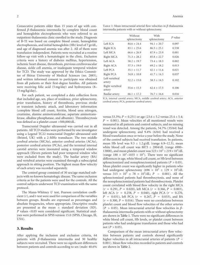
2 Anemia
Consecutive patients older than 15 years of age with con-firmed β-thalassemia intermedia by complete blood countand hemoglobin electrophoresis who were referred to anoutpatient thalassemia clinic enrolled in the study. Diagnosisof B-TI was based on complete blood count, hemoglobinelectrophoresis, and initial hemoglobin (Hb) level of 7 gr/dL,and age of diagnosed anemia was after 2. All of them weretransfusion independent. Patients were recruited at a routinefollow-up visit with a hematologist in the clinic. Exclusioncriteria were a history of diabetes mellitus, hypertension,ischemic heart disease, thrombosis, previous cerebrovasculardisease, sickle cell anemia, or inadequate temporal windowfor TCD. The study was approved by the Ethics Commit-tee of Shiraz University of Medical Sciences (no. 2885),and written informed consent to participate was obtainedfrom all patients or their first-degree families. All patientswere receiving folic acid (5 mg/day) and hydroxyurea (8–15 mg/kg/day).
For each patient, we completed a data collection formthat included age, sex, place of residence, prior splenectomy,prior transfusion, history of thrombosis, previous strokeor transient ischemic attack, and laboratory information(complete blood cell count, ferritin, blood urea nitrogen,creatinine, alanine aminotransferase, aspartate aminotrans-ferase, alkaline phosphatase, and albumin). Thrombocytosiswas defined as a platelet count >500,000/dL.
Transcranial Doppler ultrasound was carried out in allpatients. All TCD studies were performed by one investigatorusing a Legend TC22 transcranial Doppler ultrasound unit(Bristol, UK) with a 2-MHz transducer. The two middlecerebral arteries (MCAs), anterior cerebral arteries (ACAs),posterior cerebral arteries (PCAs), and the terminal internalcarotid arteries were insonated using a temporal windowapproach (Eleven patients had poor temporal window whowere excluded from the study). The basilar artery (BA)and vertebral arteries were examined through a suboccipitalapproach in sitting position. The highest mean flow velocityof each artery was recorded separately.
The control group consisted of 30 sex/age-matched sub-jects with no known hematologic disease. The same exclusioncriteria as for the patients were used for the controls. All thecontrol subjects underwent TCD examination with the sameprotocol.
The Mann-Whitney U test, Pearson correlation coeffi-cient (r), and t-test were used for comparison of the variablesbetween groups. Results are expressed as percentages andabsolute frequencies, where appropriate. Descriptive resultsare presented as the mean ± standard deviation (SD).P values <0.05 were considered significant. Statistical anal-yses were performed in SPSS version 15.0 (SPSS, Chicago, Ill,USA).
3. Results
After applying the inclusion and exclusion criteria, 64patients with β-thalassemia intermedia and 30 healthysubjects were recruited. There were no significant differencesbetween patients and controls according to sex (male: 40.6%
Table 1: Mean intracranial arterial flow velocities in β-thalassemiaintermedia patients with or without splenectomy.
Withoutsplenectomy
Withsplenectomy
P value
Left ICA 84.6± 24.4 94.1± 20.7 0.097
Right ICA 83.1± 25.6 86.5± 25.1 0.538
Left MCA 66.6± 26.9 87.9± 23.9 0.001
Right MCA 71.3± 28.2 85.8± 22.7 0.026
Left ACA 58.2± 19.7 73.4± 18.3 0.002
Right ACA 57.3± 19.0 69.2± 18.2 0.013
Left PCA 35.1± 11.7 42.1± 11.6 0.021
Right PCA 34.8± 10.8 41.7± 14.3 0.037
Left vertebralartery
52.2± 15.0 58.3± 14.5 0.102
Right vertebralartery
55.6± 15.3 62.4± 17.5 0.106
Basilar artery 66.1± 17.2 76.7± 16.6 0.016
ICA, internal carotid artery; MCA, middle cerebral artery; ACA, anteriorcerebral artery; PCA, posterior cerebral artery.
versus 53.3%; P = 0.251) or age (23.6± 5.2 versus 25.4± 5.4;P = 0.001). Mean velocities of all mentioned vessels weremeasured in all patients and control subjects, and no missingvessel was detected. Among the patients, 54.7% (35/64) hadundergone splenectomy, and 9.4% (6/64) had received ablood transfusion once or twice a year before the study. Noneof the control subjects had received transfusion. In patients,mean Hb level was 9.3 ± 1.2 g/dL (range 6.9–12.3), meanwhite blood cell count was 8873 ± 2004/dL (range 4900–13800), and mean platelet count was 523×103±219×103/dL(range 188 × 103–1035 × 103). There were no significantdifferences in age, white blood cell count, or Hb level betweensplenectomized and nonsplenectomized patients (P > 0.05).Mean platelet count was significantly higher in patients whohad undergone splenectomy (696 × 103 ± 129 × 103/dLversus 315 × 103 ± 78 × 103/dL; P = 0.001). All thesplenectomized patients had thrombocytosis, and none ofthe nonsplenectomized patients had thrombocytosis. Plateletcount correlated with blood flow velocity in the right MCA(r = 0.291, P = 0.020), left MCA (r = 0.366, P = 0.003),left ACA (r = 0.258, P = 0.040), right PCA (r = 0.270,P = 0.031), left PCA (r = 0.267, P = 0.033), and BA(r = 0.300, P = 0.016). There were no correlations betweenplatelet count and blood flow velocities of the other arteries(P > 0.05). Mean intracranial arterial flow velocities in β-thalassemia intermedia patients with or without splenectomyare shown in Table 1. There were no significant differences inwhite blood cell count, Hb levels, or platelet count betweenpatients who had undergone transfusion and those who hadnot (P > 0.05).
Comparison of the mean intracranial artery flow veloc-ities between patients and controls showed significantlyhigher velocities in all intracranial arteries of patients (P =0.001). Mean flow velocities recorded in patients and controlsare shown in Table 2.
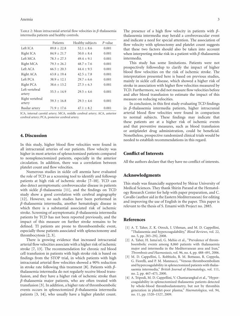
Anemia 3
Table 2: Mean intracranial arterial flow velocities in β-thalassemiaintermedia patients and healthy controls.
Patients Healthy subjects P value
Left ICA 89.8± 22.8 52.1± 8.6 0.001
Right ICA 84.9± 21.7 50.0± 8.4 0.001
Left MCA 78.3± 27.3 49.4± 9.1 0.001
Right MCA 79.3± 26.2 48.7± 7.6 0.001
Left ACA 66.5± 20.3 44.4± 9.5 0.001
Right ACA 63.8± 19.4 42.5± 7.8 0.001
Left PCA 38.9± 12.1 29.7± 6.6 0.001
Right PCA 38.6± 13.2 27.3± 6.3 0.001
Left vertebralartery
55.5± 14.9 29.5± 4.6 0.001
Right vertebralartery
59.3± 16.8 29.3± 4.6 0.001
Basilar artery 71.9± 17.6 47.1± 8.2 0.001
ICA, internal carotid artery; MCA, middle cerebral artery; ACA, anteriorcerebral artery; PCA, posterior cerebral artery.
4. Discussion
In this study, higher blood flow velocities were found inall intracranial arteries of our patients. Flow velocity washigher in most arteries of splenectomized patients comparedto nonsplenectomized patients, especially in the anteriorcirculation. In addition, there was a correlation betweenplatelet count and flow velocities.
Numerous studies in sickle cell anemia have evaluatedthe role of TCD as a screening tool to identify and followuppatients at high risk of ischemic stroke [7–10]. TCD canalso detect asymptomatic cerebrovascular disease in patientswith sickle β-thalassemia [11], and the findings on TCDstudy show a good correlation with cerebral angiography[12]. However, no such studies have been performed inβ-thalassemia intermedia, another hematologic disease inwhich there is a substantial associated risk of ischemicstroke. Screening of asymptomatic β-thalassemia intermediapatients by TCD has not been reported previously, and theimpact of this measure on further stroke remains to bedefined. TI patients are prone to thromboembolic event,especially those patients associated with splenectonomy andthrombocytosis [2, 3].
There is growing evidence that increased intracranialarterial flow velocities associate with a higher risk of ischemicstroke [7, 13]. The recommendation for chronic red bloodcell transfusion in patients with high stroke risk is based onfindings from the STOP trial, in which patients with highintracranial arterial flow velocities showed a 90% reductionin stroke rate following this treatment [8]. Patients with β-thalassemia intermedia do not regularly receive blood trans-fusion, and they have a higher risk of ischemic stroke thanβ-thalassemia major patients, who are often treated withtransfusion [3]. In addition, a higher rate of thromboembolicevents occurs in splenectomized β-thalassemia intermediapatients [3, 14], who usually have a higher platelet count.
The presence of a high flow velocity in patients with β-thalassemia intermedia may herald a cerebrovascular eventand indicate a need for special attention. The association offlow velocity with splenectomy and platelet count suggeststhat these two factors should also be taken into accountwhen interpreting stroke risk in a patient with β-thalassemiaintermedia.
This study has some limitations. Patients were notprospectively followedup to clarify the impact of higherblood flow velocities on the risk of ischemic stroke. Theinterpretation presented here is based on previous studies,mainly in sickle cell disease, which showed a higher risk ofstroke in association with higher flow velocities measured byTCD. Furthermore, we did not measure flow velocities beforeand after blood transfusion to estimate the impact of thismeasure on reducing velocities.
In conclusion, in this first study evaluating TCD findingsin β-thalassemia intermedia patients, higher intracranialarterial blood flow velocities were found in comparisonto normal subjects. These findings may indicate thatthese patients are at a higher risk of ischemic eventsand that preventive measures, such as blood transfusionor antiplatelet drug administration, could be beneficial.Nonetheless, prospective randomized clinical trials would beneeded to establish recommendations in this regard.
Conflict of Interests
All the authors declare that they have no conflict of interests.
Acknowledgments
This study was financially supported by Shiraz University ofMedical Sciences. They thank Shirin Parand at the Hematol-ogy Research Center for help with paper preparation, and C.Cavallo (author aid in the Eastern Mediterranean) for editingand improving the use of English in the paper. This paper isrelevant to the thesis of S. Emami with Project no. 2885.
References
[1] A. T. Taher, Z. K. Otrock, I. Uthman, and M. D. Cappellini,“Thalassemia and hypercoagulability,” Blood Reviews, vol. 22,no. 5, pp. 283–292, 2008.
[2] A. Taher, H. Isma’eel, G. Mehio et al., “Prevalence of throm-boembolic events among 8,860 patients with thalassaemiamajor and intermedia in the Mediterranean area and Iran,”Thrombosis and Haemostasis, vol. 96, no. 4, pp. 488–491, 2006.
[3] M. D. Cappellini, L. Robbiolo, B. M. Bottasso, R. Coppola,G. Fiorelli, and P. M. Mannucci, “Venous thromboembolismand hypercoagulability in splenectomized patients with thalas-saemia intermedia,” British Journal of Haematology, vol. 111,no. 2, pp. 467–473, 2000.
[4] A. Tripodi, M. D. Cappellini, V. Chantarangkul et al., “Hyper-coagulability in splenectomized thalassemic patients detectedby whole-blood thromboelastometry, but not by thrombingeneration in platelet-poor plasma,” Haematologica, vol. 94,no. 11, pp. 1520–1527, 2009.
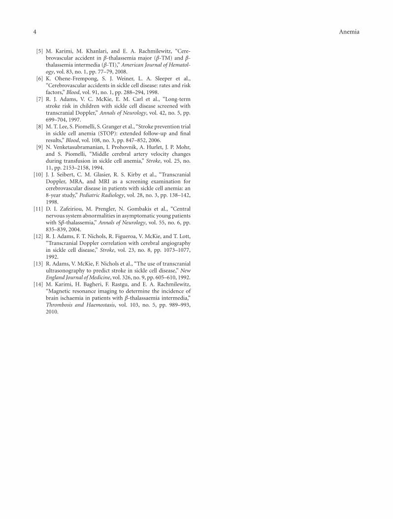
4 Anemia
[5] M. Karimi, M. Khanlari, and E. A. Rachmilewitz, “Cere-brovascular accident in β-thalassemia major (β-TM) and β-thalassemia intermedia (β-TI),” American Journal of Hematol-ogy, vol. 83, no. 1, pp. 77–79, 2008.
[6] K. Ohene-Frempong, S. J. Weiner, L. A. Sleeper et al.,“Cerebrovascular accidents in sickle cell disease: rates and riskfactors,” Blood, vol. 91, no. 1, pp. 288–294, 1998.
[7] R. J. Adams, V. C. McKie, E. M. Carl et al., “Long-termstroke risk in children with sickle cell disease screened withtranscranial Doppler,” Annals of Neurology, vol. 42, no. 5, pp.699–704, 1997.
[8] M. T. Lee, S. Piomelli, S. Granger et al., “Stroke prevention trialin sickle cell anemia (STOP): extended follow-up and finalresults,” Blood, vol. 108, no. 3, pp. 847–852, 2006.
[9] N. Venketasubramanian, I. Prohovnik, A. Hurlet, J. P. Mohr,and S. Piomelli, “Middle cerebral artery velocity changesduring transfusion in sickle cell anemia,” Stroke, vol. 25, no.11, pp. 2153–2158, 1994.
[10] J. J. Seibert, C. M. Glasier, R. S. Kirby et al., “TranscranialDoppler, MRA, and MRI as a screening examination forcerebrovascular disease in patients with sickle cell anemia: an8-year study,” Pediatric Radiology, vol. 28, no. 3, pp. 138–142,1998.
[11] D. I. Zafeiriou, M. Prengler, N. Gombakis et al., “Centralnervous system abnormalities in asymptomatic young patientswith Sβ-thalassemia,” Annals of Neurology, vol. 55, no. 6, pp.835–839, 2004.
[12] R. J. Adams, F. T. Nichols, R. Figueroa, V. McKie, and T. Lott,“Transcranial Doppler correlation with cerebral angiographyin sickle cell disease,” Stroke, vol. 23, no. 8, pp. 1073–1077,1992.
[13] R. Adams, V. McKie, F. Nichols et al., “The use of transcranialultrasonography to predict stroke in sickle cell disease,” NewEngland Journal of Medicine, vol. 326, no. 9, pp. 605–610, 1992.
[14] M. Karimi, H. Bagheri, F. Rastgu, and E. A. Rachmilewitz,“Magnetic resonance imaging to determine the incidence ofbrain ischaemia in patients with β-thalassaemia intermedia,”Thrombosis and Haemostasis, vol. 103, no. 5, pp. 989–993,2010.