-Secretase Pharmacology: What Pharmacology Will...
56
International Journal of Alzheimer’s Disease Guest Editors: Jeremy H. Toyn, Adele Rowley, Yasuji Matsuoka, Taisuke Tomita, and Bruno P. Imbimbo γ-Secretase Pharmacology: What Pharmacology Will Work for Alzheimer’s Disease?
Transcript of -Secretase Pharmacology: What Pharmacology Will...
International Journal of Alzheimer’s Disease
Guest Editors: Jeremy H. Toyn, Adele Rowley, Yasuji Matsuoka, Taisuke Tomita, and Bruno P. Imbimbo
γ-Secretase Pharmacology: What Pharmacology Will Work for Alzheimer’s Disease?

γ-Secretase Pharmacology:What Pharmacology Will Work forAlzheimer’s Disease?
International Journal of Alzheimer’s Disease
γ-Secretase Pharmacology:What Pharmacology Will Work forAlzheimer’s Disease?
Guest Editors: Jeremy H. Toyn, Adele Rowley,Yasuji Matsuoka, Taisuke Tomita, and Bruno P. Imbimbo

Copyright © 2013 Hindawi Publishing Corporation. All rights reserved.
This is a special issue published in “International Journal of Alzheimer’s Disease.” All articles are open access articles distributed underthe Creative Commons Attribution License, which permits unrestricted use, distribution, and reproduction in any medium, providedthe original work is properly cited.
Editorial Board
D. Allsop, UKCraig S. Atwood, USABrian Austen, UKJess Avila, SpainB. J. Bacskai, USAAndrew Budson, USARoger A. Bullock, UKAshley I. Bush, AustraliaGemma Casadesus, USARudolph J. Castellani, USAJames R. Connor, USASuzanne M. de la Monte, USAJusto G. de Yebenes, SpainS. M. Debanne, USASteven D. Edland, USACheng-Xin Gong, USAPaula Grammas, USA
George T. Grossberg, USAHarald J. Hampel, GermanyKurt A. Jellinger, AustriaMark Kindy, USAAmos D. Korczyn, IsraelJeff Kuret, USAAndrew J. Larner, UKHyoung-Gon Lee, USAJerzy Leszek, PolandSeth Love, UKMichelangelo Mancuso, ItalyJames G. McLarnon, CanadaPatrizia Mecocci, ItalyKenichi Meguro, JapanJudith Miklossy, CanadaPaula I. Moreira, PortugalRicardo Nitrini, Brazil
Michal Novak, SlovakiaLeonardo Pantoni, ItalyFrancesco Panza, ItalyLucilla Parnetti, ItalyGeorge Perry, USAM. C. Polidori, GermanyJeffrey R. Powell, USAJohn Powell, UKMarcella Reale, ItalyVincenzo Solfrizzi, ItalyAkihiko Takashima, JapanMatti Viitanen, SwedenB. Winblad, SwedenDavid Yew, Hong KongHenrik Zetterberg, Sweden
Contents
γ-Secretase Pharmacology: What Pharmacology Will Work for Alzheimer’s Disease?, Jeremy H. Toyn,Adele Rowley, Yasuji Matsuoka, Taisuke Tomita, and Bruno P. ImbimboVolume 2013, Article ID 849128, 2 pages
In Vivo Characterization of a Novel γ-Secretase Inhibitor SCH 697466 in Rodents and Investigation ofStrategies for Managing Notch-Related Side Effects, Lynn A. Hyde, Qi Zhang, Robert A. Del Vecchio,Prescott T. Leach, Mary E. Cohen-Williams, Lei Chen, Gwendolyn T. Wong, Nansie A. McHugh,Joseph Chen, Guy A. Higgins, Theodros Asberom, Wei Li, Dmitri Pissarnitski, Diane Levitan,Amin A. Nomeir, John W. Clader, Lili Zhang, and Eric M. ParkerVolume 2013, Article ID 823528, 14 pages
γ-Secretase-Dependent Proteolysis of Transmembrane Domain of Amyloid Precursor Protein: SuccessiveTri- and Tetrapeptide Release in Amyloid β-Protein Production, Mako Takami and Satoru FunamotoVolume 2012, Article ID 591392, 7 pages
Modulation of Gamma-Secretase for the Treatment of Alzheimer’s Disease, Barbara Tate,Timothy D. McKee, Robyn M. B. Loureiro, Jo Ann Dumin, Weiming Xia, Kevin Pojasek, Wesley F. Austin,Nathan O. Fuller, Jed L. Hubbs, Ruichao Shen, Jeff Jonker, Jeff Ives, and Brian S. BronkVolume 2012, Article ID 210756, 10 pages
Morphologic and Functional Effects of Gamma Secretase Inhibition on Splenic Marginal Zone B Cells,Maria Cristina de Vera Mudry, Franziska Regenass-Lechner, Laurence Ozmen, Bernd Altmann,Matthias Festag, Thomas Singer, Lutz Muller, Helmut Jacobsen, and Alexander FlohrVolume 2012, Article ID 289412, 7 pages
γ-Secretase Modulators: Can We Combine Potency with Safety?, Harrie J. M. Gijsen and Marc MerckenVolume 2012, Article ID 295207, 10 pages
Hindawi Publishing CorporationInternational Journal of Alzheimer’s DiseaseVolume 2013, Article ID 849128, 2 pageshttp://dx.doi.org/10.1155/2013/849128
Editorial𝛾-Secretase Pharmacology: What Pharmacology Will Work forAlzheimer’s Disease?
Jeremy H. Toyn,1 Adele Rowley,2 Yasuji Matsuoka,3
Taisuke Tomita,4 and Bruno P. Imbimbo5
1 Neuroscience Biology, Bristol-Myers Squibb Research and Development, 5 Research Parkway, Wallingford, CT 06492, USA2 TopiVert Pharma Ltd., Imperial College Incubator, Bessemer Building Level 1, Imperial College, London SW7 2AZ, UK3Neural Pathways Discovery, GlaxoSmithKline, Singapore Research Center, Singapore 1386674Department of Neuropathology and Neuroscience, Graduate School of Pharmaceutical Sciences, The University of Tokyo,Tokyo 113-0033, Japan
5 Research & Development, Chiesi Farmaceutici, Largo Francesco Belloli, 11/a, 43122 Parma, Italy
Correspondence should be addressed to Jeremy H. Toyn; [email protected]
Received 19 March 2013; Accepted 19 March 2013
Copyright © 2013 Jeremy H. Toyn et al.This is an open access article distributed under the Creative Commons Attribution License,which permits unrestricted use, distribution, and reproduction in any medium, provided the original work is properly cited.
This special issue focuses on 𝛾-secretase modulators (GSMs)and inhibitors (GSIs), two classes of small molecules withthe potential to test the amyloid hypothesis of Alzheimer’sdisease. Recent clinical trials of GSI and GSM, includingsemagacestat, avagacestat, and R-flurbiprofen, have beendiscontinued for lack of efficacy and/or side effects, themechanisms of which have not been elucidated. Detrimentaleffects of GSIs on cognition observed in AD patients may belinked to the accumulation of C-terminal fragment of APP(C99 or CTF𝛽).The stimulating effects of GSIs on skin cancerin AD patients have been linked to their inhibition of Notchprocessing. The lack of efficacy of the GSM R-flurbiprofenin AD patients has been explained with its low potency andpoor ability to cross the blood-brain barrier. The two reviewarticles and three research articles address key issues forGSI and GSM, namely, Notch-related side effects and drug-like properties, respectively. Although other amyloid-relatedapproaches are continuing in clinical trials, including anti-A𝛽 antibodies and 𝛽-site amyloid precursor protein cleavingenzyme (BACE) inhibitors, it still remains to be seen whetheror not they can decrease amyloid or A𝛽 for a sufficient periodof time at tolerable doses in patients. Therefore, renewedefforts toward GSIs and GSMs appear justified.
The review by M. Takami and S. Funamoto providesa succinct but comprehensive introduction to the stepwiseproteolytic mechanism of 𝛾-secretase. They describe the
experimental evidence for the multiple cleavages of the amy-loid precursor protein (APP) and provide some particularlyuseful illustrations showing how the enzyme processes itssubstrates to make a series of A𝛽 peptides and tripeptides.They further propose that 𝛾-secretase cleavage of Notchmay be mechanistically different from the cleavage of APP,implying an untapped potential to discover APP-selective orNotch-selective inhibitors.
H. J. M. Gijsen and M. Mercken review the currentstatus of GSM drug discovery, identify the limitations ofcurrent molecules, and present an approach to the futureoptimization of GSMs. The benchmark GSMs are succinctlyreviewed and common trends are evaluated. They give athoughtful and accessible explanation of the “conflict betweenthe physicochemical properties required for highly effica-ciousGSMand those (properties) required for drug-likeness.”This contrasts with GSI, where high potency and optimalphysical properties have been achieved for some molecules,but the biological mechanism imposes inbuilt Notch-relatedside effects. Thus, benchmark GSMs suffer from low qualityphysical properties, whereas benchmark GSIs are of highdrug-like quality but suffer from a limitation of the biologicalmechanism. The quantitative use of biological and physicalproperties in drug optimization is reviewed in a straightfor-wardway, and illustrations are used to showwhere the currentGSM and GSI molecules stand in this quantitative analysis.

2 International Journal of Alzheimer’s Disease
Also included is new data relating to a novel GSM illustratingrecent progress.
B. Tate et al. review the benchmark GSIs and GSMswhile providing data describing the novel natural product-derived GSMs discovered by Satori. GSMs typically decreaseA𝛽42 and increase A𝛽38 production; however, the Satoricompounds decrease both A𝛽42 and A𝛽38, while increasingA𝛽37 and A𝛽39. These different effects on A𝛽 peptides arewell illustrated by straightforward MALDI-TOF scans. Anexpanded definition of GSM is therefore given as compoundsthat cause “a shift of the A𝛽 pool to shorter, but variablelength, A𝛽 peptides.” In contrast to GSI, for which potencyis sensitive to APP expression level, GSMs exhibit a relativelyconstant level of potency. Nevertheless, GSMs require highplasma concentrations for associated brain A𝛽 lowering, anobservation that cannot be fully explained by properties suchas nonspecific protein binding and brain penetrance.
The original research article by L. A. Hyde et al. describesapproaches to mitigate the risk of Notch-related toxicity inrodents given the GSI SCH697466. Notch-related side effectswere evaluated in the intestine and thymus, and Notch-related biomarkers were monitored in white blood cells.Either decreased frequency of dosing, or lower doses whichcaused a partial lowering of A𝛽, were shown to decreaseNotch inhibition. The results show that appropriate choicesof the extent and duration of dosing can facilitate significantA𝛽 lowering without evidence of Notch-related side effects.
In the original research article by M. C. de Vera Mudryet al., the GSI RO4929097 was given to rats and mice toexplore the effect on immune responses. One of the mostcharacteristic Notch-related side effects of GSI is spleenmarginal zone atrophy, which raises the possibility of aconsequence for immune responses. Remarkably, despite theatrophy and decreased B cell numbers caused by chronicdosing of the GSI, the effect on immune responses was mildand reversible.
In summary, the main theme of this special issue is aboutrecent approaches to address the limitations of the two mainclasses of 𝛾-secretase-targeted small molecules. For GSIs, thefocus is on the mitigation of side effects related to Notchand other non-APP substrates. Extent and duration of GSIdosing could be varied to decrease effects on Notch, andthe consequences of decreased B cell numbers on immuneresponses were shown to be mild, thereby diminishing a per-ceived risk. Furthermore, the mechanisms of the 𝛾-secretasecleavages of APP and Notch, while not well understood, maybe sufficiently different to facilitate improved APP- or Notch-selectivity of GSIs in the future. For GSMs, the focus is onimproving the drug-like physicochemical properties whilemaintaining high potency. The future of GSMs thereforeappears to be a continuation of a challenging multiparameterdrug optimization. We hope readers will derive intellectualbenefit from these papers and enjoy them as much as we did.
Jeremy H. ToynAdele Rowley
Yasuji MatsuokaTaisuke Tomita
Bruno P. Imbimbo
Hindawi Publishing CorporationInternational Journal of Alzheimer’s DiseaseVolume 2013, Article ID 823528, 14 pageshttp://dx.doi.org/10.1155/2013/823528
Research ArticleIn Vivo Characterization of a Novel 𝛾-Secretase Inhibitor SCH697466 in Rodents and Investigation of Strategies for ManagingNotch-Related Side Effects
Lynn A. Hyde,1 Qi Zhang,1 Robert A. Del Vecchio,1 Prescott T. Leach,1
Mary E. Cohen-Williams,1 Lei Chen,2 Gwendolyn T. Wong,1 Nansie A. McHugh,1
Joseph Chen,1 Guy A. Higgins,1 Theodros Asberom,3 Wei Li,3 Dmitri Pissarnitski,3
Diane Levitan,2 Amin A. Nomeir,4 John W. Clader,3 Lili Zhang,1 and Eric M. Parker1
1 Department of Neuroscience, Merck Research Laboratories, Kenilworth, NJ 07033, USA2Department of Molecular Biomarkers, Merck Research Laboratories, Kenilworth, NJ 07033, USA3Department of Medicinal Chemistry, Merck Research Laboratories, Kenilworth, NJ 07033, USA4Department of Drug Metabolism and Pharmoackinetics, Merck Research Laboratories, Kenilworth, NJ 07033, USA
Correspondence should be addressed to Lynn A. Hyde; [email protected]
Received 29 August 2012; Accepted 27 November 2012
Academic Editor: Jeremy Toyn
Copyright © 2013 Lynn A. Hyde et al. This is an open access article distributed under the Creative Commons Attribution License,which permits unrestricted use, distribution, and reproduction in any medium, provided the original work is properly cited.
Substantial evidence implicates 𝛽-amyloid (A𝛽) peptides in the etiology of Alzheimer’s disease (AD). A𝛽 is produced by theproteolytic cleavage of the amyloid precursor protein by 𝛽- and 𝛾-secretase suggesting that 𝛾-secretase inhibition may providetherapeutic benefit for AD. Although many 𝛾-secretase inhibitors have been shown to be potent at lowering A𝛽, some have alsobeen shown to have side effects following repeated administration. All of these side effects can be attributed to altered Notchsignaling, another 𝛾-secretase substrate. Here we describe the in vivo characterization of the novel 𝛾-secretase inhibitor SCH697466 in rodents. Although SCH 697466 was effective at lowering A𝛽, Notch-related side effects in the intestine and thymus wereobserved following subchronic administration at doses that provided sustained and complete lowering of A𝛽. However, additionalstudies revealed that both partial but sustained lowering of A𝛽 and complete but less sustained lowering of A𝛽 were successfulapproaches for managing Notch-related side effects. Further, changes in several Notch-related biomarkers paralleled the side effectobservations. Taken together, these studies demonstrated that, by carefully varying the extent and duration of A𝛽 lowering by𝛾-secretase inhibitors, it is possible to obtain robust and sustained lowering of A𝛽 without evidence of Notch-related side effects.
1. Introduction
Alzheimer’s disease (AD) is a progressive age-related neu-rodegenerative disease characterized clinically by memoryloss and cognitive dysfunction followed by a disruption ofnormal daily functions, organ system failure, and, ultimately,death. However, a diagnosis of AD can only be confirmedpostmortem by the presence of distinct neuroanatomicalhallmarks including senile plaques consisting primarily of𝛽-amyloid (A𝛽) peptides, neurofibrillary tangles consistingof hyperphosphorylated tau, and substantial neuronal loss,
particularly in the hippocampus, an area of the brain whichplays a key role in memory.
Substantial genetic and neuroanatomical evidence impli-cates A𝛽 peptides in the etiology of Alzheimer’s disease(e.g., [1–3]). Therefore, it is thought that a reduction inA𝛽 production or an increase in A𝛽 clearance will have abeneficial, and potentially disease modifying, effect on thedisease.
A𝛽 is produced by sequential cleavage of amyloid precur-sor protein (APP) by 𝛽-site APP cleaving enzyme 1 (BACE1)followed by 𝛾-secretase. Thus, inhibiting 𝛾-secretase should
2 International Journal of Alzheimer’s Disease
N
F
O
SO O
O
N
N
F
Cl
OH
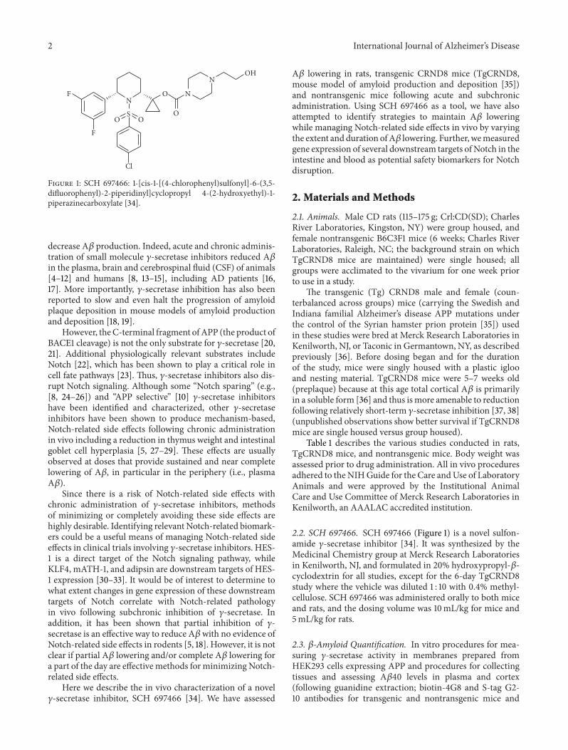
Figure 1: SCH 697466: 1-[cis-1-[(4-chlorophenyl)sulfonyl]-6-(3,5-difluorophenyl)-2-piperidinyl]cyclopropyl 4-(2-hydroxyethyl)-1-piperazinecarboxylate [34].
decrease A𝛽 production. Indeed, acute and chronic adminis-tration of small molecule 𝛾-secretase inhibitors reduced A𝛽in the plasma, brain and cerebrospinal fluid (CSF) of animals[4–12] and humans [8, 13–15], including AD patients [16,17]. More importantly, 𝛾-secretase inhibition has also beenreported to slow and even halt the progression of amyloidplaque deposition in mouse models of amyloid productionand deposition [18, 19].
However, theC-terminal fragment of APP (the product ofBACE1 cleavage) is not the only substrate for 𝛾-secretase [20,21]. Additional physiologically relevant substrates includeNotch [22], which has been shown to play a critical role incell fate pathways [23]. Thus, 𝛾-secretase inhibitors also dis-rupt Notch signaling. Although some “Notch sparing” (e.g.,[8, 24–26]) and “APP selective” [10] 𝛾-secretase inhibitorshave been identified and characterized, other 𝛾-secretaseinhibitors have been shown to produce mechanism-based,Notch-related side effects following chronic administrationin vivo including a reduction in thymus weight and intestinalgoblet cell hyperplasia [5, 27–29]. These effects are usuallyobserved at doses that provide sustained and near completelowering of A𝛽, in particular in the periphery (i.e., plasmaA𝛽).
Since there is a risk of Notch-related side effects withchronic administration of 𝛾-secretase inhibitors, methodsof minimizing or completely avoiding these side effects arehighly desirable. Identifying relevant Notch-related biomark-ers could be a useful means of managing Notch-related sideeffects in clinical trials involving 𝛾-secretase inhibitors. HES-1 is a direct target of the Notch signaling pathway, whileKLF4, mATH-1, and adipsin are downstream targets of HES-1 expression [30–33]. It would be of interest to determine towhat extent changes in gene expression of these downstreamtargets of Notch correlate with Notch-related pathologyin vivo following subchronic inhibition of 𝛾-secretase. Inaddition, it has been shown that partial inhibition of 𝛾-secretase is an effective way to reduce A𝛽with no evidence ofNotch-related side effects in rodents [5, 18]. However, it is notclear if partial A𝛽 lowering and/or complete A𝛽 lowering fora part of the day are effective methods for minimizing Notch-related side effects.
Here we describe the in vivo characterization of a novel𝛾-secretase inhibitor, SCH 697466 [34]. We have assessed
A𝛽 lowering in rats, transgenic CRND8 mice (TgCRND8,mouse model of amyloid production and deposition [35])and nontransgenic mice following acute and subchronicadministration. Using SCH 697466 as a tool, we have alsoattempted to identify strategies to maintain A𝛽 loweringwhile managing Notch-related side effects in vivo by varyingthe extent and duration ofA𝛽 lowering. Further, wemeasuredgene expression of several downstream targets ofNotch in theintestine and blood as potential safety biomarkers for Notchdisruption.
2. Materials and Methods
2.1. Animals. Male CD rats (115–175 g; Crl:CD(SD); CharlesRiver Laboratories, Kingston, NY) were group housed, andfemale nontransgenic B6C3F1 mice (6 weeks; Charles RiverLaboratories, Raleigh, NC; the background strain on whichTgCRND8 mice are maintained) were single housed; allgroups were acclimated to the vivarium for one week priorto use in a study.
The transgenic (Tg) CRND8 male and female (coun-terbalanced across groups) mice (carrying the Swedish andIndiana familial Alzheimer’s disease APP mutations underthe control of the Syrian hamster prion protein [35]) usedin these studies were bred at Merck Research Laboratories inKenilworth, NJ, or Taconic in Germantown, NY, as describedpreviously [36]. Before dosing began and for the durationof the study, mice were singly housed with a plastic iglooand nesting material. TgCRND8 mice were 5–7 weeks old(preplaque) because at this age total cortical A𝛽 is primarilyin a soluble form [36] and thus ismore amenable to reductionfollowing relatively short-term 𝛾-secretase inhibition [37, 38](unpublished observations show better survival if TgCRND8mice are single housed versus group housed).
Table 1 describes the various studies conducted in rats,TgCRND8 mice, and nontransgenic mice. Body weight wasassessed prior to drug administration. All in vivo proceduresadhered to theNIHGuide for the Care andUse of LaboratoryAnimals and were approved by the Institutional AnimalCare and Use Committee of Merck Research Laboratories inKenilworth, an AAALAC accredited institution.
2.2. SCH 697466. SCH 697466 (Figure 1) is a novel sulfon-amide 𝛾-secretase inhibitor [34]. It was synthesized by theMedicinal Chemistry group at Merck Research Laboratoriesin Kenilworth, NJ, and formulated in 20% hydroxypropyl-𝛽-cyclodextrin for all studies, except for the 6-day TgCRND8study where the vehicle was diluted 1 : 10 with 0.4% methyl-cellulose. SCH 697466 was administered orally to both miceand rats, and the dosing volume was 10mL/kg for mice and5mL/kg for rats.
2.3. 𝛽-Amyloid Quantification. In vitro procedures for mea-suring 𝛾-secretase activity in membranes prepared fromHEK293 cells expressing APP and procedures for collectingtissues and assessing A𝛽40 levels in plasma and cortex(following guanidine extraction; biotin-4G8 and S-tag G2-10 antibodies for transgenic and nontransgenic mice and
International Journal of Alzheimer’s Disease 3
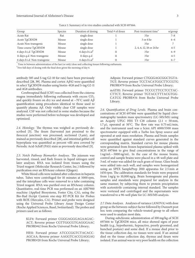
Table 1: Summary of in vivo studies conducted with SCH 697466.
Group Species Duration of dosing Total # of doses Post-treatment time∗ 𝑛/groupAcute Rat Rat single dose 1 3 hr 7–8Acute TgCRND8 Mouse single dose 1 3 or 6 hr 4–6Acute Non-transgenic Mouse single dose 1 3 hr 7Time course TgCRND8 Mouse single dose 1 2, 4, 6, 12, 18 or 24 hr 4-56 days b.i.d. TgCRND8 Mouse 6 days 𝑏.𝑖.𝑑† 11 3 hr 4–911 days q.d. Non-transgenic Mouse 11 days q.d. 11 3 hr 4-56 days b.i.d. Non-transgenic Mouse 6 days 𝑏.𝑖.𝑑† 11 3 hr 4–7∗
Time in between administration of the last (or only) dose and collecting tissues following euthanasia.†Five full days of dosing with the final dose given the morning of day 6.
antibody 585 and S-tag G2-10 for rats) have been previouslydescribed [28, 39]. Plasma and cortex A𝛽42 were quantifiedin select TgCRND8 studies using biotin-4G8 and S-tag G2-11and 4G8 antibodies.
Cerebrospinal fluid (CSF) was collected from the cisternamagna immediately following euthanasia with excess CO
2
and quickly frozen on dry ice and stored at −70∘C until A𝛽quantification using procedures identical to those used toquantify plasma A𝛽. Only visibly clear CSF samples wereanalyzed. CSF was not collected in some studies because thestudies were performed before technique was developed andoptimized.
2.4. Histology. The thymus was weighed as previously de-scribed [5]. The ileum (harvested just proximal to theileocecal junction) was processed, sectioned (3 𝜇m), andstained as previously described [28], and intestinal goblet cellhyperplasia was quantified as percent villi area covered byPeriodic Acid-Schiff (PAS) stain as previously described [5].
2.5. Notch Pathway Biomarker Analyses. The jejunum washarvested, rinsed, and flash frozen in liquid nitrogen untillater analyses. RNA was isolated from tissues using theTrizol reagent (Molecular Research Center, Inc.) followed bypurification over an RNAeasy column (Qiagen).
White blood cells were isolated after collection in heparintubes. Tubes were centrifuged for 10 minutes at 5000 rpm,and the interphase cells were removed to a tube containingTrizol reagent. RNA was purified over an RNAeasy column.Quantitative, real-time PCR was performed on an ABI7900machine (Applied Biosystems, Foster City, CA), using theBio-Rad iScript Custom one-step RT-PCR Kit for Probeswith ROX (Hercules, CA). Primer and probe were designedusing the Universal Probe Library Assay Design Center(Roche Applied Sciences, Basel, Switzerland).The probes andprimers used are as follows:
KLF4: Forward primer CGGGAGGGAGAAGAC-ACT; Reverse primer CGTTGGCGTGAGGGAACPROBE#62 from Roche Universal Probe Library;
HES1: Forward primer ATCCCGGTCTACACC-AGCAA; Reverse primer AAGGTCACCGAGGAGPROBE#20 from Roche Universal Probe Library;
Adipsin: Forward primer CTGGGAGCGGCTGTA-TGT; Reverse primer TCCTACATGGCTTCCGTGPROBE#79 from Roche Universal Probe Library;mATH1: Forward primer TCCCCTTCCTCCTAC-CTTCT; Reverse primer TGTACCTTTACGTGG-CATCG PROBE#34 from Roche Universal ProbeLibrary.
2.6. Quantification of Drug Levels. Plasma and brain con-centrations of SCH 697466 were quantified by liquid chro-matography tandem mass spectrometry (LC-MS/MS) usingan Acquity UPLC HSS T3 C18 column (2.1 × 50mm,1.7 𝜇), operated at 50∘C. The flow rate was 0.75mL/min,and the instrument used was a Sciex API 4000/5500 massspectrometer equipped with a Turbo Ion Spray source andoperated at unit mass resolution. Plasma and brain sampleswere quantified against standard curves generated in thecorresponding matrix. Standard curves for mouse plasmawere generated from frozen heparinized plasma spiked withSCH 697466 to give final concentrations in the range of1 ng/mL—5000 ng/mL. In the case of the mouse brains,control and sample brains were placed in a 48-well plate and3mL of water was added for each gram of tissue. Glass beadswere added into each well, and samples were homogenizedusing an SPEX SamplePrep 2010 apparatus for 2.5min at1450 rpm. The calibration standards for brain were preparedfrom 1 ng/g to 10,000 ng/g. Brain homogenate and plasmasamples and standards were prepared for analysis in thesame manner, by subjecting them to protein precipitationwith acetonitrile containing internal standard. The sampleswere vortexed and centrifuged and the supernatants weretransferred to a 96-well plate for LC-MS/MS analysis.
2.7. Data Analyses. Analyses of variance (ANOVA) with dosegroup as the between-subject factor followed byDunnett posthoc tests comparing the vehicle-treated group to all otherswere used to analyze most data.
During subchronic administration of 100mg/kg of SCH697466 to TgCRND8 mice, all mice showed signs of poorhealth (e.g., hypothermic, hypoactive, poorly groomed, andhunched posture) and some died. If a mouse died prior tothe tissue collection day, no tissues were used. If an animaldied on the tissue collection day, thymus and ileum wereisolated. If an animalwas in very poor health on the collection
4 International Journal of Alzheimer’s Disease
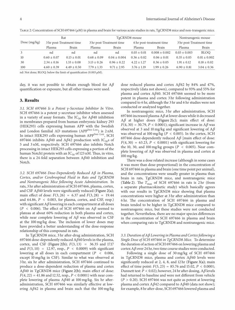
Table 2: Concentration of SCH 697466 (𝜇M) in plasma and brain for various acute studies in rats, TgCRND8mice and non-transgenic mice.
Dose (mg/kg)Rat TgCRND8 mouse Nontransgenic mouse
3 hr post-Treatment time 3 hr post-Treatment time 6 hr post-treatment time 3 hr post-Treatment timePlasma Brain Plasma Brain Plasma Brain Plasma Brain
3 nd nd nd nd 0.05 ± 0.01 0.008 ± 0.002 0.03 ± 0.003 BLOQ10 0.60 ± 0.07 0.13 ± 0.01 0.68 ± 0.09 0.04 ± 0.004 0.36 ± 0.02 0.06 ± 0.01 0.35 ± 0.05 0.01 ± 0.00230 2.34 ± 0.16 1.33 ± 0.08 3.13 ± 0.26 0.96 ± 0.22 4.22 ± 1.27 0.36 ± 0.05 1.91 ± 0.12 0.18 ± 0.02100 4.60 ± 0.39 4.49 ± 0.50 7.79 ± 1.33 9.71 ± 2.95 3.76 ± 1.19 1.99 ± 0.26 6.90 ± 0.81 3.04 ± 0.36nd: Not done; BLOQ: below the limit of quantification (0.003 𝜇M).
day, it was not possible to obtain enough blood for A𝛽quantification or exposure, but all other tissues were used.
3. Results
3.1. SCH 697466 Is a Potent 𝛾-Secretase Inhibitor In Vitro.SCH 697466 is a potent 𝛾-secretase inhibitor when assessedin a variety of assay formats. The IC
50for A𝛽40 inhibition
in membranes prepared from human embryonic kidney 293(HEK293) cells expressing human APP with the Swedishand London familial AD mutations (APPSwe-Lon) is 2 nM.In intact HEK293 cells expressing human APPSwe-Lon, SCH697466 inhibits A𝛽40 and A𝛽42 production with IC
50s of
5 and 3 nM, respectively. SCH 697466 also inhibits Notchprocessing in intact HEK293 cells expressing a portion of thehuman Notch1 protein with an IC
50of 123 nM.Thus, in vitro,
there is a 24-fold separation between A𝛽40 inhibition andNotch.
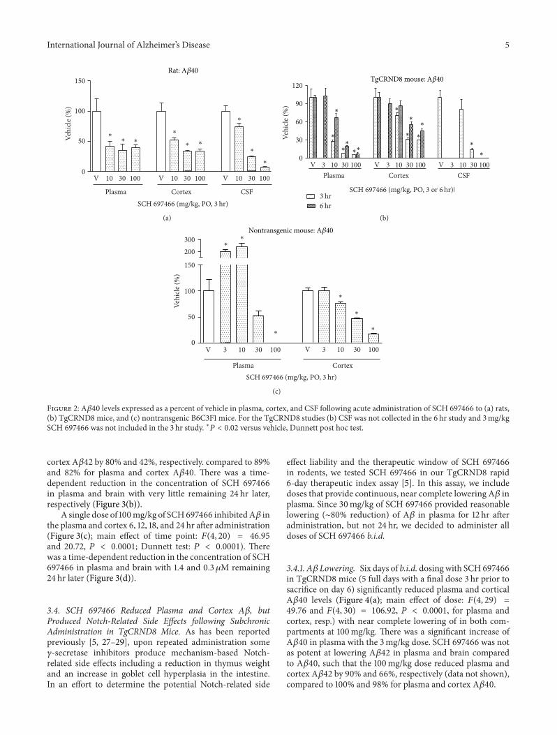
3.2. SCH 697466 Dose-Dependently Reduced A𝛽 in Plasma,Cortex, and/or Cerebrospinal Fluid in Rats and TgCRND8and Nontransgenic Mice following Acute Administration. Inrats, 3 hr after administration of SCH 697466, plasma, cortex,and CSF A𝛽40 levels were significantly reduced (Figure 2(a);main effect of dose: 𝐹(3, 26) = 6.37 and 𝐹(3, 28) = 19.44and 64.86, 𝑃 < 0.003, for plasma, cortex, and CSF, resp.)with significantA𝛽 lowering in each compartment at all doses(𝑃 < 0.006). The effect of SCH 697466 on A𝛽 seemed toplateau at about 60% reduction in both plasma and cortex,while near complete lowering of A𝛽 was observed in CSFat the 100mg/kg dose. The inclusion of lower doses mayhave provided a better understanding of the dose-responserelationship of this compound in rats.
In TgCRND8 mice, 3 hr after-drug administration, SCH697466 dose-dependently reduced A𝛽40 levels in the plasma,cortex, and CSF (Figure 2(b); 𝐹(3, 13) = 36.35 and 17.17and 𝐹(3, 10) = 12.97, resp., 𝑃 < 0.0009) with significantlowering at all doses in each compartment (𝑃 < 0.006,except 10mg/kg in CSF). Similar to what was observed at3 hr, six hr after administration, SCH 697466 continued toproduce a dose-dependent reduction of plasma and cortexA𝛽40 in TgCRND8 mice (Figure 2(b); main effect of dose:𝐹(4, 22) = 41.86 and 12.52, resp., 𝑃 < 0.0001) with near com-plete lowering of plasma A𝛽40 at 100mg/kg. Six hr after-administration, SCH 697466 was similarly effective at low-ering A𝛽42 in plasma and brain such that the 100mg/kg
dose reduced plasma and cortex A𝛽42 by 84% and 47%,respectively (data not shown), compared to 93% and 55% forplasma and cortex A𝛽40. SCH 697466 seemed to be morepotent in plasma and cortex 3 hr following administrationcompared to 6 hr, although the 3 hr and 6 hr studies were notconducted or analyzed together.
In nontransgenic mice, 3 hr after administration, SCH697466 increased plasmaA𝛽 at lower doses while it decreasedA𝛽 at higher doses (Figure 2(c); main effect of dose:𝐹(4, 30) = 30.79, 𝑃 < 0.0001); significant increase in A𝛽 wasobserved at 3 and 10mg/kg and significant lowering of A𝛽was observed at 100mg/kg (𝑃 < 0.005). In the cortex, SCH697466 dose-dependently reduced A𝛽 [main effect of dose:𝐹(4, 30) = 63.23, 𝑃 < 0.0001) with significant lowering forthe 10, 30, and 100mg/kg groups (𝑃 < 0.005). Near com-plete lowering of A𝛽 was observed in plasma and cortex at100mg/kg.
There was a dose related increase (although in some casesit was more than dose-proportional) in the concentration ofSCH697466 in plasma and brain (one time point per animal),and the concentrations were usually greater in plasma thanbrain in rats, TgCRND8 mice, and nontransgenic mice(Table 2). The 𝑇max of SCH 697466 in rats is 2 hr (froma separate pharmacokinetic study) which basically agreeswith our results in TgCRND8 mice showing that plasmaconcentrations were higher at 3 hr after administration than6 hr. The concentration of SCH 697466 in plasma andbrain tended to be higher in TgCRND8 mice compared tonontransgenic mice, but these studies were not conductedtogether. Nevertheless, there are no major species differencesin the concentration of SCH 697466 in plasma and brainwhen comparing rats to TgCRND8 and nontransgenic mice.
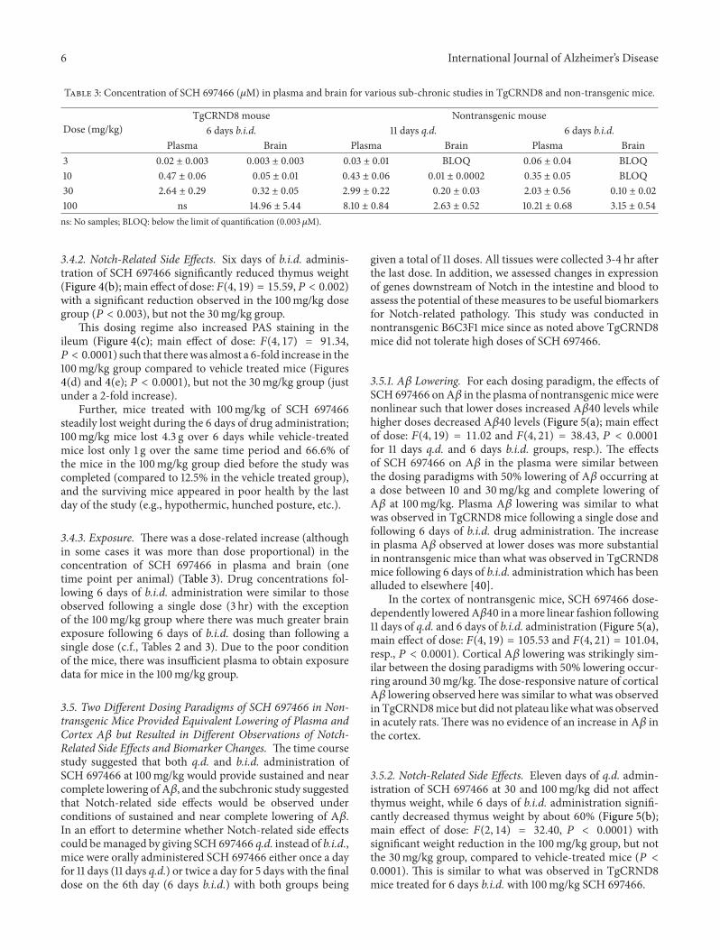
3.3. Duration of A𝛽 Lowering in Plasma andCortex following aSingle Dose of SCH 697466 in TgCRND8Mice. To determinethe duration of action of SCH697466 on lowering plasma andcortexA𝛽 over 24 hr, two time course studieswere conducted.
Following a single dose of 30mg/kg of SCH 697466in TgCRND8 mice, plasma and cortex A𝛽40 levels weresignificantly reduced at 2, 4, 8, and 12 hr (Figure 3(a); maineffect of time point: 𝐹(5, 23) = 85.76 and 15.02, 𝑃 < 0.0001;Dunnett test:𝑃 < 0.02); however, 24 hr after dosing, A𝛽 levelshad returned to baseline and were not different from vehicle(𝑃 > 0.20). SCH 697466 was not quite as potent at loweringplasma and cortex A𝛽42 compared to A𝛽40 (data not show);for example, 8 hr after-dose, SCH697466 lowered plasma and
International Journal of Alzheimer’s Disease 5
10 30 100 10 30 100 10 30 1000
50
100
150
Plasma Cortex CSF
∗∗ ∗
∗
∗ ∗
∗
∗
∗
Vehi
cle (%
)
V V V
SCH 697466 (mg/kg, PO, 3 hr)
(a)
3 hr6 hr
3 10 30 100 3 10 30 100 3 10 30 1000
30
60
90
120
Plasma Cortex CSF
Vehi
cle (%
)
V V V
SCH 697466 (mg/kg, PO, 3 or 6 hr)|
(b)
3 10 30 100 3 10 30 1000
50
100
150
200300
Plasma Cortex
∗∗
∗
∗
∗
∗
Vehi
cle (%
)
V V
SCH 697466 (mg/kg, PO, 3 hr)
(c)
Figure 2: A𝛽40 levels expressed as a percent of vehicle in plasma, cortex, and CSF following acute administration of SCH 697466 to (a) rats,(b) TgCRND8 mice, and (c) nontransgenic B6C3F1 mice. For the TgCRND8 studies (b) CSF was not collected in the 6 hr study and 3mg/kgSCH 697466 was not included in the 3 hr study. ∗𝑃 < 0.02 versus vehicle, Dunnett post hoc test.
cortex A𝛽42 by 80% and 42%, respectively. compared to 89%and 82% for plasma and cortex A𝛽40. There was a time-dependent reduction in the concentration of SCH 697466in plasma and brain with very little remaining 24 hr later,respectively (Figure 3(b)).
A single dose of 100mg/kg of SCH697466 inhibitedA𝛽 inthe plasma and cortex 6, 12, 18, and 24 hr after administration(Figure 3(c); main effect of time point: 𝐹(4, 20) = 46.95and 20.72, 𝑃 < 0.0001; Dunnett test: 𝑃 < 0.0001). Therewas a time-dependent reduction in the concentration of SCH697466 in plasma and brain with 1.4 and 0.3𝜇M remaining24 hr later (Figure 3(d)).
3.4. SCH 697466 Reduced Plasma and Cortex A𝛽, butProduced Notch-Related Side Effects following SubchronicAdministration in TgCRND8 Mice. As has been reportedpreviously [5, 27–29], upon repeated administration some𝛾-secretase inhibitors produce mechanism-based Notch-related side effects including a reduction in thymus weightand an increase in goblet cell hyperplasia in the intestine.In an effort to determine the potential Notch-related side
effect liability and the therapeutic window of SCH 697466in rodents, we tested SCH 697466 in our TgCRND8 rapid6-day therapeutic index assay [5]. In this assay, we includedoses that provide continuous, near complete lowering A𝛽 inplasma. Since 30mg/kg of SCH 697466 provided reasonablelowering (∼80% reduction) of A𝛽 in plasma for 12 hr afteradministration, but not 24 hr, we decided to administer alldoses of SCH 697466 b.i.d.
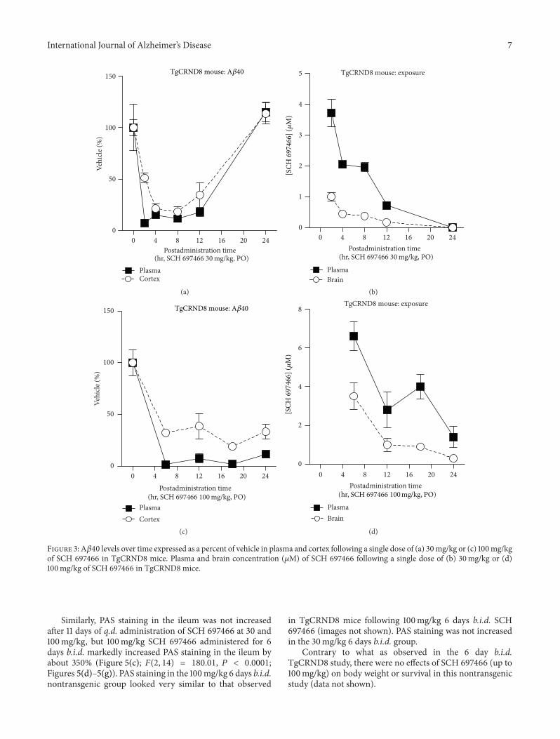
3.4.1. A𝛽 Lowering. Six days of b.i.d. dosingwith SCH697466in TgCRND8 mice (5 full days with a final dose 3 hr prior tosacrifice on day 6) significantly reduced plasma and corticalA𝛽40 levels (Figure 4(a); main effect of dose: 𝐹(4, 29) =49.76 and 𝐹(4, 30) = 106.92, 𝑃 < 0.0001, for plasma andcortex, resp.) with near complete lowering of in both com-partments at 100mg/kg. There was a significant increase ofA𝛽40 in plasma with the 3mg/kg dose. SCH 697466 was notas potent at lowering A𝛽42 in plasma and brain comparedto A𝛽40, such that the 100mg/kg dose reduced plasma andcortex A𝛽42 by 90% and 66%, respectively (data not shown),compared to 100% and 98% for plasma and cortex A𝛽40.
6 International Journal of Alzheimer’s Disease
Table 3: Concentration of SCH 697466 (𝜇M) in plasma and brain for various sub-chronic studies in TgCRND8 and non-transgenic mice.
Dose (mg/kg)TgCRND8 mouse Nontransgenic mouse
6 days b.i.d. 11 days q.d. 6 days b.i.d.Plasma Brain Plasma Brain Plasma Brain
3 0.02 ± 0.003 0.003 ± 0.003 0.03 ± 0.01 BLOQ 0.06 ± 0.04 BLOQ10 0.47 ± 0.06 0.05 ± 0.01 0.43 ± 0.06 0.01 ± 0.0002 0.35 ± 0.05 BLOQ30 2.64 ± 0.29 0.32 ± 0.05 2.99 ± 0.22 0.20 ± 0.03 2.03 ± 0.56 0.10 ± 0.02100 ns 14.96 ± 5.44 8.10 ± 0.84 2.63 ± 0.52 10.21 ± 0.68 3.15 ± 0.54ns: No samples; BLOQ: below the limit of quantification (0.003𝜇M).
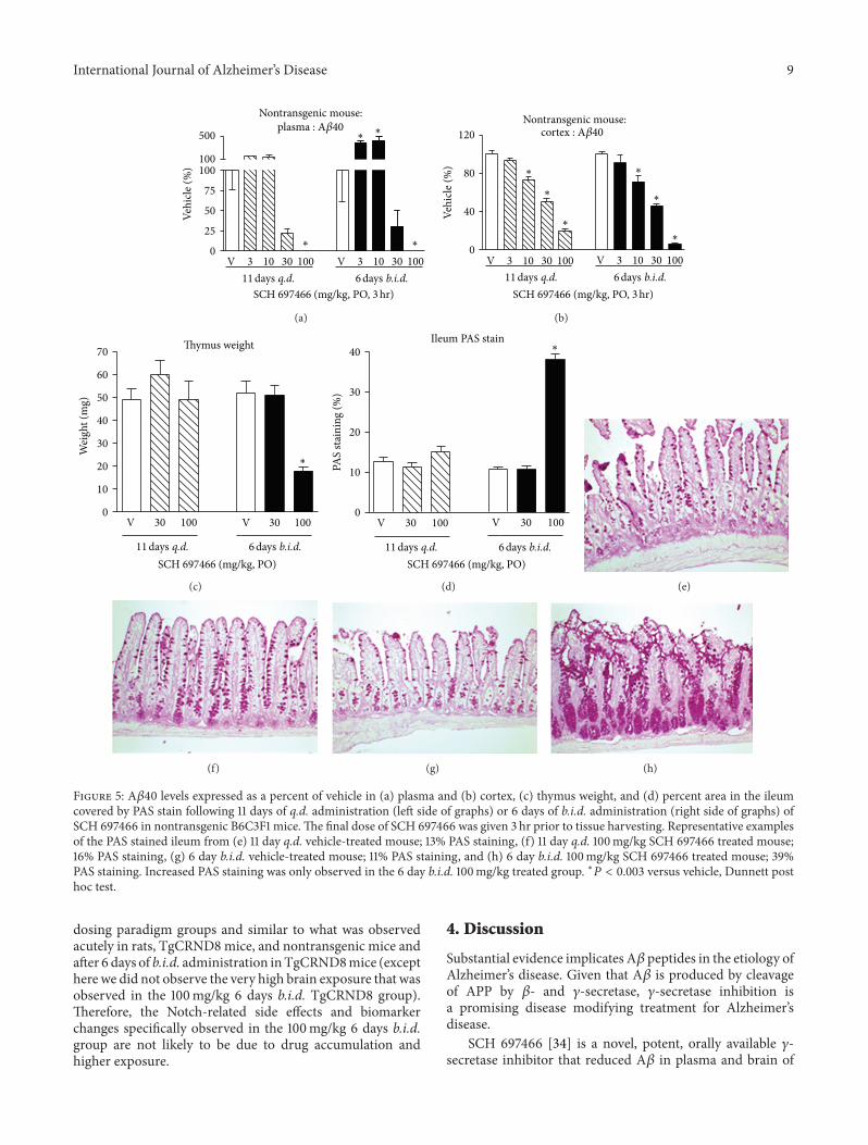
3.4.2. Notch-Related Side Effects. Six days of b.i.d. adminis-tration of SCH 697466 significantly reduced thymus weight(Figure 4(b); main effect of dose:𝐹(4, 19) = 15.59,𝑃 < 0.002)with a significant reduction observed in the 100mg/kg dosegroup (𝑃 < 0.003), but not the 30mg/kg group.
This dosing regime also increased PAS staining in theileum (Figure 4(c); main effect of dose: 𝐹(4, 17) = 91.34,𝑃 < 0.0001) such that therewas almost a 6-fold increase in the100mg/kg group compared to vehicle treated mice (Figures4(d) and 4(e); 𝑃 < 0.0001), but not the 30mg/kg group (justunder a 2-fold increase).
Further, mice treated with 100mg/kg of SCH 697466steadily lost weight during the 6 days of drug administration;100mg/kg mice lost 4.3 g over 6 days while vehicle-treatedmice lost only 1 g over the same time period and 66.6% ofthe mice in the 100mg/kg group died before the study wascompleted (compared to 12.5% in the vehicle treated group),and the surviving mice appeared in poor health by the lastday of the study (e.g., hypothermic, hunched posture, etc.).
3.4.3. Exposure. There was a dose-related increase (althoughin some cases it was more than dose proportional) in theconcentration of SCH 697466 in plasma and brain (onetime point per animal) (Table 3). Drug concentrations fol-lowing 6 days of b.i.d. administration were similar to thoseobserved following a single dose (3 hr) with the exceptionof the 100mg/kg group where there was much greater brainexposure following 6 days of b.i.d. dosing than following asingle dose (c.f., Tables 2 and 3). Due to the poor conditionof the mice, there was insufficient plasma to obtain exposuredata for mice in the 100mg/kg group.
3.5. Two Different Dosing Paradigms of SCH 697466 in Non-transgenic Mice Provided Equivalent Lowering of Plasma andCortex A𝛽 but Resulted in Different Observations of Notch-Related Side Effects and Biomarker Changes. The time coursestudy suggested that both q.d. and b.i.d. administration ofSCH 697466 at 100mg/kg would provide sustained and nearcomplete lowering of A𝛽, and the subchronic study suggestedthat Notch-related side effects would be observed underconditions of sustained and near complete lowering of A𝛽.In an effort to determine whether Notch-related side effectscould bemanaged by giving SCH 697466 q.d. instead of b.i.d.,mice were orally administered SCH 697466 either once a dayfor 11 days (11 days q.d.) or twice a day for 5 days with the finaldose on the 6th day (6 days b.i.d.) with both groups being
given a total of 11 doses. All tissues were collected 3-4 hr afterthe last dose. In addition, we assessed changes in expressionof genes downstream of Notch in the intestine and blood toassess the potential of these measures to be useful biomarkersfor Notch-related pathology. This study was conducted innontransgenic B6C3F1 mice since as noted above TgCRND8mice did not tolerate high doses of SCH 697466.
3.5.1. A𝛽 Lowering. For each dosing paradigm, the effects ofSCH 697466 onA𝛽 in the plasma of nontransgenicmice werenonlinear such that lower doses increased A𝛽40 levels whilehigher doses decreased A𝛽40 levels (Figure 5(a); main effectof dose: 𝐹(4, 19) = 11.02 and 𝐹(4, 21) = 38.43, 𝑃 < 0.0001for 11 days q.d. and 6 days b.i.d. groups, resp.). The effectsof SCH 697466 on A𝛽 in the plasma were similar betweenthe dosing paradigms with 50% lowering of A𝛽 occurring ata dose between 10 and 30mg/kg and complete lowering ofA𝛽 at 100mg/kg. Plasma A𝛽 lowering was similar to whatwas observed in TgCRND8 mice following a single dose andfollowing 6 days of b.i.d. drug administration. The increasein plasma A𝛽 observed at lower doses was more substantialin nontransgenic mice than what was observed in TgCRND8mice following 6 days of b.i.d. administration which has beenalluded to elsewhere [40].
In the cortex of nontransgenic mice, SCH 697466 dose-dependently loweredA𝛽40 in amore linear fashion following11 days of q.d. and 6 days of b.i.d. administration (Figure 5(a),main effect of dose: 𝐹(4, 19) = 105.53 and 𝐹(4, 21) = 101.04,resp., 𝑃 < 0.0001). Cortical A𝛽 lowering was strikingly sim-ilar between the dosing paradigms with 50% lowering occur-ring around 30mg/kg.The dose-responsive nature of corticalA𝛽 lowering observed here was similar to what was observedin TgCRND8mice but did not plateau like what was observedin acutely rats. There was no evidence of an increase in A𝛽 inthe cortex.
3.5.2. Notch-Related Side Effects. Eleven days of q.d. admin-istration of SCH 697466 at 30 and 100mg/kg did not affectthymus weight, while 6 days of b.i.d. administration signifi-cantly decreased thymus weight by about 60% (Figure 5(b);main effect of dose: 𝐹(2, 14) = 32.40, 𝑃 < 0.0001) withsignificant weight reduction in the 100mg/kg group, but notthe 30mg/kg group, compared to vehicle-treated mice (𝑃 <0.0001). This is similar to what was observed in TgCRND8mice treated for 6 days b.i.d. with 100mg/kg SCH 697466.
International Journal of Alzheimer’s Disease 7
0 4 8 12 16 20 240
50
100
150
PlasmaCortex
Vehi
cle (%
)
Postadministration time(hr, SCH 697466 30 mg/kg, PO)
(a)
TgCRND8 mouse: exposure
0 4 8 12 16 20 240
1
2
3
4
5
PlasmaBrain
Postadministration time(hr, SCH 697466 30 mg/kg, PO)
(b)
0 4 8 12 16 20 240
50
100
150
PlasmaCortex
Postadministration time
Vehi
cle (%
)
(hr, SCH 697466 100 mg/kg, PO)
(c)
TgCRND8 mouse: exposure
0 4 8 12 16 20 240
2
4
6
8
PlasmaBrain
Postadministration time(hr, SCH 697466 100 mg/kg, PO)
(d)
Figure 3: A𝛽40 levels over time expressed as a percent of vehicle in plasma and cortex following a single dose of (a) 30mg/kg or (c) 100mg/kgof SCH 697466 in TgCRND8 mice. Plasma and brain concentration (𝜇M) of SCH 697466 following a single dose of (b) 30mg/kg or (d)100mg/kg of SCH 697466 in TgCRND8 mice.
Similarly, PAS staining in the ileum was not increasedafter 11 days of q.d. administration of SCH 697466 at 30 and100mg/kg, but 100mg/kg SCH 697466 administered for 6days b.i.d. markedly increased PAS staining in the ileum byabout 350% (Figure 5(c); 𝐹(2, 14) = 180.01, 𝑃 < 0.0001;Figures 5(d)–5(g)). PAS staining in the 100mg/kg 6 days b.i.d.nontransgenic group looked very similar to that observed
in TgCRND8 mice following 100mg/kg 6 days b.i.d. SCH697466 (images not shown). PAS staining was not increasedin the 30mg/kg 6 days b.i.d. group.
Contrary to what as observed in the 6 day b.i.d.TgCRND8 study, there were no effects of SCH 697466 (up to100mg/kg) on body weight or survival in this nontransgenicstudy (data not shown).
8 International Journal of Alzheimer’s Disease
3 10 30 100 3 10 30 1000
25
50
75
100
125
150
175
Plasma Cortex
Veh
icle
(%
)
V V
∗
∗
∗
∗
∗
∗
SCH 697466 (mg/kg, PO, b.i.d., 6 days)
(a)
Thymus weight
3 10 30 1000
10
20
30
40
50
60
Wei
ght
(mg)
V
∗
SCH 697466 (mg/kg, PO, b.i.d., 6 days)
(b)
Ileum PAS stain
3 10 30 1000
10
20
30
40
PAS
stai
nin
g (%
)
V
∗
SCH 697466 (mg/kg, PO, b.i.d., 6 days)
(c)
Figure 4: (a) A𝛽40 levels expressed as a percent of vehicle in plasma and cortex, (b) thymus weight, and (c) percent area in the ileum coveredby PAS stain following 6 days of b.i.d. administration of SCH 697466 to TgCRND8 mice. The final dose of SCH 697466 was given on day six3 hr prior to tissue harvesting. ∗𝑃 < 0.0006 versus vehicle, Dunnett post hoc test.
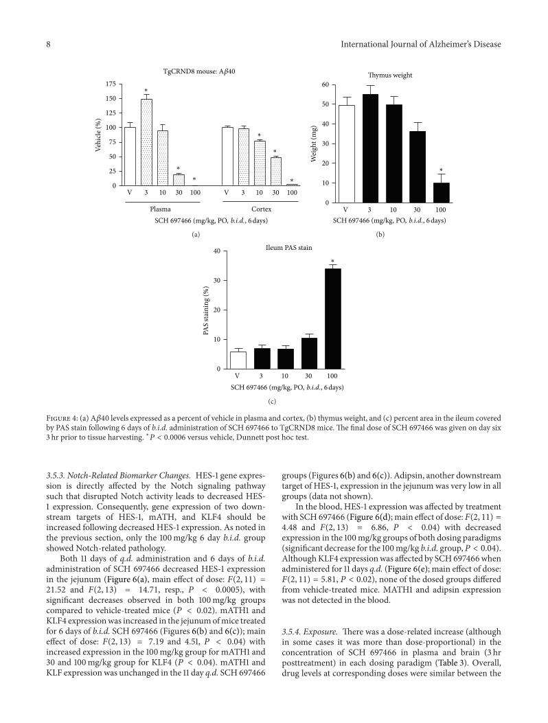
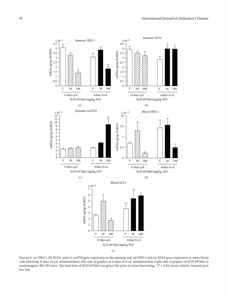
3.5.3. Notch-Related Biomarker Changes. HES-1 gene expres-sion is directly affected by the Notch signaling pathwaysuch that disrupted Notch activity leads to decreased HES-1 expression. Consequently, gene expression of two down-stream targets of HES-1, mATH, and KLF4 should beincreased following decreased HES-1 expression. As noted inthe previous section, only the 100mg/kg 6 day b.i.d. groupshowed Notch-related pathology.
Both 11 days of q.d. administration and 6 days of b.i.d.administration of SCH 697466 decreased HES-1 expressionin the jejunum (Figure 6(a), main effect of dose: 𝐹(2, 11) =21.52 and 𝐹(2, 13) = 14.71, resp., 𝑃 < 0.0005), withsignificant decreases observed in both 100mg/kg groupscompared to vehicle-treated mice (𝑃 < 0.02). mATH1 andKLF4 expressionwas increased in the jejunumofmice treatedfor 6 days of b.i.d. SCH 697466 (Figures 6(b) and 6(c)); maineffect of dose: 𝐹(2, 13) = 7.19 and 4.51, 𝑃 < 0.04) withincreased expression in the 100mg/kg group for mATH1 and30 and 100mg/kg group for KLF4 (𝑃 < 0.04). mATH1 andKLF expression was unchanged in the 11 day q.d. SCH 697466
groups (Figures 6(b) and 6(c)). Adipsin, another downstreamtarget of HES-1, expression in the jejunumwas very low in allgroups (data not shown).
In the blood, HES-1 expression was affected by treatmentwith SCH697466 (Figure 6(d);main effect of dose:𝐹(2, 11) =4.48 and 𝐹(2, 13) = 6.86, 𝑃 < 0.04) with decreasedexpression in the 100mg/kg groups of both dosing paradigms(significant decrease for the 100mg/kg b.i.d. group,𝑃 < 0.04).AlthoughKLF4 expressionwas affected by SCH697466whenadministered for 11 days q.d. (Figure 6(e); main effect of dose:𝐹(2, 11) = 5.81, 𝑃 < 0.02), none of the dosed groups differedfrom vehicle-treated mice. MATH1 and adipsin expressionwas not detected in the blood.
3.5.4. Exposure. There was a dose-related increase (althoughin some cases it was more than dose-proportional) in theconcentration of SCH 697466 in plasma and brain (3 hrposttreatment) in each dosing paradigm (Table 3). Overall,drug levels at corresponding doses were similar between the
International Journal of Alzheimer’s Disease 9
Nontransgenic mouse:
3 10 30 100 3 10 30 1000
25
50
75
100100
500
Veh
icle
(%
)
V V
SCH 697466 (mg/kg, PO, 3 hr)
11 days q.d. 6 days b.i.d.
∗∗
∗ ∗
(a)
Nontransgenic mouse:
3 10 30 100 3 10 30 1000
40
80
120
Veh
icle
(%
)
V V
SCH 697466 (mg/kg, PO, 3 hr)
11 days q.d. 6 days b.i.d.
∗
∗
∗
∗
∗
∗
(b)
Thymus weight
30 100 30 1000
10
20
30
40
50
60
70
Wei
ght
(mg)
V V
11 days q.d. 6 days b.i.d.
∗
SCH 697466 (mg/kg, PO)
(c)
Ileum PAS stain
30 100 30 1000
10
20
30
40
PAS
stai
nin
g (%
)
V V
11 days q.d. 6 days b.i.d.
∗
SCH 697466 (mg/kg, PO)
(d) (e)
(f) (g) (h)
Figure 5: A𝛽40 levels expressed as a percent of vehicle in (a) plasma and (b) cortex, (c) thymus weight, and (d) percent area in the ileumcovered by PAS stain following 11 days of q.d. administration (left side of graphs) or 6 days of b.i.d. administration (right side of graphs) ofSCH 697466 in nontransgenic B6C3F1 mice.The final dose of SCH 697466 was given 3 hr prior to tissue harvesting. Representative examplesof the PAS stained ileum from (e) 11 day q.d. vehicle-treated mouse; 13% PAS staining, (f) 11 day q.d. 100mg/kg SCH 697466 treated mouse;16% PAS staining, (g) 6 day b.i.d. vehicle-treated mouse; 11% PAS staining, and (h) 6 day b.i.d. 100mg/kg SCH 697466 treated mouse; 39%PAS staining. Increased PAS staining was only observed in the 6 day b.i.d. 100mg/kg treated group. ∗𝑃 < 0.003 versus vehicle, Dunnett posthoc test.
dosing paradigm groups and similar to what was observedacutely in rats, TgCRND8mice, and nontransgenic mice andafter 6 days of b.i.d. administration inTgCRND8mice (excepthere we did not observe the very high brain exposure that wasobserved in the 100mg/kg 6 days b.i.d. TgCRND8 group).Therefore, the Notch-related side effects and biomarkerchanges specifically observed in the 100mg/kg 6 days b.i.d.group are not likely to be due to drug accumulation andhigher exposure.
4. Discussion
Substantial evidence implicates A𝛽 peptides in the etiology ofAlzheimer’s disease. Given that A𝛽 is produced by cleavageof APP by 𝛽- and 𝛾-secretase, 𝛾-secretase inhibition isa promising disease modifying treatment for Alzheimer’sdisease.
SCH 697466 [34] is a novel, potent, orally available 𝛾-secretase inhibitor that reduced A𝛽 in plasma and brain of
10 International Journal of Alzheimer’s Disease
Jejunum: HES-1
30 100 30 1000
0.5
1
1.5
2
2.5
3
3.5
4
4.5
VV
11 days q.d. 6 days b.i.d.
∗
∗
SCH 697466 (mg/kg, PO)
× 10−4
mR
NA
ng/
ng
18 s
RN
A
(a)
Jejunum: KLF4
4.5
30 100 30 1000
0.5
1
1.5
2
2.5
3
3.5
4
VV
11 days q.d. 6 days b.i.d.
∗∗
SCH 697466 (mg/kg, PO)
× 10−4
mR
NA
ng/
ng
18 s
RN
A
(b)
Jejunum: mATH1
0123456789
101112
30 100 30 100VV
11 days q.d. 6 days b.i.d.
∗
SCH 697466 (mg/kg, PO)
× 10−4
mR
NA
ng/
ng
18 s
RN
A
(c)
Blood: HES-1
0
2.5
5
7.5
10
∗
30 100 30 100VV
11 days q.d. 6 days b.i.d.SCH 697466 (mg/kg, PO)
× 10− 6
mR
NA
ng/
ng
18 s
RN
A
(d)
Blood: KLF4
0
1
2
3
4
5
6
7
30 100 30 100VV
11 days q.d. 6 days b.i.d.SCH 697466 (mg/kg, PO)
× 10−6
mR
NA
ng/
ng
18 s
RN
A
(e)
Figure 6: (a) HES-1, (b) KLF4, and (c) mATH gene expression in the jejunum and (d) HES-1 and (e) KLF4 gene expression in white bloodcells following 11 days of q.d. administration (left side of graphs) or 6 days of b.i.d. administration (right side of graphs) of SCH 697466 innontransgenic B6C3F1 mice. The final dose of SCH 697466 was given 3 hr prior to tissue harvesting. ∗𝑃 < 0.04 versus vehicle, Dunnett posthoc test.
International Journal of Alzheimer’s Disease 11
rats, preplaque TgCRND8 mice, and nontransgenic mice.The acute effects of SCH 697466 on A𝛽 levels were relativelysimilar between the rodent species and genotypes with about50% lowering of A𝛽 in plasma, cortex, and CSF occurringbetween 10 and 30mg/kg (or between ∼0.4 and 3 𝜇M plasmaconcentration of SCH 697466) and near complete loweringof A𝛽 occurring at 100mg/kg (or about 5–8 𝜇M plasmaconcentration). Therefore, as has been reported for other 𝛾-secretase inhibitors, SCH697466was effective at reducingA𝛽in vivo.
Although 𝛾-secretase inhibitors reduceA𝛽well in periph-eral and central compartments, some 𝛾-secretase inhibitorsalso produce mechanism-based side effects including areduction in thymus size and an increase in intestinal gobletcell hyperplasia [5, 27–29]. Both of these effects are likelymediated by a disruption of Notch processing [30, 41, 42].Based on our experience in rodents, these Notch-relatedside effects seem to be typically observed at doses that givecontinuous, near complete lowering A𝛽 in plasma for at least3 days [5]. Alternatively, we and others have shown that“partial inhibition” doses of these same compounds providereasonable lowering of A𝛽 (about >50% reduction) withoutNotch-related side effects [5, 18]. However, it is not clear howpartial inhibition can best be applied in the managementof Notch-related side effects, that is, is it preferable to aimfor incomplete inhibition for a sustained period of time orcomplete inhibition for a short period of time?Therefore, weconducted additional subchronic studies with SCH 697466to address this question as well as to assess the in vivotherapeutic window of this compound.
As was observed acutely in TgCRND8 mice, subchronicb.i.d. administration of SCH 697466 dose-dependently low-ered A𝛽 in plasma and cortex with about 50% inhibitionoccurring between 10 and 30mg/kg (or between 2–10 𝜇Mplasma exposure). SCH 697466 given at 100mg/kg b.i.d.provided sustained and almost complete lowering of plasmaA𝛽, but also produced Notch-related side effects, includinga reduction in thymus size and increased goblet cell hyper-plasia. Alternatively, SCH 697466 given at 30mg/kg b.i.d. toTgCRND8 mice provided sustained, although not complete,lowering of A𝛽 (48% reduction in cortex) without Notch-related side effects. Similar results were previously reportedwith another 𝛾-secretase inhibitor, LY-411,575 [5]. Thus,although both the 30 and 100mg/kg doses of SCH 697466provided sustained lowering of A𝛽, only the dose that alsoprovided nearly complete lowering (100mg/kg) producedNotch-related side effects, while the “partial” inhibition dose(30mg/kg) did not. In other words, a short duration of actiondoes not seem to be required to mitigate Notch-related sideeffects, just incomplete inhibition.
Interestingly, although SCH 697466 had a reasonablein vitro separation between IC
50s for A𝛽 lowering and
Notch processing (∼24-fold), this did not translate in vivo.After subchronic 6 day b.i.d. administration of SCH 697466,the ED
50for reducing brain A𝛽40 was about 30mg/kg
and Notch-related side effects were observed at 100mg/kg.Therefore, although there was a clear therapeutic window forSCH 697466 in vivo, it was small (∼3-fold based on dose and∼5-fold based on plasma concentrations) and significantly
less than the therapeutic window predicted by in vitroexperiments. It is important to note that SCH 697466 wasless potent at lowering the more amyloidogenic A𝛽42 speciesespecially in brain suggesting that the in vivo therapeuticindex will likely be smaller if A𝛽42 data were used instead.
Given that the duration of action of SCH 697466 at100mg/kg is relatively long (>24 hr), both b.i.d. and q.d.administration would provide sustained and near completelowering of A𝛽. It was hypothesized, however, that q.d.administration of SCH 697466 might decrease the incidenceofNotch-related side effects compared to those observedwithb.i.d. administration while maintaining excellent lowering ofA𝛽 (>80% reduction). Indeed, in a study with nontransgenicmice, although lowering of plasma and brain A𝛽 was verysimilar between the b.i.d. and q.d. dosing paradigms, theNotch-related side effects were strikingly different such thatNotch-related pathology was only observed at 100mg/kgwhen administered b.i.d. and not q.d. This did not seem tobe due to large increases in exposure or drug accumulationin the b.i.d. group. Of note, although nontransgenic micetolerated high doses of SCH 697466 better than TgCRND8mice as measured by body weight loss and survival, bothgroups of mice showed similar reduction in thymus weightand increase in intestinal goblet cell hyperplasia at 100mg/kgb.i.d. Thus, Notch-related side effects can be managed evenwith sustained and near complete lowering of A𝛽 by opti-mizing the frequency of dosing of 𝛾-secretase inhibitors.However, it cannot be completely excluded that longerterm administration of 𝛾-secretase inhibitors even with anoptimized frequency of dosing might eventually result inNotch-related side effects; further studies would be requiredto address this possibility.
Recent data suggest that there may be additional optionsfor mitigating Notch-related side effects with 𝛾-secretaseinhibition. A study by Das et al. [43] showed that treatingtransgenic mice with the 𝛾-secretase inhibitor LY-411,575 fora short period of time during a key, early phase of amyloidaccumulation resulted in long-lasting beneficial effects onbrain A𝛽 without the need for continuous drug treatment.These data along with our own suggest that there may beways to safely and effectively lower brain A𝛽with 𝛾-secretaseinhibition. Further studies would be required to determinehow useful these approaches would be in the clinic.
Based on our findings that Notch-related side effectscan be managed with sustained but incomplete loweringof A𝛽, or near complete lowering of A𝛽 but with moreintermittent administration, it is possible that some of the“Notch-sparing” and “APP-selective” 𝛾-secretase inhibitorsreported in the literature may be managing Notch-relatedside effects by using these strategies instead of the moleculesreally being “Notch sparing” in vivo. For example, after 7 daysof b.i.d. dosing of ELN 475516, an “APP-selective” 𝛾-secretaseinhibitor, there was no evidence of Notch-related toxicity asmeasured by thymus weight and goblet cell hyperplasia in theileum and the doses and frequency of drug administrationutilized provided sustained but incomplete (∼74%) loweringof A𝛽 [10]. Perhaps if this compound was able to obtainnear complete lowering of A𝛽 in a sustained fashion, Notch-related side effects may have indeed been observed.

12 International Journal of Alzheimer’s Disease
It is important to note that in these studies, we wereusing reduction in thymus size and intestinal goblet cellhyperplasia as representativemeasures of “Notch-related sideeffects.” However, it is possible that there are other toxicitiesor effects related to 𝛾-secretase inhibition and/or alteredNotch signaling that we did not measure and that maynot be mitigated or avoided with partial inhibition and/orintermittent dosing paradigms. Further, the translatabilityof these treatment approaches or mitigation strategies inrodents to humans is not clear.
Although some Notch-related side effects are reversible[5], it would be useful to have a clinically tractable biomarkerto monitor potential effects of 𝛾-secretase inhibitors onNotch processing in the clinic. HES-1 gene expression isdirectly affected by the Notch signaling pathway such thatdisruptedNotch activity leads to decreasedHES-1 expression.Consequently, gene expression of two downstream targets ofHES-1, mATH and KLF4, is increased following decreasedHES-1 expression [32, 33]. In certain cases, the gene expres-sion profiles of these Notch-related biomarkers paralleledthe Notch-related pathology observed. For example, the100mg/kg dose of SCH 697466 when given b.i.d. inducedNotch-related pathology and also decreased HES-1 in thejejunum and in the blood and increased mATH1 and KLF4expression in the jejunum. The biomarker data did notcompletely align with the in vivo data though, for example,HES-1 gene expression was decreased in the jejunum of micetreated with 100mg/kg q.d. when no pathology was observedand KLF4 gene expression was increased inmice treated with30mg/kg b.i.d. when no pathology was observed and whenHES-1 expression (upstream of KLF4) was not decreased. It isalso possible that the biomarker changes that were observedin the absence of pathology reflect relevant changes in Notchsignaling that after a longer period of dosing would indeedresult in observations of Notch-related pathology. In general,however, when clear Notch-related pathology was observed,changes in Notch-related biomarkers were simultaneouslyobserved. This is in agreement with other reports usingexpression of these same genes as potential biomarkers forNotch-related pathology [10].
Interestingly and perhaps paradoxically in several studiesan increase in plasma A𝛽was observed at lower doses of SCH697466 (e.g., 3 and 10mg/kg). This increase in plasma A𝛽 or“A𝛽 rise” has been frequently observed with other 𝛾-secretaseinhibitors in vivo, including in humans, and occurs at lowconcentrations likely under conditions of partial enzymeoccupancy [15, 40, 44, 45]. In addition, the A𝛽 rise was moreprominent in nontransgenic mice than transgenic mice ashas been supported elsewhere [40, 44]. Importantly, we neverobserved an increase in A𝛽 levels in the cortex with SCH697466.
To summarize, SCH 697466 is a novel, potent 𝛾-secretaseinhibitor that is effective at reducing A𝛽 in plasma, CSF,and brain in rodents. SCH 697466 has a small, but distinct,in vivo therapeutic index between A𝛽 lowering and Notch-related side effects in the ileum and thymus in mice. Apartial inhibition dose of SCH 697466 provided reasonablereduction of A𝛽 (∼50%) that was sustained over 24 hr whengiven b.i.d. without evidence of Notch-related side effects.
Although the reasons are not entirely clear, q.d. dosingprovided another way to maintain excellent, continuousinhibition of A𝛽 (>80%) with SCH 697466, but withoutNotch-related side effects. Therefore, by utilizing partial,but sustained, inhibition doses or optimizing the frequencyof administration, it may be possible to obtain robust andsustained lowering of A𝛽 without Notch-related side effectsusing 𝛾-secretase inhibitors. Finally, HES-1 expression inblood may be a useful Notch-related biomarker in the clinic.
Acknowledgments
The authors would like to thank Marco Baptista, CarinaBleickardt, Benjamin Eschle, Nicholas Jones, Sherry Lu, andJulie Strain for help with the tissue collections; Howard Jonesand Marie Sondey for their help with some of the histology;RonaldManning for breeding and genotyping the TgCRND8mice; and Greg Tucker, Lucy Xu, Jing Lan, James Jean, andLynn Rogers for analyzing the plasma and brain samples.
References
[1] D. J. Selkoe, “The molecular pathology of Alzheimer’s disease,”Neuron, vol. 6, no. 4, pp. 487–498, 1991.
[2] J. A. Hardy andG. A. Higgins, “Alzheimer’s disease: the amyloidcascade hypothesis,” Science, vol. 256, no. 5054, pp. 184–185,1992.
[3] J. Hardy and D. J. Selkoe, “The amyloid hypothesis ofAlzheimer’s disease: progress and problems on the road totherapeutics,” Science, vol. 297, no. 5580, pp. 353–356, 2002.
[4] D. M. Barten, J. E. Meredith Jr., R. Zaczek, J. G. Houston, andC. F. Albright, “𝛾-secretase inhibitors for Alzheimer’s disease:balancing efficacy and toxicity,” Drugs in R and D, vol. 7, no. 2,pp. 87–97, 2006.
[5] L. A. Hyde, N. A. McHugh, J. Chen et al., “Studies to investigatethe in vivo therapeutic window of the 𝛾-secretase inhibitor N2-[(2S)-2-(3,5-difluorophenyl)-2-hydroxyethanoyl]-N1-[(7S)-5-methyl-6-oxo-6,7-dihydro-5H-dibenzo[b,d] azepin-7-yl]-L-alaninamide (LY411,575) in the CRND8 mouse,” Journal ofPharmacology and Experimental Therapeutics, vol. 319, no. 3,pp. 1133–1143, 2006.
[6] J. D. Best, M. T. Jay, F. Otu et al., “In vivo characterizationof A𝛽(40) changes in brain and cerebrospinal fluid usingthe novel 𝛾-secretase inhibitor N-[cis-4-[(4-chlorophenyl)sul-fonyl]-4-(2,5-difluorophenyl)cyclohexyl]-1,1,1-trifluoromethan-esulfonamide (MRK-560) in the rat,” Journal of Pharmacologyand Experimental Therapeutics, vol. 317, no. 2, pp. 786–790,2006.
[7] C. V. C. Prasad, M. Zheng, S. Vig et al., “Discovery of (S)-2-((S)-2-(3,5-difluorophenyl)-2-hydroxyacetamido)-N-((S,Z)-3-methyl-4-oxo-4,5-dihydro-3H-benzo[d][1,2]diazepin-5-yl)propanamide (BMS-433796): a 𝛾-secretase inhibitor with A𝛽lowering activity in a transgenic mouse model of Alzheimer’sdisease,” Bioorganic and Medicinal Chemistry Letters, vol. 17,no. 14, pp. 4006–4011, 2007.
[8] R. L. Martone, H. Zhou, K. Atchison- et al., “Begacestat (GSI-953): a novel, selective thiophene sulfonamide inhibitor of amy-loid precursor protein gamma-secretase for the treatment ofAlzheimer’s disease,” Journal of Pharmacology and ExperimentalTherapeutics, vol. 331, no. 2, pp. 598–608, 2009.
International Journal of Alzheimer’s Disease 13
[9] K. W. Gillman, J. E. Starrett, M. F. Parker et al., “Discoveryand evaluation of BMS-708163, a potent, selective and orallybioavailable I3-Secretase Inhibitor,” ACS Medicinal ChemistryLetters, vol. 1, no. 3, p. 120, 2010.
[10] G. S. Basi, S. Hemphill, E. F. Brigham et al., “Amyloid precursorprotein selective gamma-secretase inhibitors for treatment ofAlzheimer’s disease,”Alzheimer’s Research &Therapy, vol. 2, no.6, p. 36, 2010.
[11] T. A. Lanz, K. M. Wood, K. E. G. Richter et al., “Pharmacody-namics and pharmacokinetics of the 𝛾-secretase inhibitor PF-3084014,” Journal of Pharmacology and ExperimentalTherapeu-tics, vol. 334, no. 1, pp. 269–277, 2010.
[12] Y. Mitani, J. Yarimizu, K. Saita et al., “Differential effectsbetween gamma-secretase inhibitors and modulators on cog-nitive function in amyloid precursor protein-transgenic andnontransgenic mice,” Journal of Neuroscience, vol. 32, no. 6, pp.2037–2050, 2012.
[13] D. B. Henley, P. C. May, R. A. Dean, and E. R. Siemers,“Development of semagacestat (LY450139), a functional 𝛾-secretase inhibitor, for the treatment of Alzheimer’s disease,”Expert Opinion on Pharmacotherapy, vol. 10, no. 10, pp. 1657–1664, 2009.
[14] R. J. Bateman, E. R. Siemers, K. G. Mawuenyega et al., “A𝛾-secretase inhibitor decreases amyloid-𝛽 production in thecentral nervous system,” Annals of Neurology, vol. 66, no. 1, pp.48–54, 2009.
[15] G. Tong, J. S. Wang, O. Sverdlov et al., “Multicenter, ran-domized, double-blind, placebo-controlled, single-ascendingdose study of the oral gamma-secretase inhibitor BMS-708163(Avagacestat): tolerability profile, pharmacokinetic parameters,and pharmacodynamic markers,” Clinical Therapeutics, vol. 34,no. 3, pp. 654–667, 2012.
[16] A. S. Fleisher, R. Raman, E. R. Siemers et al., “Phase 2 safetytrial targeting amyloid beta production with a gamma-secretaseinhibitor in Alzheimer disease,” Archives of Neurology, vol. 65,no. 8, pp. 1031–1038, 2008.
[17] E. R. Siemers, J. F. Quinn, J. Kaye et al., “Effects of a 𝛾-secretaseinhibitor in a randomized study of patients with Alzheimerdisease,” Neurology, vol. 66, no. 4, pp. 602–604, 2006.
[18] J. D. Best, D. W. Smith, M. A. Reilly et al., “The novel 𝛾secretase inhibitor N-[cis-4-[(4-chlorophenyl)sulfonyl]-4-(2,5-difluorophenyl)cyclohexyl]-1,1,1-trifluoromethanesulfonamide(MRK-560) reduces amyloid plaque deposition withoutevidence of notch-related pathology in the Tg2576 mouse,”Journal of Pharmacology and Experimental Therapeutics, vol.320, no. 2, pp. 552–558, 2007.
[19] D. Abramowski, K. H. Wiederhold, U. Furrer et al., “Dynamicsof A𝛽 turnover and deposition in different 𝛽-amyloid precursorprotein transgenic mouse models following 𝛾-secretase inhibi-tion,” Journal of Pharmacology and Experimental Therapeutics,vol. 327, no. 2, pp. 411–424, 2008.
[20] S. J. Pollack and H. Lewis, “𝛾-Secretase inhibitors forAlzheimer’s disease: challenges of a promiscuous protease,”Current Opinion in Investigational Drugs, vol. 6, no. 1, pp. 35–47,2005.
[21] A. Lleo and C. A. Saura, “gamma-secretase substrates andtheir implications for drug development inAlzheimer’s disease,”Current Topics in Medicinal Chemistry, vol. 11, no. 12, pp. 1513–1527, 2011.
[22] B. De Strooper, W. Annaert, P. Cupers et al., “A presenilin-1-dependent 𝛾-secretase-like protease mediates release of notch
intracellular domain,” Nature, vol. 398, no. 6727, pp. 518–522,1999.
[23] S. Artavanis-Tsakonas, M. D. Rand, and R. J. Lake, “Notch sig-naling: cell fate control and signal integration in development,”Science, vol. 284, no. 5415, pp. 770–776, 1999.
[24] D. C. Cole, J. R. Stock, A. F. Kreft et al., “(S)-N-(5-Chlo-rothiophene-2-sulfonyl)-𝛽,𝛽-diethylalaninol a Notch-1-sparing𝛾-secretase inhibitor,” Bioorganic and Medicinal Chemistry Let-ters, vol. 19, no. 3, pp. 926–929, 2009.
[25] A. Kreft, B. Harrison, S. Aschmies et al., “Discovery of a novelseries of Notch-sparing 𝛾-secretase inhibitors,” Bioorganic andMedicinal Chemistry Letters, vol. 18, no. 14, pp. 4232–4236, 2008.
[26] S. C. Mayer, A. F. Kreft, B. Harrison et al., “Discovery of begace-stat, a Notch-1-sparing 𝛾-secretase inhibitor for the treatment ofAlzheimer’s disease,” Journal of Medicinal Chemistry, vol. 51, no.23, pp. 7348–7351, 2008.
[27] G. H. Searfoss, W. H. Jordan, D. O. Calligaro et al., “Adipsin, abiomarker of gastrointestinal toxicity mediated by a functionalgamma-secretase inhibitor,” Journal of Biological Chemistry, vol.278, no. 46, pp. 46107–46116, 2003.
[28] G. T. Wong, D. Manfra, F. M. Poulet et al., “Chronic treatmentwith the gamma-secretase inhibitor LY-411,575 inhibits beta-amyloid peptide production and alters lymphopoiesis andintestinal cell differentiation,” Journal of Biological Chemistry,vol. 279, no. 13, pp. 12876–12882, 2004.
[29] J. Milano, J. McKay, C. Dagenais et al., “Modulation of Notchprocessing by 𝛾-secretase inhibitors causes intestinal gobletcell metaplasia and induction of genes known to specify gutsecretory lineage differentiation,” Toxicological Sciences, vol. 82,no. 1, pp. 341–358, 2004.
[30] J. H. Van Es, M. E. Van Gijn, O. Riccio et al., “Notch/𝛾-secretase inhibition turns proliferative cells in intestinal cryptsand adenomas into goblet cells,” Nature, vol. 435, no. 7044, pp.959–963, 2005.
[31] M. Katoh and M. Katoh, “Notch signaling in gastrointestinaltract (Review),” International Journal of Oncology, vol. 30, no.1, pp. 247–251, 2007.
[32] H. Zheng,D.M. Pritchard, X. Yang et al., “KLF4 gene expressionis inhibited by the notch signaling pathway that controls gobletcell differentiation in mouse gastrointestinal tract,” AmericanJournal of Physiology, vol. 296, no. 3, pp. G490–G498, 2009.
[33] C. A. Richmond and D. T. Breault, “Regulation of gene expres-sion in the intestinal epithelium,” Progress in Molecular Biologyand Translational Science, vol. 96, pp. 207–229, 2010.
[34] T. Asberom, Z. Zhao, T. A. Bara et al., “Discovery of 𝛾-secretase inhibitors efficacious in a transgenic animal modelof Alzheimer’s disease,” Bioorganic and Medicinal ChemistryLetters, vol. 17, no. 2, pp. 511–516, 2007.
[35] M. A. Chishti, D. S. Yang, C. Janus et al., “Early-onset amyloiddeposition and cognitive deficits in transgenicmice expressing adouble mutant form of amyloid precursor protein 695,” Journalof Biological Chemistry, vol. 276, no. 24, pp. 21562–21570, 2001.
[36] L. A. Hyde, T. M. Kazdoba, M. Grilli et al., “Age-progressingcognitive impairments and neuropathology in transgenicCRND8 mice,” Behavioural Brain Research, vol. 160, no. 2, pp.344–355, 2005.
[37] T. A. Lanz, C. S. Himes, G. Pallante et al., “The 𝛾-secre-tase inhibitor N-[N-(3,5-difluorophenacetyl)-lalanyl]-S-phe-nylglycine t-butyl ester reduces A𝛽 levels in vivo in plasma andcerebrospinal fluid in young (plaque-free) and aged (plaque-bearing) Tg2576 mice,” Journal of Pharmacology and Experi-mental Therapeutics, vol. 305, no. 3, pp. 864–871, 2003.
14 International Journal of Alzheimer’s Disease
[38] D. M. Barten, V. L. Guss, J. A. Corsa et al., “Dynamics of 𝛽-amyloid reductions in brain, cerebrospinal fluid, and plasma of𝛽-amyloid precursor protein transgenic mice treated with a 𝛾-secretase inhibitor,” Journal of Pharmacology and ExperimentalTherapeutics, vol. 312, no. 2, pp. 635–643, 2005.
[39] L. Zhang, L. Song, G. Terracina, Y. Liu, B. Pramanik, and E.Parker, “Biochemical characterization of the 𝛾-secretase activitythat produces 𝛽-amyloid peptides,” Biochemistry, vol. 40, no. 16,pp. 5049–5055, 2001.
[40] C. R. Burton, J. E. Meredith, D. M. Barten et al., “The amyloid-𝛽 rise and 𝛾-secretase inhibitor potency depend on the level ofsubstrate expression,” Journal of Biological Chemistry, vol. 283,no. 34, pp. 22992–23003, 2008.
[41] J. Jensen, E. E. Pedersen, P. Galante et al., “Control of endoder-mal endocrine development by Hes-1,” Nature Genetics, vol. 24,no. 1, pp. 36–44, 2000.
[42] S. Fre, M. Huyghe, P. Mourikis, S. Robine, D. Louvard, and S.Artavanis-Tsakonas, “Notch signals control the fate of immatureprogenitor cells in the intestine,” Nature, vol. 435, no. 7044, pp.964–968, 2005.
[43] P. Das, C. Verbeeck, L. Minter et al., “Transient pharmacologiclowering of Abeta production prior to deposition results insustained reduction of amyloid plaque pathology,” MolecularNeurodegeneration, vol. 7, p. 39, 2012.
[44] T. A. Lanz, M. J. Karmilowicz, K. M. Wood et al., “Con-centration-dependent modulation of amyloid-𝛽 in vivo andin vitro using the 𝛾-secretase inhibitor, LY-450139,” Journal ofPharmacology and ExperimentalTherapeutics, vol. 319, no. 2, pp.924–933, 2006.
[45] E. Siemers,M. Skinner, R. A.Dean et al., “Safety, tolerability, andchanges in amyloid𝛽 concentrations after administration of a 𝛾-secretase inhibitor in volunteers,” Clinical Neuropharmacology,vol. 28, no. 3, pp. 126–132, 2005.
Hindawi Publishing CorporationInternational Journal of Alzheimer’s DiseaseVolume 2012, Article ID 591392, 7 pagesdoi:10.1155/2012/591392
Review Article
γ-Secretase-Dependent Proteolysis of TransmembraneDomain of Amyloid Precursor Protein: Successive Tri- andTetrapeptide Release in Amyloid β-Protein Production
Mako Takami1, 2 and Satoru Funamoto1
1 Department of Neuropathology, Graduate School of Life and Medical Sciences, Doshisha University, Kyotanabe,Kyoto 610-0934, Japan
2 Pharma Eight Co. Ltd., Kyoto, Kyoto 602-0841, Japan
Correspondence should be addressed to Satoru Funamoto, [email protected]
Received 13 September 2012; Revised 27 November 2012; Accepted 12 December 2012
Academic Editor: Jeremy Toyn
Copyright © 2012 M. Takami and S. Funamoto. This is an open access article distributed under the Creative CommonsAttribution License, which permits unrestricted use, distribution, and reproduction in any medium, provided the original work isproperly cited.
γ-Secretase cleaves the carboxyl-terminal fragment (βCTF) of APP not only in the middle of the transmembrane domain (γ-cleavage), but also at sites close to the membrane/cytoplasm boundary (ε-cleavage), to produce the amyloid β protein (Aβ) andthe APP intracellular domain (AICD), respectively. The AICD49–99 and AICD50–99 species were identified as counterparts ofthe long Aβ species Aβ48 and Aβ49, respectively. We found that Aβ40 and AICD50–99 were the predominant species in cellsexpressing wild-type APP and presenilin, whereas the production of Aβ42 and AICD49–99 was enhanced in cells expressingfamilial Alzheimer’s disease mutants of APP and presenilin. These long Aβ species were identified in cell lysates and mouse brainextracts, which suggests that ε-cleavage is the first cleavage of βCTF to produce Aβ by γ-secretase. Here, we review the progress ofresearch on the mechanism underlying the proteolysis of the APP transmembrane domain based on tri- and tetrapeptide release.
1. Introduction
The amyloid precursor protein (APP) is a type I membraneprotein. After ectodomain shedding by β-secretase, thecarboxyl-terminal fragment (βCTF) of APP becomes a directsubstrate of γ-secretase and is processed into the amyloidβ protein (Aβ) and the APP intracellular domain (AICD)[1–5]. γ-secretase is an enigmatic protease composed ofpresenilin 1/2, nicastrin, Aph-1, and Pen-2 that catalyzesproteolysis in the hydrophobic environment of the lipidbilayer [6–15]. Currently, over 50 molecules are reportedas γ-secretase substrates, which reflects the physiologicalimportance of this enzyme [16]. For instance, the Notchreceptor on the plasma membrane is cleaved by γ-secretaseupon ligand binding and the liberated Notch intracellulardomain (NICD) translocates into the nucleus and activatesthe expression of transcription factors to suppress neu-ronal differentiation [17, 18]. This indicates that inhibitionof γ-secretase for suppression of Aβ production causes
harmful side effects. To avoid this risk in anti-Alzheimer’sdisease (AD) therapeutics, it is very important to elucidatethe molecular mechanism underlying γ-secretase-dependentproteolysis. Recently, it was revealed that γ-secretase forms ahydrophilic pore and three water-accessible cavities [19–23].Here, we review the progress of research on the mechanismunderlying the proteolysis of the transmembrane domain ofβCTF.
2. Discovery of ε-Cleavage duringAPP Processing
After the β-secretase-dependent cleavage of APP, the ecto-domain of APP is released into the extracellular space andβCTF (as a stub in the lipid bilayer) is the direct substrate ofγ-secretase [2, 3, 24]. βCTF is composed of 99 amino acidsand is eventually processed into the 38–43-residue-long Aβ,suggesting that the counterparts of those Aβ species should
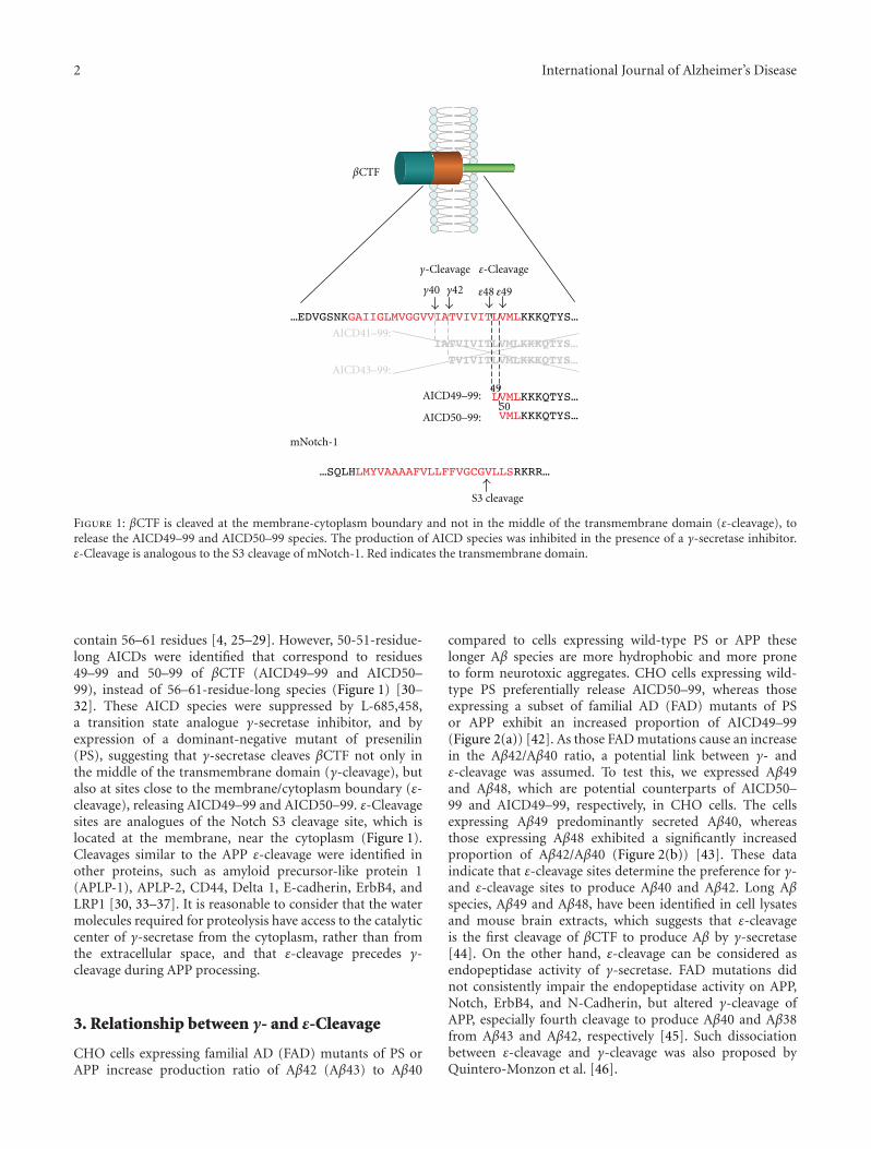
2 International Journal of Alzheimer’s Disease
βCTF
γ-Cleavage ε-Cleavage
γ40 γ42 ε48 ε49
AICD41–99:
AICD43–99:
mNotch-1
S3 cleavage
49
50AICD49–99:
AICD50–99:
Figure 1: βCTF is cleaved at the membrane-cytoplasm boundary and not in the middle of the transmembrane domain (ε-cleavage), torelease the AICD49–99 and AICD50–99 species. The production of AICD species was inhibited in the presence of a γ-secretase inhibitor.ε-Cleavage is analogous to the S3 cleavage of mNotch-1. Red indicates the transmembrane domain.
contain 56–61 residues [4, 25–29]. However, 50-51-residue-long AICDs were identified that correspond to residues49–99 and 50–99 of βCTF (AICD49–99 and AICD50–99), instead of 56–61-residue-long species (Figure 1) [30–32]. These AICD species were suppressed by L-685,458,a transition state analogue γ-secretase inhibitor, and byexpression of a dominant-negative mutant of presenilin(PS), suggesting that γ-secretase cleaves βCTF not only inthe middle of the transmembrane domain (γ-cleavage), butalso at sites close to the membrane/cytoplasm boundary (ε-cleavage), releasing AICD49–99 and AICD50–99. ε-Cleavagesites are analogues of the Notch S3 cleavage site, which islocated at the membrane, near the cytoplasm (Figure 1).Cleavages similar to the APP ε-cleavage were identified inother proteins, such as amyloid precursor-like protein 1(APLP-1), APLP-2, CD44, Delta 1, E-cadherin, ErbB4, andLRP1 [30, 33–37]. It is reasonable to consider that the watermolecules required for proteolysis have access to the catalyticcenter of γ-secretase from the cytoplasm, rather than fromthe extracellular space, and that ε-cleavage precedes γ-cleavage during APP processing.
3. Relationship between γ- and ε-Cleavage
CHO cells expressing familial AD (FAD) mutants of PS orAPP increase production ratio of Aβ42 (Aβ43) to Aβ40
compared to cells expressing wild-type PS or APP theselonger Aβ species are more hydrophobic and more proneto form neurotoxic aggregates. CHO cells expressing wild-type PS preferentially release AICD50–99, whereas thoseexpressing a subset of familial AD (FAD) mutants of PSor APP exhibit an increased proportion of AICD49–99(Figure 2(a)) [42]. As those FAD mutations cause an increasein the Aβ42/Aβ40 ratio, a potential link between γ- andε-cleavage was assumed. To test this, we expressed Aβ49and Aβ48, which are potential counterparts of AICD50–99 and AICD49–99, respectively, in CHO cells. The cellsexpressing Aβ49 predominantly secreted Aβ40, whereasthose expressing Aβ48 exhibited a significantly increasedproportion of Aβ42/Aβ40 (Figure 2(b)) [43]. These dataindicate that ε-cleavage sites determine the preference for γ-and ε-cleavage sites to produce Aβ40 and Aβ42. Long Aβspecies, Aβ49 and Aβ48, have been identified in cell lysatesand mouse brain extracts, which suggests that ε-cleavageis the first cleavage of βCTF to produce Aβ by γ-secretase[44]. On the other hand, ε-cleavage can be considered asendopeptidase activity of γ-secretase. FAD mutations didnot consistently impair the endopeptidase activity on APP,Notch, ErbB4, and N-Cadherin, but altered γ-cleavage ofAPP, especially fourth cleavage to produce Aβ40 and Aβ38from Aβ43 and Aβ42, respectively [45]. Such dissociationbetween ε-cleavage and γ-cleavage was also proposed byQuintero-Monzon et al. [46].
International Journal of Alzheimer’s Disease 3
βCTF
γ40 γ42
Aβ40
Aβ40
βCTF
Aβ42
Aβ42
Aβ40
Aβ42
AICD50–99
AICD49–99
AICD50–99
AICD49–99
AICD49–99
γ40 γ42 ε48 ε49ε48 ε49
(a)
Aβ40
Aβ49
Aβ42
Aβ48
γ40 γ42 ε49 γ40 γ42 ε48
Aβ40/Aβ42 Aβ40/Aβ42
(b)
Figure 2: Relationship between γ- and ε-cleavage. (a) Cells expressing wild-type PS or APP predominantly produce Aβ40 and AICD50–99,while cells expressing a FAD mutant of PS or APP exhibited increased proportion of Aβ42 and AICD49–99. (b) Expression of Aβ49 resultsin an increase in Aβ40/Aβ42 ratio, whereas expression of Aβ48 leads to opposite results. ↗ increase, ↘ decrease.
βCTF1
1Aβ42
1Aβ40
42
40 50
49
494643
45 48
γ40 γ42 ε48 ε49
AICD50–99
AICD49–99
99
99
99
Aβ40 product line
Aβ42 product line
(a)
βCTF1
1Aβ38 AICD49–99
99
99
γ38 γ42 ε48
38 4942 45 48
(b)
Figure 3: Tri- and tetrapeptide release from βCTF. (a) Upon ε-cleavage at ε48, γ-secretase releases the VIT and TVI tripeptides successivelyto produce Aβ42. (b) In the Aβ40 product line, after ε-cleavage at ε49, βCTF is converted into Aβ40 by releasing ITL, VIV, and IAT. Aβ42 isa direct substrate during Aβ38 production, which acts by releasing the VVIA tetrapeptide.
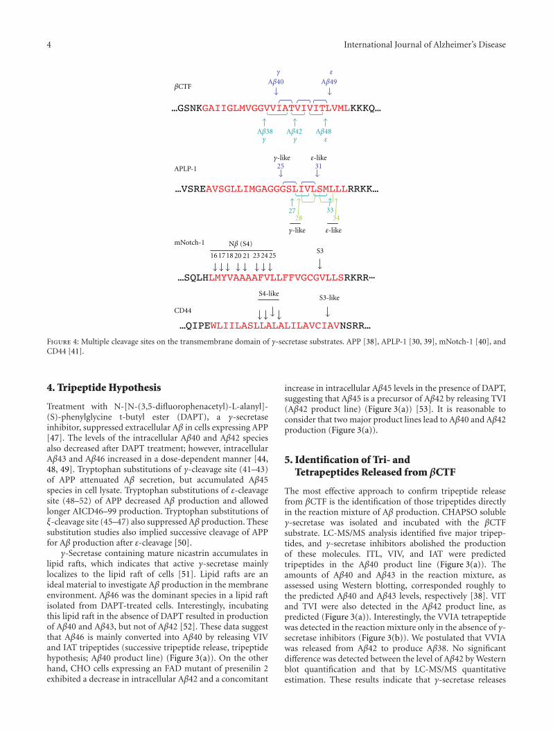
4 International Journal of Alzheimer’s Disease
16 17 18 20 21 23 24 25
S4-likeS3-like
Nβ (S4)S3
γ-like
γ-like
2827
25 31
3334
ε-like
ε-like
CD44
mNotch-1
Aβ38 Aβ42
Aβ40 Aβ49
Aβ48
γ
γ γ
ε
ε
APLP-1
βCTF
Figure 4: Multiple cleavage sites on the transmembrane domain of γ-secretase substrates. APP [38], APLP-1 [30, 39], mNotch-1 [40], andCD44 [41].
4. Tripeptide Hypothesis
Treatment with N-[N-(3,5-difluorophenacetyl)-L-alanyl]-(S)-phenylglycine t-butyl ester (DAPT), a γ-secretaseinhibitor, suppressed extracellular Aβ in cells expressing APP[47]. The levels of the intracellular Aβ40 and Aβ42 speciesalso decreased after DAPT treatment; however, intracellularAβ43 and Aβ46 increased in a dose-dependent manner [44,48, 49]. Tryptophan substitutions of γ-cleavage site (41–43)of APP attenuated Aβ secretion, but accumulated Aβ45species in cell lysate. Tryptophan substitutions of ε-cleavagesite (48–52) of APP decreased Aβ production and allowedlonger AICD46–99 production. Tryptophan substitutions ofξ-cleavage site (45–47) also suppressed Aβ production. Thesesubstitution studies also implied successive cleavage of APPfor Aβ production after ε-cleavage [50].
γ-Secretase containing mature nicastrin accumulates inlipid rafts, which indicates that active γ-secretase mainlylocalizes to the lipid raft of cells [51]. Lipid rafts are anideal material to investigate Aβ production in the membraneenvironment. Aβ46 was the dominant species in a lipid raftisolated from DAPT-treated cells. Interestingly, incubatingthis lipid raft in the absence of DAPT resulted in productionof Aβ40 and Aβ43, but not of Aβ42 [52]. These data suggestthat Aβ46 is mainly converted into Aβ40 by releasing VIVand IAT tripeptides (successive tripeptide release, tripeptidehypothesis; Aβ40 product line) (Figure 3(a)). On the otherhand, CHO cells expressing an FAD mutant of presenilin 2exhibited a decrease in intracellular Aβ42 and a concomitant
increase in intracellular Aβ45 levels in the presence of DAPT,suggesting that Aβ45 is a precursor of Aβ42 by releasing TVI(Aβ42 product line) (Figure 3(a)) [53]. It is reasonable toconsider that two major product lines lead to Aβ40 and Aβ42production (Figure 3(a)).
5. Identification of Tri- andTetrapeptides Released from βCTF
The most effective approach to confirm tripeptide releasefrom βCTF is the identification of those tripeptides directlyin the reaction mixture of Aβ production. CHAPSO solubleγ-secretase was isolated and incubated with the βCTFsubstrate. LC-MS/MS analysis identified five major tripep-tides, and γ-secretase inhibitors abolished the productionof these molecules. ITL, VIV, and IAT were predictedtripeptides in the Aβ40 product line (Figure 3(a)). Theamounts of Aβ40 and Aβ43 in the reaction mixture, asassessed using Western blotting, corresponded roughly tothe predicted Aβ40 and Aβ43 levels, respectively [38]. VITand TVI were also detected in the Aβ42 product line, aspredicted (Figure 3(a)). Interestingly, the VVIA tetrapeptidewas detected in the reaction mixture only in the absence of γ-secretase inhibitors (Figure 3(b)). We postulated that VVIAwas released from Aβ42 to produce Aβ38. No significantdifference was detected between the level of Aβ42 by Westernblot quantification and that by LC-MS/MS quantitativeestimation. These results indicate that γ-secretase releases
International Journal of Alzheimer’s Disease 5
tri- and tetrapeptides successively upon ε-cleavage of βCTF,to produce Aβ species. These tri- and tetrapeptides releasedfrom βCTF were detected even in the lipid raft fraction(Takami, unpublished observation).
6. Is Tripeptide Release a GeneralProperty of Substrate Cleavage byγ-Secretase?
Successive tripeptide release was observed in βCTF process-ing by γ-secretase. We also found that γ-secretase released tri-and tetrapeptides successively from αCTF substrate (Takami,unpublished observation). Recently, tripeptide spacing ofendoproteolysis on presenilin has been reported [54]. Thesesuggest that successive tri- and tetrapeptide release is ageneral property of γ-secretase-mediated intramembraneproteolysis.
Yanagida et al. reported that APLP-1 was also cleavedinto three Aβ-like peptides [39]. As three ε-like cleavagesare known, it is likely that APLP-1 is processed in threeproduct lines by successive tripeptide release [30] (Figure 4).The transmembrane domain of mNotch-1 is cleaved byγ-secretase after ectodomain shedding to liberate NICD(S3 cleavage). NICD containing V1744 was found as theprominent species produced by S3 cleavage [55]. To date, itseems reasonable to suppose that there is a single cleavagesite in S3. γ-Secretase also cleaves mNotch-1 at the lumen-membrane boundary (S4 cleavage) to release Notch βpeptides (Nβ) (Figure 4) [40, 56, 57]. Fenofibrate treatmentincreased the proportion of Nβ25, but not that of Nβ21,which implies that Nβ25 and Nβ21 correspond to Aβ42 andAβ40, respectively [57]. However, it is unlikely that severalNβ product lines exist in Notch processing because of thesingle S3 site. The production of Nβ species may not fit thetripeptide-processing model (Figure 4). CD44 is cleaved notonly at the membrane-cytoplasm boundary, but also at themiddle of the transmembrane domain, which results in therelease of Aβ-like peptides [33, 41]. Similar to Notch, theprocessing of the CD44 transmembrane domain may not fitthe tripeptide-processing model (Figure 4).
7. Conclusion and Perspectives
The tripeptide hypothesis was confirmed in the processingof the APP transmembrane domain, which accounts forthe production of Aβ species. Although the physiologicalsignificance of the multiple cleavage of the transmembranedomain is unknown, it is important to illustrate the cleavagemechanisms of other γ-secretase substrates, because the lim-itation of this stepwise mechanism would help to elucidatethe substrate-specific inhibition of Aβ production. As shownin Figure 4, APLP-1 may be cleaved by tripeptide release;however, Notch and CD44 do not fit this processing model[40, 41]. γ-Secretase is widely believed to be a promiscuousprotease; however, the cleavage mechanisms of APP andNotch, at least, seem to be different (Figure 4), whichindicates that γ-secretase distinguishes substrates duringproteolysis. Perhaps absence of helix breaker glycine residues
in mid-portion of transmembrane domain allows multipleS4 cleavages even after single S3 cleavage in Notch. Fromthis point of view, uncovering the mechanisms underlyingγ-secretase-dependent cleavage offers a basis for new ther-apeutic approaches that are aimed at substrate-specific Aβinhibition.
Abbreviations
Aβ: Amyloid β proteinAICD: APP intracellular domainAPP: Amyloid precursor proteinβCTF: Carboxyl terminal fragment of APPDAPT: N-[N-(3,5-difluorophenacetyl)-L-alanyl]-(S)-
phenylglycine t-butylesterFAD: Familial Alzheimer’s diseaseLC-MS/MS: Liquid chromatography-tandem mass
spectrometryNβ: Notch β peptideNICD: Notch intracellular domainPS: Presenilin.
Acknowledgments
The authors thank Nobuto Kakuda for critical comments onthis paper. Work in the authors’ laboratories is supportedin part by the Core Research for Evolutional Science andTechnology of JST and by MEXT-Supported Program for theStrategic Research Foundation at Private Universities 2012–2016.
References
[1] C. Haass, E. H. Koo, A. Mellon, A. Y. Hung, and D. J.Selkoe, “Targeting of cell-surface β-amyloid precursor proteinto lysosomes: alternative processing into amyloid-bearingfragments,” Nature, vol. 357, no. 6378, pp. 500–503, 1992.
[2] M. Citron, D. B. Teplow, and D. J. Selkoe, “Generation ofamyloid β protein from its precursor is sequence specific,”Neuron, vol. 14, no. 3, pp. 661–670, 1995.
[3] R. Vassar, B. D. Bennett, S. Babu-Khan et al., “β-Secretasecleavage of Alzheimer’s amyloid precursor protein by thetransmembrane aspartic protease BACE,” Science, vol. 286, no.5440, pp. 735–741, 1999.
[4] I. Pinnix, U. Musunuru, H. Tun et al., “A novel γ-secretaseassay based on detection of the putative C-terminal fragment-γ of amyloid β protein precursor,” Journal of BiologicalChemistry, vol. 276, no. 1, pp. 481–487, 2001.
[5] D. J. Selkoe, “Alzheimer’s disease: genes, proteins, and ther-apy,” Physiological Reviews, vol. 81, no. 2, pp. 741–766, 2001.
[6] B. De Strooper, P. Saftig, K. Craessaerts et al., “Deficiency ofpresenilin-1 inhibits the normal cleavage of amyloid precursorprotein,” Nature, vol. 391, no. 6665, pp. 387–390, 1998.
[7] B. De Strooper and G. Konig, “Alzheimer’s disease. A firm basefor drug development,” Nature, vol. 402, no. 6761, pp. 471–472, 1999.
[8] M. S. Wolfe, W. Xia, B. L. Ostaszewski, T. S. Diehl, W. T.Kimberly, and D. J. Selkoe, “Two transmembrane aspartatesin presenilin-1 required for presenilin endoproteolysis and γ-secretase activity,” Nature, vol. 398, no. 6727, pp. 513–517,1999.
6 International Journal of Alzheimer’s Disease
[9] G. Struhl and I. Greenwald, “Presenilin is required for activityand nuclear access of notch in drosophila,” Nature, vol. 398,no. 6727, pp. 522–525, 1999.
[10] G. Yu, M. Nishimura, S. Arawaka et al., “Nicastrin modu-lates presenilin-mediated notch/glp-1 signal transduction andβAPP processing,” Nature, vol. 407, no. 6800, pp. 48–54, 2000.
[11] C. Goutte, “Genetics leads the way to the accomplices ofpresenilins,” Developmental Cell, vol. 3, no. 1, pp. 6–7, 2002.
[12] R. Francis, G. McGrath, J. Zhang et al., “aph-1 and pen-2 arerequired for Notch pathway signaling, γ-secretase cleavage ofβAPP, and presenilin protein accumulation,” DevelopmentalCell, vol. 3, no. 1, pp. 85–97, 2002.
[13] D. Edbauer, E. Winkler, J. T. Regula, B. Pesold, H. Steiner, andC. Haass, “Reconstitution of γ-secretase activity,” Nature CellBiology, vol. 5, no. 5, pp. 486–488, 2003.
[14] W. T. Kimberly, M. J. LaVoie, B. L. Ostaszewski, W. Ye, M. S.Wolfe, and D. J. Selkoe, “γ-Secretase is a membrane proteincomplex comprised of presenilin, nicastrin, aph-1, and pen-2,” Proceedings of the National Academy of Sciences of the UnitedStates of America, vol. 100, no. 11, pp. 6382–6387, 2003.
[15] N. Takasugi, T. Tomita, I. Hayashi et al., “The role of presenilincofactors in the γ-secratase complex,” Nature, vol. 422, no.6930, pp. 438–441, 2003.
[16] A. J. Beel and C. R. Sanders, “Substrate specificity of γ-secretase and other intramembrane proteases,” Cellular andMolecular Life Sciences, vol. 65, no. 9, pp. 1311–1334, 2008.
[17] M. S. Brown, J. Ye, R. B. Rawson, and J. L. Goldstein,“Regulated intramembrane proteolysis: a control mechanismconserved from bacteria to humans,” Cell, vol. 100, no. 4, pp.391–398, 2000.
[18] M. S. Wolfe and R. Kopan, “Intramembrane proteolysis:theme and variations,” Science, vol. 305, no. 5687, pp. 1119–1123, 2004.
[19] A. Tolia, K. Horre, and B. De Strooper, “Transmembranedomain 9 of presenilin determines the dynamic conformationof the catalytic site of γ-secretase,” Journal of BiologicalChemistry, vol. 283, no. 28, pp. 19793–19803, 2008.
[20] S. Takagi, A. Tominaga, C. Sato, T. Tomita, and T. Iwatsubo,“Participation of transmembrane domain 1 of presenilin 1in the catalytic pore structure of the γ-secretase,” Journal ofNeuroscience, vol. 30, no. 47, pp. 15943–15950, 2010.
[21] C. Sato, S. Takagi, T. Tomita, and T. Iwatsubo, “The C-terminalPAL motif and transmembrane domain 9 of presenilin 1 areinvolved in the formation of the catalytic pore of the γ-secretase,” Journal of Neuroscience, vol. 28, no. 24, pp. 6264–6271, 2008.
[22] K. Takeo, N. Watanabe, T. Tomita, and T. Iwatsubo, “Contri-bution of the γ-secretase subunits to the formation of catalyticpore of presenilin 1 protein,” Journal of Biological Chemistry,vol. 287, no. 31, pp. 25834–25843, 2012.
[23] P. Osenkowski, H. Li, W. Ye et al., “Cryoelectron microscopystructure of purified γ-secretase at 12 A resolution,” Journal ofMolecular Biology, vol. 385, no. 2, pp. 642–652, 2009.
[24] M. Citron, T. Oltersdorf, C. Haass et al., “Mutation of theβ-amyloid precursor protein in familial Alzheimer’s diseaseincreases β-protein production,” Nature, vol. 360, no. 6405,pp. 672–674, 1992.
[25] J. T. Jarrett, E. P. Berger, and P. T. Lansbury, “The carboxyterminus of the β amyloid protein is critical for the seedingof amyloid formation: implications for the pathogenesis ofAlzheimer’s disease,” Biochemistry, vol. 32, no. 18, pp. 4693–4697, 1993.
[26] R. Wang, D. Sweeney, S. E. Gandy, and S. S. Sisodia,“The profile of soluble amyloid β protein in cultured cell
media. Detection and quantification of amyloid β protein andvariants by immunoprecipitation-mass spectrometry,” Journalof Biological Chemistry, vol. 271, no. 50, pp. 31894–31902,1996.
[27] M. Citron, D. Westaway, W. Xia et al., “Mutant presenilins ofAlzheimer’s disease increase production of 42-residue amyloidβ-protein in both transfected cells and transgenic mice,”Nature Medicine, vol. 3, no. 1, pp. 67–72, 1997.
[28] N. J. Clarke, A. J. Tomlinson, Y. Ohyagi, S. Younkin, and S.Naylor, “Detection and quantitation of cellularly derived amy-loid β peptides by immunoprecipitation-HPLC-MS,” FEBSLetters, vol. 430, no. 3, pp. 419–423, 1998.
[29] D. Beher, J. D. J. Wrigley, A. P. Owens, and M. S. Shearman,“Generation of C-terminally truncated amyloid-β peptides isdependent on γ-secretase activity,” Journal of Neurochemistry,vol. 82, no. 3, pp. 563–575, 2002.
[30] Y. Gu, H. Misonou, T. Sato, N. Dohmae, K. Takio, and Y.Ihara, “Distinct intramembrane cleavage of the beta-amyloidprecursor protein family resembling gamma-secretase-likecleavage of Notch,” Journal of Biological Chemistry, vol. 276,no. 38, pp. 35235–35238, 2001.
[31] M. Sastre, H. Steiner, K. Fuchs et al., “Presenilin-dependent γ-secretase processing of β-amyloid precursor protein at a sitecorresponding to the S3 cleavage of Notch,” EMBO Reports,vol. 2, no. 9, pp. 835–841, 2001.
[32] A. Weidemann, S. Eggert, F. B. M. Reinhard et al., “Anovel ε-cleavage within the transmembrane domain of theAlzheimer amyloid precursor protein demonstrates homologywith notch processing,” Biochemistry, vol. 41, no. 8, pp. 2825–2835, 2002.
[33] I. Okamoto, Y. Kawano, D. Murakami et al., “Proteolyticrelease of CD44 intracellular domain and its role in the CD44signaling pathway,” Journal of Cell Biology, vol. 155, no. 5, pp.755–762, 2001.
[34] H. J. Lee, K. M. Jung, Y. Z. Huang et al., “Presenilin-dependentγ-secretase-like intramembrane cleavage of ErbB4,” Journal ofBiological Chemistry, vol. 277, no. 8, pp. 6318–6323, 2002.
[35] P. Marambaud, J. Shioi, G. Serban et al., “A presenilin-1/γ-secretase cleavage releases the E-cadherin intracellular domainand regulates disassembly of adherens junctions,” EMBOJournal, vol. 21, no. 8, pp. 1948–1956, 2002.
[36] P. May, Y. Krishna Reddy, and J. Herz, “Proteolytic processingof low density lipoprotein receptor-related protein mediatesregulated release of its intracellular domain,” Journal ofBiological Chemistry, vol. 277, no. 21, pp. 18736–18743, 2002.
[37] T. Ikeuchi and S. S. Sisodia, “The Notch ligands, Delta1 andJagged2, are substrates for presenilin-dependent “γ-secretase”cleavage,” Journal of Biological Chemistry, vol. 278, no. 10, pp.7751–7754, 2003.
[38] M. Takami, Y. Nagashima, Y. Sano et al., “γ-Secretase:successive tripeptide and tetrapeptide release from the trans-membrane domain of β-carboxyl terminal fragment,” Journalof Neuroscience, vol. 29, no. 41, pp. 13042–13052, 2009.
[39] K. Yanagida, M. Okochi, S. Tagami et al., “The 28-aminoacid form of an APLPl-derived Aβ-like peptide is a surrogatemarker for Aβ42 production in the central nervous system,”EMBO Molecular Medicine, vol. 1, no. 4, pp. 223–235, 2009.
[40] J. Wanngren, J. Ottervald, S. Parpal et al., “Second generationγ-secretase modulators exhibit different modulation of notchβ and Aβ production,” Journal of Biological Chemistry, vol. 287,no. 39, pp. 32640–32650, 2012.
[41] S. Lammich, M. Okochi, M. Takeda et al., “Presenilin-dependent intramembrane proteolysis of CD44 leads to theliberation of its intracellular domain and the secretion of an
International Journal of Alzheimer’s Disease 7
Aβ-like peptide,” Journal of Biological Chemistry, vol. 277, no.47, pp. 44754–44759, 2002.
[42] T. Sato, N. Dohmae, Y. Qi et al., “Potential link betweenamyloid beta-protein 42 and C-terminal fragment gamma 49-99 of beta-amyloid precursor protein,” Journal of BiologicalChemistry, vol. 278, no. 27, pp. 24294–20301, 2003.
[43] S. Funamoto, M. Morishima-Kawashima, Y. Tanimura, N.Hirotani, T. C. Saido, and Y. Ihara, “Truncated carboxyl-terminal fragments of β-amyloid precursor protein are pro-cessed to amyloid β-proteins 40 and 42,” Biochemistry, vol. 43,no. 42, pp. 13532–13540, 2004.
[44] Y. Qi-Takahara, M. Morishima-Kawashima, Y. Tanimura etal., “Longer forms of amyloid β protein: implications forthe mechanism of intramembrane cleavage by γ-secretase,”Journal of Neuroscience, vol. 25, no. 2, pp. 436–445, 2005.
[45] L. Chevez-Gutierrez, L. Bammens, I. Benilova et al., “Themechanism of γ-Secretase dysfunction in familial Alzheimerdisease,” EMBO Journal, vol. 31, no. 10, pp. 2261–2274, 2012.
[46] O. Quintero-Monzon, M. M. Martin, M. A. Fernandez, C.A. Cappello, P. Osenkowski, and M. S. Wolfe, “Dissociationbetween the processivity and total activity of γ-secretase:implications for the mechanism of Alzheimer’s disease-causing presenilin mutations,” Biochemistry, vol. 50, no. 42,pp. 9023–9035, 2011.
[47] H. F. Dovey, V. John, J. P. Anderson et al., “Functional gamma-secretase inhibitors reduce beta-amyloid peptide levels inbrain,” Journal of Neurochemistry, vol. 76, no. 1, pp. 173–181,2001.
[48] G. Zhao, G. Mao, J. Tan et al., “Identification of a newpresenilin-dependent ζ-cleavage site within the transmem-brane domain of amyloid precursor protein,” Journal ofBiological Chemistry, vol. 279, no. 49, pp. 50647–50650, 2004.
[49] G. Zhao, M. Z. Cui, G. Mao et al., “γ-cleavage is dependenton ζ-cleavage during the proteolytic processing of amyloidprecursor protein within its transmembrane domain,” Journalof Biological Chemistry, vol. 280, no. 45, pp. 37689–37697,2005.
[50] T. Sato, Y. Tanimura, N. Hirotani, T. C. Saido, M. Morishima-Kawashima, and Y. Ihara, “Blocking the cleavage at mid-portion between γ- and ε-sites remarkably suppresses thegeneration of amyloid β-protein,” FEBS Letters, vol. 579, no.13, pp. 2907–2912, 2005.
[51] K. S. Vetrivel, H. Cheng, W. Lin et al., “Association ofγ-secretase with lipid rafts in post-golgi and endosomemembranes,” Journal of Biological Chemistry, vol. 279, no. 43,pp. 44945–44954, 2004.
[52] S. Yagishita, M. Morishima-Kawashima, S. Ishiura, and Y.Ihara, “Aβ46 is processed to Aβ40 and Aβ43, but not to Aβ42,in the low density membrane domains,” Journal of BiologicalChemistry, vol. 283, no. 2, pp. 733–738, 2008.
[53] S. Yagishita, M. Morishima-Kawashima, Y. Tanimura, S.Ishiura, and Y. Ihara, “DAPT-induced intracellular accumu-lations of longer amyloid β-proteins: further implications forthe mechanism of intramembrane cleavage by γ-secretase,”Biochemistry, vol. 45, no. 12, pp. 3952–3960, 2006.
[54] A. Fukumori, R. Fluhrer, H. Steiner, and C. Haass, “Three-amino acid spacing of presenilin endoproteolysis suggests ageneral stepwise cleavage of γ-secretase-mediated intramem-brane proteolysis,” Journal of Neuroscience, vol. 30, no. 23, pp.7853–7862, 2010.
[55] S. Osawa, S. Funamoto, M. Nobuhara et al., “Phosphoinosi-tides suppress γ-secretase in both the detergent-soluble and-insoluble states,” Journal of Biological Chemistry, vol. 283, no.28, pp. 19283–19292, 2008.
[56] M. Okochi, H. Steiner, A. Fukumori et al., “Presenilinsmediate a dual intramembranous γ-secretase cleavage ofNotch-1,” EMBO Journal, vol. 21, no. 20, pp. 5408–5416, 2002.
[57] M. Okochi, A. Fukumori, J. Jiang et al., “Secretion of theNotch-1 Aβ-like peptide during Notch signaling,” Journal ofBiological Chemistry, vol. 281, no. 12, pp. 7890–7898, 2006.
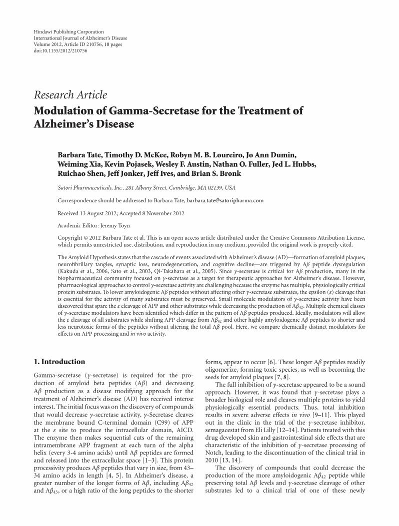
Hindawi Publishing CorporationInternational Journal of Alzheimer’s DiseaseVolume 2012, Article ID 210756, 10 pagesdoi:10.1155/2012/210756
Research Article
Modulation of Gamma-Secretase for the Treatment ofAlzheimer’s Disease
Barbara Tate, Timothy D. McKee, Robyn M. B. Loureiro, Jo Ann Dumin,Weiming Xia, Kevin Pojasek, Wesley F. Austin, Nathan O. Fuller, Jed L. Hubbs,Ruichao Shen, Jeff Jonker, Jeff Ives, and Brian S. Bronk
Satori Pharmaceuticals, Inc., 281 Albany Street, Cambridge, MA 02139, USA
Correspondence should be addressed to Barbara Tate, [email protected]
Received 13 August 2012; Accepted 8 November 2012
Academic Editor: Jeremy Toyn
Copyright © 2012 Barbara Tate et al. This is an open access article distributed under the Creative Commons Attribution License,which permits unrestricted use, distribution, and reproduction in any medium, provided the original work is properly cited.
The Amyloid Hypothesis states that the cascade of events associated with Alzheimer’s disease (AD)—formation of amyloid plaques,neurofibrillary tangles, synaptic loss, neurodegeneration, and cognitive decline—are triggered by Aβ peptide dysregulation(Kakuda et al., 2006, Sato et al., 2003, Qi-Takahara et al., 2005). Since γ-secretase is critical for Aβ production, many in thebiopharmaceutical community focused on γ-secretase as a target for therapeutic approaches for Alzheimer’s disease. However,pharmacological approaches to control γ-secretase activity are challenging because the enzyme has multiple, physiologically criticalprotein substrates. To lower amyloidogenic Aβ peptides without affecting other γ-secretase substrates, the epsilon (ε) cleavage thatis essential for the activity of many substrates must be preserved. Small molecule modulators of γ-secretase activity have beendiscovered that spare the ε cleavage of APP and other substrates while decreasing the production of Aβ42. Multiple chemical classesof γ-secretase modulators have been identified which differ in the pattern of Aβ peptides produced. Ideally, modulators will allowthe ε cleavage of all substrates while shifting APP cleavage from Aβ42 and other highly amyloidogenic Aβ peptides to shorter andless neurotoxic forms of the peptides without altering the total Aβ pool. Here, we compare chemically distinct modulators foreffects on APP processing and in vivo activity.
1. Introduction
Gamma-secretase (γ-secretase) is required for the pro-duction of amyloid beta peptides (Aβ) and decreasingAβ production as a disease modifying approach for thetreatment of Alzheimer’s disease (AD) has received intenseinterest. The initial focus was on the discovery of compoundsthat would decrease γ-secretase activity. γ-Secretase cleavesthe membrane bound C-terminal domain (C99) of APPat the ε site to produce the intracellular domain, AICD.The enzyme then makes sequential cuts of the remainingintramembrane APP fragment at each turn of the alphahelix (every 3-4 amino acids) until Aβ peptides are formedand released into the extracellular space [1–3]. This proteinprocessivity produces Aβ peptides that vary in size, from 43–34 amino acids in length [4, 5]. In Alzheimer’s disease, agreater number of the longer forms of Aβ, including Aβ42
and Aβ43, or a high ratio of the long peptides to the shorter
forms, appear to occur [6]. These longer Aβ peptides readilyoligomerize, forming toxic species, as well as becoming theseeds for amyloid plaques [7, 8].
The full inhibition of γ-secretase appeared to be a soundapproach. However, it was found that γ-secretase plays abroader biological role and cleaves multiple proteins to yieldphysiologically essential products. Thus, total inhibitionresults in severe adverse effects in vivo [9–11]. This playedout in the clinic in the trial of the γ-secretase inhibitor,semagacestat from Eli Lilly [12–14]. Patients treated with thisdrug developed skin and gastrointestinal side effects that arecharacteristic of the inhibition of γ-secretase processing ofNotch, leading to the discontinuation of the clinical trial in2010 [13, 14].
The discovery of compounds that could decrease theproduction of the more amyloidogenic Aβ42 peptide whilepreserving total Aβ levels and γ-secretase cleavage of othersubstrates led to a clinical trial of one of these newly
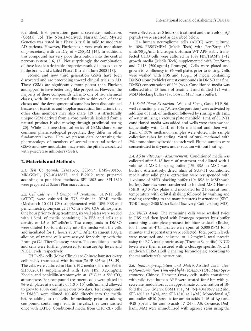
2 International Journal of Alzheimer’s Disease
identified, first generation gamma-secretase modulators(GSMs) [15]. The NSAID-derived, Flurizan from MyriadGenetics was tested in a Phase 3 trial in mild to moderateAD patients. However, Flurizan is a very weak modulatorof γ-secretase, with an IC50 of ∼250 μM [16]. In addition,this compound has very poor distribution into the centralnervous system [16, 17]. Not surprisingly, the combinationof these less than desirable properties resulted in no exposurein the brain, and a failed Phase III trial in June 2008 [18].
Second and now third generation GSMs have beendiscovered and are proceeding toward clinical trials in AD.These GSMs are significantly more potent than Flurizanand appear to have better drug-like properties. However, themajority of these compounds fall into one of two chemicalclasses, with little structural diversity within each of theseclasses and the development of some has been discontinuedbecause of toxicities and biopharmaceutical limitations thatother class members may also share [19]. A structurallyunique GSM derived from a core molecule isolated from anatural product is also moving through preclinical testing[20]. While all three chemical series of GSMs share somecommon pharmacological properties, they differ in otherfundamental ways. Here we present data contrasting thepharmacology of members of several structural series ofGSMs and how modulation may avoid the pitfalls associatedwith γ-secretase inhibitors (GSIs).
2. Materials and Methods
2.1. Test Compounds. LY411575, GSI-953, BMS-708163,MK-GSM1, JNJ-40418677, and E-2012 were preparedaccording to published methods. SPI-1802 and SPI-1810were prepared at Satori Pharmaceuticals.
2.2. Cell Culture and Compound Treatment. SUP-T1 cells(ATCC) were cultured in T75 flasks in RPMI media(Mediatech 10-041-CV) supplemented with 10% FBS andpenicillin/streptomycin at 37◦C in a 5% CO2 atmosphere.One hour prior to drug treatment, six well plates were seededwith 1.5 mL of media containing 2% FBS and cells at adensity of 1.5 × 10⁶ cells/mL. Test compounds in DMSOwere diluted 100-fold directly into the media with the cellsand incubated for 18 hours at 37◦C. After treatment 100 μLaliquots of treated cells were assayed for viability with thePromega Cell Titer Glo assay system. The conditioned mediaand cells were further processed to measure Aβ levels andNICD levels, respectively.
CHO-2B7 cells (Mayo Clinic) are Chinese hamster ovarycells stably transfected with human βAPP 695 wt [38, 39].The cells were cultured in Ham’s F12 media (Thermo FisherSH30026.01) supplemented with 10% FBS, 0.25 mg/mLZeocin and penicillin/streptomycin at 37◦C in a 5% CO2
atmosphere. For compound treatment, cells were plated in96-well plates at a density of 1.0 × 10⁵ cells/mL and allowedto grow to 100% confluence over two days. Test compoundsin DMSO were diluted 100-fold directly into the mediabefore adding to the cells. Immediately prior to addingcompound-containing media to the cells, they were washedonce with 1XPBS. Conditioned media from CHO-2B7 cells
were collected after 5 hours of treatment and the levels of Aβpeptides were assessed as described below.
H4 human neuroglioma cells (ATCC) were culturedin 10% FBS/DMEM (Media Tech) with Pen/Strep (50units/50 μg/mL; Invitrogen). Human WT APP stably trans-fected CHO cells were cultured in 10% FBS/HAM’S F-12growth media (Media Tech) supplemented with Pen/Strepand G418 (500 μg/mL; Promega). Cells were plated andgrown to confluency in 96-well plates prior to dosing. Cellswere washed with PBS and 100 μL of media containingDMSO alone (vehicle) or test compounds in DMSO at a finalDMSO concentration of 1% (v/v). Conditioned media wascollected after 18 hours of treatment and diluted 1 : 1 withMSD blocking buffer (1% BSA in MSD wash buffer).
2.3. Solid Phase Extraction. Wells of 30 mg Oasis HLB 96-well extraction plates (Waters Corporation) were activated byaddition of 1 mL of methanol followed by rinsing with 1 mLof water utilizing a vacuum plate manifold. 1 mL of SUP-T1conditioned media was added and wells were then washedsequentially with 2 mL of 10% methanol and then with2 mL of 30% methanol. Samples were eluted into samplecollection tubes by adding 250 μL of 90% methanol with2% ammonium hydroxide to each well. Eluted samples wereconcentrated to dryness under vacuum without heating.
2.4. Aβ In Vitro Assay Measurement. Conditioned media wascollected after 5–18 hours of treatment and diluted with 1volume of MSD blocking buffer (1% BSA in MSD washbuffer). Alternatively, dried films of SUP-T1 conditionedmedia after solid phase extraction were resuspended with1 volume of MSD blocking buffer (1% BSA in MSD washbuffer). Samples were transferred to blocked MSD Human(6E10) Aβ 3-Plex plates and incubated for 2 hours at roomtemperature with orbital shaking followed by washing andreading according to the manufacturer’s instructions (SEC-TOR Imager 2400 Meso Scale Discovery, Gaithersburg MD).
2.5. NICD Assay. The remaining cells were washed twicein PBS and then lysed with Promega reporter lysis buffercontaining a complete protease inhibitor cocktail (Roche)for 1 hour at 4◦C. Lysates were spun at 5,000 RPM for 5minutes and supernatants were collected. Total protein levelswere measured and adjusted to 1-2 mg/mL total proteinusing the BCA total protein assay (Thermo Scientific). NICDlevels were then measured with a cleavage specific Notch1sandwich ELISA (Cell Signaling Technologies) according tothe manufacturer’s instructions.
2.6. Immunoprecipitation and Matrix-Assisted Laser Des-orption/Ionization Time-of-Flight (MALDI-TOF) Mass Spec-trometry. Chinese Hamster Ovary cells stably transfectedwith wild-type human APP were treated for 6 hrs with γ-secretase modulators at an approximate concentration of 10-fold the IC50 (Merck GSM1 at 1 μM, JNJ-40418677 at 2 μM,SPI-1802 at 3 μM, and SPI-1810 at 2 μM.) Monoclonal Aβantibodies 6E10 (specific for amino acids 1–16 of Aβ) and4G8 (specific for amino acids 17–24 of Aβ; Covance, Ded-ham, MA) were immobilized with agarose resin using the
International Journal of Alzheimer’s Disease 3
AminoLink Plus reagents (Thermo Scientific, Rockport, IL).Conditioned media from treated cells was precleared withagarose resin overnight, and the supernatant was incubatedwith agarose-conjugated 6E10/4G8 for 6 hrs. Immunopre-cipitates were washed extensively prior to analysis.
Analyses were performed on a Shimadzu Biotech AximaTOF2 (Shimadzu Instruments) matrix-assisted-laser des-orption/ionization time-of-flight (MALDI-TOF) mass spec-trometer. Peptides were analyzed in positive ion linear mode.For intact peptide mass measurement the instrument wasset with a mass range extending up to 6000 m/z using apulsed extraction setting of 3500. An average mass externalstandard was used which consisted of angiotensin II (1047.2),P14R (1534.86) and ACTH clip 18–39 (2465.20), insulin B(3496.67), and insulin (5734.51). For sample preparation,5 μL aliquots of Aβ containing immunoprecipitates werediluted with 10 μL of 0.1% TFA and then desalted using a C18Zip Tip (Millipore, Corp.). Samples were directly depositedfrom the Zip Tip onto the MALDI sample target andthen mixed with 0.5 μL of matrix solution which consistedof 5 mg/mL of alpha cyano-4-hydroxy cinnamic acid inacetonitrile: 0.1% TFA (50 : 50). Data was acquired manuallyusing a set laser power and averaging 1500–2000 laser shots.
2.7. In Vivo Study Methods. All animal handling and pro-cedures were conducted in full compliance to AAALACInternational and NIH regulations and guidelines regardinganimal care and welfare.
Either transgenic mice (Tg2576, 3 mos; N = 21) orwild-type Sprague Dawley rats (200–225 g; N = 8) wereutilized to assess in vivo efficacy. All animals were acclimatedto the test facility for a minimum of two days prior toinitiation of the study. Compounds were dosed orally in10 : 20 : 70 Ethanol/Solutol/Water via oral gavage. Sampleswere harvested at 6 hrs after dose for Aβ and compoundexposure levels. Blood samples were collected into K2EDTAand stored on wet ice until processed to plasma by centrifu-gation (3500 rpm at 5◦C) within 30 minutes of collection.Each brain was dissected into three parts: left and righthemispheres and cerebellum. Brain tissues were rinsed withice cold phosphate buffered saline (without Mg2+ or Ca2+),blotted dry and weighed. Plasma and cerebella were analyzedfor parent drug via LC/MS/MS. Parent drug levels were com-pared to a standard curve to establish the unknown levels.
2.8. Rodent Aβ Determination. This protocol is a modi-fication of protocols described by Lanz et al. [40] andRogers et al. [41]. Frozen hemispheres were weighed intotared homogenization tubes (MP Biomedicals#6933050 forrat; MP Biomedicals, Solon, OH) and (Simport#T501-4AT;Simport, Beloeil, Qc, Canada) containing one 5 mm stainlesssteel bead (Qiagen#69989) for mouse). For every gram ofbrain, 10 mLs of 6 M guanidine hydrochloride (wild-typerat) or 0.2% diethyl amine in 50 mM NaCl (transgenicmouse) was added to the brain-containing tubes on wetice. Rat hemispheres were homogenized for one minuteand mouse hemispheres were homogenized for 30 secondsat the 6.5 setting using the FastPrep-24 Tissue and Cellhomogenizer (MP Biomedicals#116004500). Homogenates
were rocked for two hours at 4◦C, then precleared by ultra-centrifugation at 100,000 ×g for one hour at 4◦C. Preclearedwild-type rat homogenates were concentrated over solidphase extraction (SPE) columns (Oasis HLB 96-well SPEplate 30 um, Waters#WAT058951; Waters Corp., Milford,MA). Briefly, SPE columns were prepared by wetting with1 mL of 100% methanol followed by dH2O using vacuumto pull liquids through. Brain homogenates were then addedto the prepared columns (1.0 mL from rat). Columns werewashed twice with 10% methanol followed by two washeswith 30% methanol. Labeled eluent collection tubes (Costarcluster tubes #4413; Corning Inc., Corning, NY) were placedunder SPE columns and samples were eluted under very mildvacuum with 300 μL of 2% NH4OH/90% methanol. Eluentswere dried to films under vacuum with no heat in a speedvacuum microcentrifuge. Films were resuspended in 150 μLof Meso Scale Discovery (MSD, Gaithersburg MD) blockingbuffer (1% BSA in MSD wash buffer) for one hour at roomtemperature with occasional vortexing. A volume of 45 μL ofprecleared transgenic mouse brain homogenates were dilutedinto 450 μL of blocking buffer and were neutralized with 5 μLof 0.5 M Tris pH 6.8. For Aβ38, 40, and 42 measurements,MSD 96 well multispot Human/Rodent (4G8) Aβ triplexultrasensitive ELISA plates were blocked with MSD blockingbuffer for 1 hour at room temperature with orbital shaking.A volume of 25 μL of neat resuspended wild-type ratbrain homogenate films or diluted transgenic mouse brainhomogenates were added in duplicates to the blocked 3-plex Aβ MSD plates with SULFO-TAG 4G8 antibody (MSD).The Aβ 3-Plex plates were incubated for 2 hours at roomtemperature with orbital shaking followed by washing andreading according to the manufacturer’s instructions (SEC-TOR Imager 2400, MSD). The average Aβ concentrationsfrom duplicate measurements of each animal were convertedto percent vehicle values and the treatment group averageswere statistically compared by ANOVA analysis.
3. Results and Discussion
γ-Secretase is a complex enzyme with multiple substrates andmultiple cleavage sites on at least some of these substrates,including APP. Complete inhibition of γ-secretase activityby targeting the ε cleavage site prevents the processing ofmultiple physiologically relevant proteins, leading to thesevere side effects reported in AD patients [12, 13]. On theother hand, chemically modulating the enzyme with a GSMis a more precise mechanism to enhance certain cleavageevents while preventing the cut that yields the amyloidogenicpeptide, Aβ42 which is linked to the pathophysiological ini-tiation of AD (Figure 6). In preclinical toxicological testing,GSMs appear to be free of the mechanism-based toxicitiesattributed to the inhibition of Notch processing that haveplagued the enzyme inhibitors. In vitro data demonstrate thatNotch cleavage to NICD is not inhibited by any of severalrepresented GSMs at concentrations that do not disruptcell viability (Figure 1). Treatment with GSMs in rodentshave not shown the classical Notch-related toxicities that areassociated with GSIs, suggesting that the complete processingof Notch to NICD can occur in the presence of GSMs.
4 International Journal of Alzheimer’s Disease
−14 −13 −12 −11 −10 −9 −8 −7 −6 −5
0
20
40
60
80
100
120
GSI
log (mol/L)
Veh
icle
(%
)
LY411575, IC50 = 1.3 nMGSI-953, IC50 = 225 nMBMS-706163, IC50 = 23 nM
(a)
−10 −9 −8 −7 −6 −50
20
40
60
80
100
120
Merck GSM1 E-2012
JNJ-40418677 SPI-1802
GSM
log (mol/L)
Veh
icle
(%
)
(b)
Figure 1: Multiple GSI and GSMs were examined in for their ability to inhibit NOTCH cleavage using the SUP-T1 cellular assay.
3.1. GSMs Do Not Show a Potency Shift with Changes inSubstrate Concentration. γ-Secretase inhibitors can show asignificant shift in potency in APP transfected cell-basedassays depending on the level of expression of APP. Table 1compares the IC50 for Aβ42 lowering of different GSIs inmultiple cell lines with varying levels of APP expression. Inthe highest expressing cell lines, the GSIs assayed here appearmore potent (lower IC50s in the high expressing, transfectedCHO-2B7 cell line) than when the same compounds aretested in assays using lower expressing cell lines (nativeH4 cell line). This potency shift seen with GSIs has beenattributed to a shift in the enzyme/substrate ratio [42], wherethere is a higher ratio in wild-type cells versus those thatoverexpress APP. This higher ratio in turn requires morecompounds to inhibit the enzymatic reaction. Interestingly,the potency shift due to substrate concentration that occurswith GSIs does not occur with GSMs. As seen in Table 1, theIC50 for all the GSMs shown are consistent across cell lines,regardless of the APP expression levels. While this potencyshift is not well understood, these data do suggest that GSIsand GSMs affect the enzyme in fundamentally different ways.
3.2. Structural Diversity of GSMs. Many of the secondgeneration GSMs were inspired by the NSAIDs (Figure 2).For instance, all compounds shown in the figure wereinitially derived from the aryl acetic acid motif found in1 and similar NSAIDs [21]. The similarity between thesecompounds makes for a crowded patent landscape, withsome compounds potentially covered in multiple patentapplications from different sponsors. In addition, for thisstructural series, the physicochemical properties indicate thatall of these compounds carry an increased safety risk due tothe high logP, low PSA [43–45], and high degree of aromatic-ity [46, 47]. Compounds 7–9 are designed to improve uponthese properties, but are still considered high risk due to
these same physicochemical based in silico models [27–29].In vivo preclinical toxicity testing will ultimately be needed toassess the safety profile of these similar structures. However,the lack of progression of compounds from this class intoand through clinical development suggests that this scaffoldmay have challenges that will continue to slow or preventsuccessful conduct of the studies required for registration.
A second class of GSMs that has received signifi-cant attention is summarized in Figure 3. Following initialdisclosure of 10 and 11, a number of pharmaceuticalcompanies pursued structurally related chemical series [30,48]. Although this class offers clear distinction from theNSAID-inspired compounds above, their physicochemicalproperties also reside outside of the molecular space mostfrequently affiliated with marketed agents for oral therapy.For example, the number of aromatic rings and clogP ofthe representatives shown are higher than the average fororal compounds on the market [46, 47], leading to a higherprobability of safety and biopharmaceutical challenges withthis class. Although some groups have been successful indeveloping promising structural alternatives, as exemplifiedby 16–18, little has been reported on the development of anyrepresentatives from this general scaffold [34–36].
A novel and structurally distinct chemical architecture ofa third class of GSMs has been reported by Satori Pharmaceu-ticals. This scaffold was first isolated from black cohosh,leading to the characterization of initial hit 19 (Figure 4)[20]. A combination of synthetic and medicinal chemistryoptimization led to 20, which is reported to have better drugproperties than 19 [20]. To date, the compounds reportedby Satori also fall outside of the guidelines most typicallyassociated with good in vivo disposition. The group notes,however, that the majority of marketed agents derived fromnatural products also violate these same guidelines, a trendthat has led some to conclude that molecules derived via
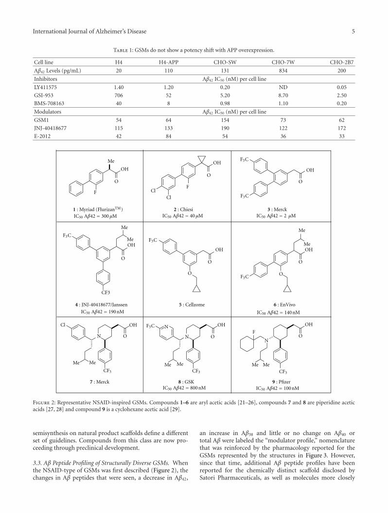
International Journal of Alzheimer’s Disease 5
Table 1: GSMs do not show a potency shift with APP overexpression.
Cell line H4 H4-APP CHO-SW CHO-7W CHO-2B7
Aβ42 Levels (pg/mL) 20 110 131 834 200
Inhibitors Aβ42 IC50 (nM) per cell line
LY411575 1.40 1.20 0.20 ND 0.05
GSI-953 706 52 5.20 8.70 2.50
BMS-708163 40 8 0.98 1.10 0.20
Modulators Aβ42 IC50 (nM) per cell line
GSM1 54 64 154 73 62
JNJ-40418677 115 133 190 122 172
E-2012 42 84 54 36 33
Me
O
OHOH
FF
O
OH
F3C
O
OH
OO
Me
Me
O
O
OH
N
Cl
Me
Me
CF3
Me Me
ClCl
F3C
F3C
OH
OH
O
O
F3C
F3C
Me Me
O
OHN
N O
F3C OH
MeMe
F
N
IC50 Aβ42 = 300 μM IC50 Aβ42 = 40 μM IC50 Aβ42 = 2 μM
IC50 Aβ42 = 190 nM IC50 Aβ42 = 140 nM
IC50 Aβ42 = 800 nM IC50 Aβ42 = 100 nM
CF3 CF3 CF3
1 : Myriad (FlurizanTM)
6 : EnVivo
3 : Merck
7 : Merck
2 : Chiesi
4 : JNJ-40418677/Janssen
9 : Pfizer8 : GSK
5 : Cellzome
Figure 2: Representative NSAID-inspired GSMs. Compounds 1–6 are aryl acetic acids [21–26], compounds 7 and 8 are piperidine aceticacids [27, 28] and compound 9 is a cyclohexane acetic acid [29].
semisynthesis on natural product scaffolds define a differentset of guidelines. Compounds from this class are now pro-ceeding through preclinical development.
3.3. Aβ Peptide Profiling of Structurally Diverse GSMs. Whenthe NSAID-type of GSMs was first described (Figure 2), thechanges in Aβ peptides that were seen, a decrease in Aβ42,
an increase in Aβ38 and little or no change on Aβ40 ortotal Aβ were labeled the “modulator profile,” nomenclaturethat was reinforced by the pharmacology reported for theGSMs represented by the structures in Figure 3. However,since that time, additional Aβ peptide profiles have beenreported for the chemically distinct scaffold disclosed bySatori Pharmaceuticals, as well as molecules more closely
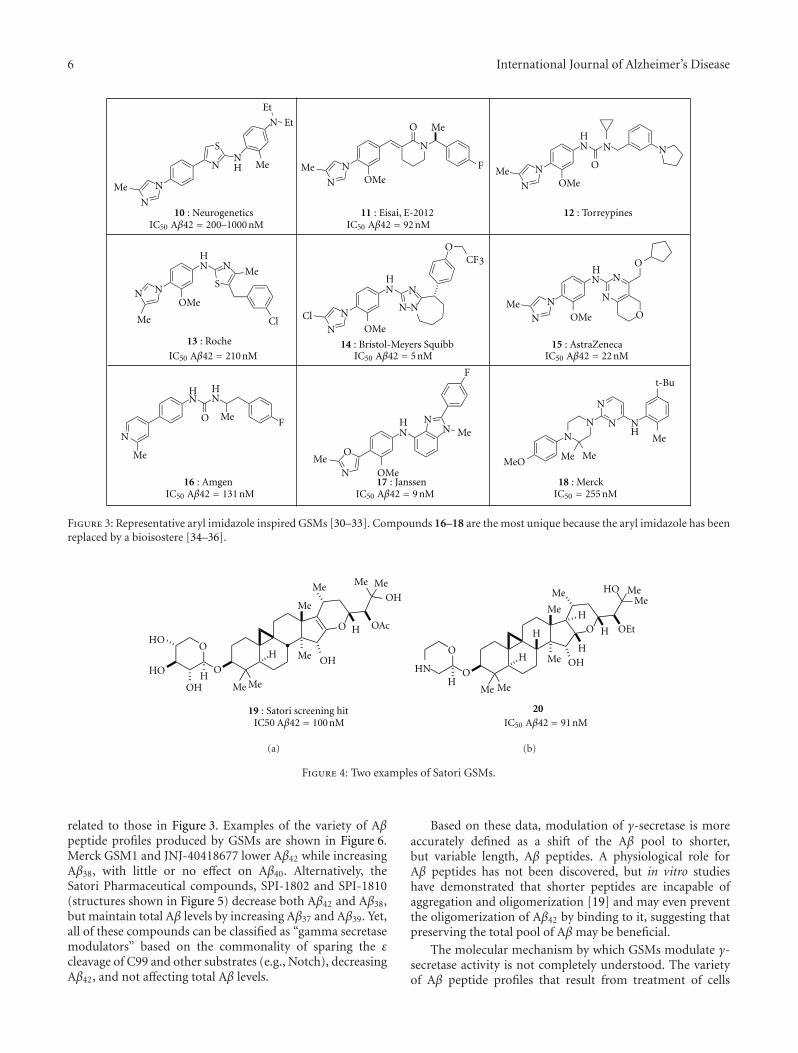
6 International Journal of Alzheimer’s Disease
N
NMe
N
SNH
N Et
Et
Me
N
NMeOMe
N
O Me
F
N
OMe
OMe
HN
ON
N
N
Me
NOMe
HN N Me
Me
SN
ClN
NCl
OMe
HN N
NN
OCF3
N
NMe
OHN N
N
O
HN N
MeO
H
N
F
Me O
NMe
OMe
HN
NN Me
F
NN
N N
N
H
Me
t-Bu
Me
Me
MeO
IC50 Aβ42 = 200–1000 nM IC50 Aβ42 = 92 nM
IC50 Aβ42 = 210 nM IC50 Aβ42 = 5 nM IC50 Aβ42 = 22 nM
IC50 Aβ42 = 131 nM IC50 Aβ42 = 9 nM IC50 = 255 nM
10 : Neurogenetics 11 : Eisai, E-2012 12 : Torreypines
13 : Roche 14 : Bristol-Meyers Squibb 15 : AstraZeneca
16 : Amgen 17 : Janssen 18 : Merck
Figure 3: Representative aryl imidazole inspired GSMs [30–33]. Compounds 16–18 are the most unique because the aryl imidazole has beenreplaced by a bioisostere [34–36].
O
Me
OH
Me
OAc
Me
Me
Me
H
Me Me
O
O
OHHO
IC50 Aβ42 = 100 nM
OH
HO
H
H
19 : Satori screening hit
(a)
O
OMe
H
H
H
HMe Me
O OEt
Me
Me
HO MeMe
OHH
H
IC50 Aβ42 = 91 nM
HN
20
(b)
Figure 4: Two examples of Satori GSMs.
related to those in Figure 3. Examples of the variety of Aβpeptide profiles produced by GSMs are shown in Figure 6.Merck GSM1 and JNJ-40418677 lower Aβ42 while increasingAβ38, with little or no effect on Aβ40. Alternatively, theSatori Pharmaceutical compounds, SPI-1802 and SPI-1810(structures shown in Figure 5) decrease both Aβ42 and Aβ38,but maintain total Aβ levels by increasing Aβ37 and Aβ39. Yet,all of these compounds can be classified as “gamma secretasemodulators” based on the commonality of sparing the εcleavage of C99 and other substrates (e.g., Notch), decreasingAβ42, and not affecting total Aβ levels.
Based on these data, modulation of γ-secretase is moreaccurately defined as a shift of the Aβ pool to shorter,but variable length, Aβ peptides. A physiological role forAβ peptides has not been discovered, but in vitro studieshave demonstrated that shorter peptides are incapable ofaggregation and oligomerization [19] and may even preventthe oligomerization of Aβ42 by binding to it, suggesting thatpreserving the total pool of Aβ may be beneficial.
The molecular mechanism by which GSMs modulate γ-secretase activity is not completely understood. The varietyof Aβ peptide profiles that result from treatment of cells
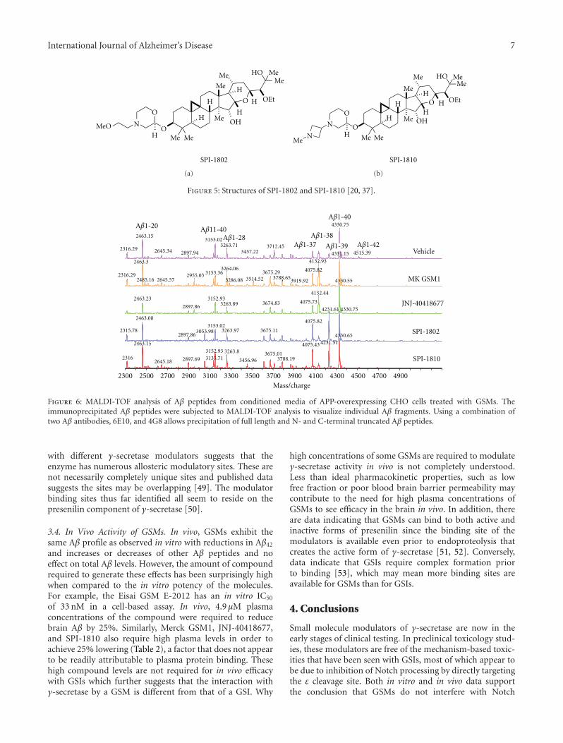
International Journal of Alzheimer’s Disease 7
O
MeMe
OHH
Me
Me
O H
H
H
H OEt
Me
HO
Me Me
O
N
H
MeO
SPI-1802
(a)
O
OHMe
Me
HH
Me
Me
O
H
H
H OEt
Me
HO
Me
O
HN
N
MeMe
SPI-1810
(b)
Figure 5: Structures of SPI-1802 and SPI-1810 [20, 37].
2300 2500 2700 2900 3100 3300 3500 3700 3900 4100 4300 4500 4700 4900Mass/charge
4331.15
4132.932463.3
4330.75
4330.55
4330.75
4330.65
4231.61
3264.06
4231.512463.15
4075.82
2463.15
4075.43
2463.08
4132.44
3153.36 4075.823675.29
3153.02
3152.93
3263.8
2955.03
2316.29
3153.02
3152.93
3263.71
2463.23
3788.65
3712.45
4075.73
3675.013135.71
3514.52
3263.973053.98
2316
3675.112315.78
2316.29
3788.19
3263.89 3674.83
2645.57
2897.69
2485.16
3456.962645.18
2897.86
3286.08 3919.92
2897.942645.34
2897.86
4515.393457.22
SPI-1810
SPI-1802
JNJ-40418677
MK GSM1
Vehicle
Aβ1-40
Aβ1-39
Aβ1-38Aβ1-37
Aβ1-20
Aβ1-42
Aβ11-40Aβ1-28
Figure 6: MALDI-TOF analysis of Aβ peptides from conditioned media of APP-overexpressing CHO cells treated with GSMs. Theimmunoprecipitated Aβ peptides were subjected to MALDI-TOF analysis to visualize individual Aβ fragments. Using a combination oftwo Aβ antibodies, 6E10, and 4G8 allows precipitation of full length and N- and C-terminal truncated Aβ peptides.
with different γ-secretase modulators suggests that theenzyme has numerous allosteric modulatory sites. These arenot necessarily completely unique sites and published datasuggests the sites may be overlapping [49]. The modulatorbinding sites thus far identified all seem to reside on thepresenilin component of γ-secretase [50].
3.4. In Vivo Activity of GSMs. In vivo, GSMs exhibit thesame Aβ profile as observed in vitro with reductions in Aβ42
and increases or decreases of other Aβ peptides and noeffect on total Aβ levels. However, the amount of compoundrequired to generate these effects has been surprisingly highwhen compared to the in vitro potency of the molecules.For example, the Eisai GSM E-2012 has an in vitro IC50
of 33 nM in a cell-based assay. In vivo, 4.9 μM plasmaconcentrations of the compound were required to reducebrain Aβ by 25%. Similarly, Merck GSM1, JNJ-40418677,and SPI-1810 also require high plasma levels in order toachieve 25% lowering (Table 2), a factor that does not appearto be readily attributable to plasma protein binding. Thesehigh compound levels are not required for in vivo efficacywith GSIs which further suggests that the interaction withγ-secretase by a GSM is different from that of a GSI. Why
high concentrations of some GSMs are required to modulateγ-secretase activity in vivo is not completely understood.Less than ideal pharmacokinetic properties, such as lowfree fraction or poor blood brain barrier permeability maycontribute to the need for high plasma concentrations ofGSMs to see efficacy in the brain in vivo. In addition, thereare data indicating that GSMs can bind to both active andinactive forms of presenilin since the binding site of themodulators is available even prior to endoproteolysis thatcreates the active form of γ-secretase [51, 52]. Conversely,data indicate that GSIs require complex formation priorto binding [53], which may mean more binding sites areavailable for GSMs than for GSIs.
4. Conclusions
Small molecule modulators of γ-secretase are now in theearly stages of clinical testing. In preclinical toxicology stud-ies, these modulators are free of the mechanism-based toxic-ities that have been seen with GSIs, most of which appear tobe due to inhibition of Notch processing by directly targetingthe ε cleavage site. Both in vitro and in vivo data supportthe conclusion that GSMs do not interfere with Notch

8 International Journal of Alzheimer’s Disease
Table 2: GSM potency versus plasma exposure in mice.
E-2012 JNJ-40418677 GSM1 SPI-1810
In vitro Aβ42 IC50 (nM) CHO-2B7 cells 33 172 62 114
Plasma exposure (μM) 4923 7764 2744 14638
Plasma fold over Aβ42 IC50 for 25% reduction 149 45 44 128
Brain exposure (μM) 2749 7497 8681 20368
Brain fold over Aβ42 IC50 for 25% reduction 83 44 140 179
GSISI GSM
Prevent the ε cleavage
All Aβ peptides and
NotchCD44Erb4
Inhibitors Modulators
Modulators alter processivity
Satori profile
37 38 39 40 42
37 38 39 40 42
NSAID-like GSMsother γ-secretase substrates
Figure 7: Diagram highlighting the differences between inhibitors and modulators.
processing, but instead, via slight shifts in the cleavage siteon APP, lower Aβ42 production to produce the other normalAβ peptide products (Figures 6 and 7). Thus, modulationof γ-secretase may represent the most selective approach totreating Alzheimer’s disease via a decrease in production ofAβ42, more selective that an inhibitor of either beta or γ-secretase . Modulators do differ both in chemical structureand in their effects on APP processing, producing the desireddecrease in Aβ42 and a variety of changes in other Aβpeptides. Understanding the molecular basis of these varyingprofiles may shed further light on the biology of γ-secretase.
Disclosures
All authors are or were employees of Satori Pharmaceuticals,Inc.
Acknowledgments
A portion of this work was presented at the AAIC Annualmeetings in 2011 and 2012. The authors would like to thankDr. John Leszyk from the University of Massachusetts forMALDI-TOF Aβ profile analysis and Kelly Ames, BusinessManager at Satori Pharmaceuticals, Inc.
References
[1] N. Kakuda, S. Funamoto, S. Yagishita et al., “Equimolar pro-duction of amyloid β-protein and amyloid precursor proteinintracellular domain from β-carboxyl-terminal fragment by γ-secretase,” Journal of Biological Chemistry, vol. 281, no. 21, pp.14776–14786, 2006.
[2] T. Sato, N. Dohmae, Y. Qi et al., “Potential link betweenamyloid β-protein 42 and C-terminal fragment γ 49-99 of β-amyloid precursor protein,” Journal of Biological Chemistry,vol. 278, no. 27, pp. 24294–20301, 2003.
[3] Y. Qi-Takahara, M. Morishima-Kawashima, Y. Tanimura etal., “Longer forms of amyloid β protein: implications for themechanism of intramembrane cleavage by γ-secretase,” Jour-nal of Neuroscience, vol. 25, no. 2, pp. 436–445, 2005.
[4] M. Takami, Y. Nagashima, Y. Sano et al., “γ-Secretase: succes-sive tripeptide and tetrapeptide release from the transmem-brane domain of β-carboxyl terminal fragment,” Journal ofNeuroscience, vol. 29, no. 41, pp. 13042–13052, 2009.
[5] T. E. Golde, Y. Ran, and K. M. Felsenstein, “Shifting a complexdebate on γ-secretase cleavage and Alzheimer’s disease,” TheEMBO Journal, vol. 31, no. 10, pp. 2237–2239, 2012.
[6] N. Suzuki, T. T. Cheung, X. D. Cai et al., “An increasedpercentage of long amyloid β protein secreted by familialamyloid β protein precursor (βAPP717) mutants,” Science, vol.264, no. 5163, pp. 1336–1340, 1994.
[7] T. Saito, T. Suemoto, N. Brouwers et al., “Potent amyloido-genicity and pathogenicity of Aβ 243,” Nature Neuroscience,vol. 14, no. 8, pp. 1023–1032, 2011.
[8] J. T. Jarrett, E. P. Berger, and P. T. Lansbury Jr., “The carboxyterminus of the β amyloid protein is critical for the seedingof amyloid formation: Implications for the pathogenesis ofAlzheimer’s disease,” Biochemistry, vol. 32, no. 18, pp. 4693–4697, 1993.
[9] J. Milano, J. McKay, C. Dagenais et al., “Modulation of Notchprocessing by γ-secretase inhibitors causes intestinal gobletcell metaplasia and induction of genes known to specify gutsecretory lineage differentiation,” Toxicological Sciences, vol.82, no. 1, pp. 341–358, 2004.
[10] G. H. Searfoss, W. H. Jordan, D. O. Calligaro et al., “Adipsin, abiomarker of gastrointestinal toxicity mediated by a functional

International Journal of Alzheimer’s Disease 9
γ-secretase inhibitor,” Journal of Biological Chemistry, vol. 278,no. 46, pp. 46107–46116, 2003.
[11] G. T. Wong, D. Manfra, F. M. Poulet et al., “Chronic treatmentwith the γ-secretase inhibitor LY-411,575 inhibits γ-amyloidpeptide production and alters lymphopoiesis and intestinalcell differentiation,” Journal of Biological Chemistry, vol. 279,no. 13, pp. 12876–12882, 2004.
[12] E. Lilly, “Lilly Halts Development of Semagacestat for Alz-heimer’s Disease Based on Preliminary Results of Phase IIIClinical Trials,” 2010, http://newsroom.lilly.com/releasedetail.cfm?releaseid=499794.
[13] D. J. Selkoe, “Resolving controversies on the path to Alzheim-er’s therapeutics,” Nature Medicine, vol. 17, no. 9, pp. 1060–1065, 2011.
[14] B. P. Imbimbo, F. Panza, V. Frisardi et al., “Therapeutic inter-vention for Alzheimer’s disease with γ-secretase inhibitors:still a viable option?” Expert Opinion on Investigational Drugs,vol. 20, no. 3, pp. 325–341, 2011.
[15] S. Weggen, J. L. Eriksen, P. Das et al., “A subset of NSAIDslower amyloidogenic Aβ42 independently of cyclooxygenaseactivity,” Nature, vol. 414, no. 6860, pp. 212–216, 2001.
[16] T. E. Golde, L. S. Schneider, and E. H. Koo, “Anti-Aβ thera-peutics in alzheimer’s disease: the need for a paradigm shift,”Neuron, vol. 69, no. 2, pp. 203–213, 2011.
[17] B. P. Imbimbo, “Why did tarenflurbil fail in alzheimer’s dis-ease?” Journal of Alzheimer’s Disease, vol. 17, no. 4, pp. 757–760, 2009.
[18] R. C. Green, L. S. Schneider, D. A. Amato et al., “Effect of tar-enflurbil on cognitive decline and activities of daily living inpatients with mild Alzheimer disease: a randomized controlledtrial,” Journal of the American Medical Association, vol. 302, no.23, pp. 2557–2564, 2009.
[19] B. Bulic, J. Ness, S. Hahn, A. Rennhack, T. Jumpertz, and S.Weggen, “Chemical biology, molecular mechanism and clin-ical perspective of γ-secretase modulators in Alzheimer’s dis-ease,” Current Neuropharmacology, vol. 9, no. 4, pp. 598–622,2011.
[20] J. L. Hubbs, N. O. Fuller, W. F. Austin et al., “Optimization of anatural product-based class of gamma-secretase modulators,”Journal of Medicinal Chemistry, vol. 55, no. 21, pp. 9270–9282,2012.
[21] E. H. M. Koo, T. E. Golde, and D. R. Galasko, “Nonsteroidalantiinflammatory drug (NSAID) and NSAID derivative amy-loid Aβ42 polypeptide-lowering agents for the treatment ofAlzheimer’s disease, and screening methods,” pp. 73, MayoFoundation for Medical Education and Research, USA, 2001.
[22] L. Raveglia, I. Peretto, S. Radaelli, B. P. Imbimbo, A. Rizzi,and G. Villetti, “Preparation of 1-phenylalkanecarboxylic acidderivatives for the treatment of neurodegenerative diseases,”pp. 91, Chiesi Farmaceutici S.p.A., Italy, 2004.
[23] P. Blurton et al., “Arylacetic acids and related compounds andtheir preparation, pharmaceutical compositions and their usefor treatment of diseases associated with the deposition of β-amyloid peptides in the brain such as Alzheimer’s disease,” pp.58, Merck Sharp & Dohme Limited, UK, 2006.
[24] C. Y. Ho, Preparation of Biphenyl Derivatives as γ-secretAseModulators, Janssen Pharmaceutica, Belg, Germany, 2009.
[25] F. Wilson, A. Reid, V. Reader et al., “Preparation of bipheny-lacetic acids as γ-secretase modulators for the treatment ofAlzheimer’s disease,” pp. 57, Cellzome, Germany, 2006.
[26] G. Shapiro and R. Chesworth, “1,3,4-Trisubstituted benzenesas γ-secretase inhibitors and their preparation and use inthe treatment of neurodegenerative diseases,” pp. 165, EnvivoPharmaceuticals, USA, 2009.
[27] J. J. Kulagowski, A. Madin, P. M. Ridgill, and M. E. Seward,“Preparation of piperidines and related compounds for treat-ment of Alzheimer’s disease,” pp. 178, Merck Sharp & DohmeLimited, UK, 2006.
[28] A. Hall, R. L. Elliott, G. M. P. Giblin et al., “Piperidine-derivedγ-secretase modulators,” Bioorganic and Medicinal ChemistryLetters, vol. 20, no. 3, pp. 1306–1311, 2010.
[29] E. C. W. Am et al., “Aminocyclohexanes and aminotetrahydro-pyrans as γ-secretase modulators and their preparation anduse for thetreatment of neurological and psychiatric diseases,”pp. 91, Pfizer, USA, 2011.
[30] S. Cheng, D. D. Comer, L. Mao, G. P. Balow, and D. Pleynet,“Aryl compounds and uses in modulating amyloid β,” pp. 178,Neurogenetics, USA 2004.
[31] K. Baumann et al., “Preparation of thiazolyl-substitutedimidazolylphenylamine derivatives and related compounds asmodulators of amyloid beta,” pp. 32, Hoffmann-La Roche,USA, 2008.
[32] L. R. Marcin et al., “Preparation of bicyclic compounds, espe-cially bicyclic triazoles, for the reduction of beta-amyloid pro-tein production,” pp. 242, Bristol-Myers Squibb Company,USA, 2010.
[33] R. Forsblom, K. Paulsen, M. Waldman, D. Rotticci, and E.Santangelo, “Preparation of 2-aminopyrimidines as amyloidbeta modulators,” pp. 227, AstraZeneca AB, Sweden, 2010.
[34] K. Biswas, J. J. Chen, J. R. Falsey et al., “Preparation of ureacompounds as gamma secretase modulators,” pp. 116, Amgen,USA, 2009.
[35] H. J. M. Gijsen, A. I. Velter, G. J. MacDonald et al., “Novel sub-stituted bicyclic heterocyclic compounds as gamma secretasemodulators and their preparation and use in the treatmentof diseases,” pp. 164, Ortho-Mcneil-Janssen Pharmaceuticals,USA, 2010.
[36] P. Blurton et al., “Preparation of heteroarylpiperazine deriva-tives for use in treatment of Alzheimer’s disease,” pp.68, Merck& Co., Merck Sharp & Dohme Limited, USA, 2008.
[37] M. A. Findeis, F. Schroeder, T. D. McKee et al., “Discovery of anovel pharmacological and structural class of gamma secretasemodulators derived from the extract of actaea racemosa,” ACSChemical Neuroscience, vol. 3, no. 11, pp. 941–951, 2012.
[38] S. J. Haugabook, D. M. Yager, E. A. Eckman, T. E. Golde, S. G.Younkin, and C. B. Eckman, “High throughput screens for theidentification of compounds that alter the accumulation of theAlzheimer’s amyloid β peptide (Aβ),” Journal of NeuroscienceMethods, vol. 108, no. 2, pp. 171–179, 2001.
[39] M. P. Murphy, S. N. Uljon, P. E. Fraser et al., “Presenilin1 regulates pharmacologically distinct γ-secretase activities:implications for the role of presenilin in γ-secretase cleavage,”Journal of Biological Chemistry, vol. 275, no. 34, pp. 26277–26284, 2000.
[40] T. A. Lanz, M. J. Karmilowicz, K. M. Wood et al., “Concen-tration-dependent modulation of amyloid-β in vivo and invitro using the γ-secretase inhibitor, LY-450139,” Journal ofPharmacology and Experimental Therapeutics, vol. 319, no. 2,pp. 924–933, 2006.
[41] K. Rogers et al., “Optimization of a natural product-basedclass of gamma secretase modulators,” in International Confer-ence on Alzheimer’s Disease, Vienna, Austria, 2009.
[42] C. R. Burton, J. E. Meredith, D. M. Barten et al., “The amyloid-β rise and γ-secretase inhibitor potency depend on the level ofsubstrate expression,” Journal of Biological Chemistry, vol. 283,no. 34, pp. 22992–23003, 2008.
[43] J. D. Hughes, J. Blagg, D. A. Price et al., “Physiochemical drugproperties associated with in vivo toxicological outcomes,”

10 International Journal of Alzheimer’s Disease
Bioorganic and Medicinal Chemistry Letters, vol. 18, no. 17, pp.4872–4875, 2008.
[44] T. T. Wager, X. Hou, P. R. Verhoest, and A. Villalobos, “Movingbeyond rules: the development of a central nervous systemmultiparameter optimization (CNS MPO) approach to enablealignment of druglike properties,” ACS Chemical Neuroscience,vol. 1, no. 6, pp. 435–449, 2010.
[45] T. T. Wager, R. Y. Chandrasekaran, X. Hou et al., “Definingdesirable central nervous system drug space through the align-ment of molecular properties, in vitro ADME, and safetyattributes,” ACS Chemical Neuroscience, vol. 1, no. 6, pp. 420–434, 2010.
[46] F. Lovering, J. Bikker, and C. Humblet, “Escape from flatland:increasing saturation as an approach to improving clinicalsuccess,” Journal of Medicinal Chemistry, vol. 52, no. 21, pp.6752–6756, 2009.
[47] T. J. Ritchie and S. J. F. Macdonald, “The impact of aromaticring count on compound developability—are too many aro-matic rings a liability in drug design?” Drug Discovery Today,vol. 14, no. 21-22, pp. 1011–1020, 2009.
[48] T. Kimura, K. Kawano, E. Doi et al., “Preparation of cin-namide, 3-benzylidenepiperidin-2-one, phenylpropynamidecompounds as amyloid β production inhibitors,” pp. 679,Eisai, Japan, 2005.
[49] A. Ebke, T. Luebbers, A. Fukumori et al., “Novel γ-secretaseenzyme modulators directly target presenilin protein,” Journalof Biological Chemistry, vol. 286, pp. 37181–37186, 2011.
[50] M. S. Wolfe, “gamma-Secretase inhibitors and modulatorsfor Alzheimer’s disease,” Journal of Neurochemistry, vol. 120,supplement 1, pp. 89–98, 2012.
[51] Y. Ohki, T. Higo, K. Uemura et al., “Phenylpiperidine-type γ-secretase modulators target the transmembrane domain 1 ofpresenilin 1,” The EMBO Journal, vol. 30, no. 23, pp. 4815–4824, 2011.
[52] S. Weggen and D. Beher, “Molecular consequences of am-yloid precursor protein and presenilin mutations causingautosomal-dominant Alzheimer’s disease,” Alzheimer’s Re-search and Therapy, vol. 4, no. 2, Article ID 9, 2012.
[53] K. Takeo, N. Watanabe, T. Tomita, and T. Iwatsubo, “Contri-bution of the γ-secretase subunits to the formation of catalyticpore of presenilin 1 protein,” Journal of Biological Chemistry,vol. 287, no. 31, pp. 25834–25843, 2012.
Hindawi Publishing CorporationInternational Journal of Alzheimer’s DiseaseVolume 2012, Article ID 289412, 7 pagesdoi:10.1155/2012/289412
Research Article
Morphologic and Functional Effects of Gamma SecretaseInhibition on Splenic Marginal Zone B Cells
Maria Cristina de Vera Mudry,1 Franziska Regenass-Lechner,1
Laurence Ozmen,2 Bernd Altmann,1 Matthias Festag,1 Thomas Singer,1
Lutz Muller,1 Helmut Jacobsen,2 and Alexander Flohr3
1 Non-Clinical Safety, Pharma Research and Early Development, F. Hoffmann-La Roche Ltd.,Grenzacherstrasse 124, 4070 Basel, Switzerland
2 Molecular Neuroscience, Pharma Research and Early Development, F. Hoffmann-La Roche Ltd.,Grenzacherstrasse 124, 4070 Basel, Switzerland
3 Medicinal Chemistry, Pharma Research and Early Development, F. Hoffmann-La Roche Ltd.,Grenzacherstrasse 124, 4070 Basel, Switzerland
Correspondence should be addressed to Maria Cristina de Vera Mudry, maria cristina.de vera [email protected]
Received 4 August 2012; Accepted 17 November 2012
Academic Editor: Yasuji Matsuoka
Copyright © 2012 Maria Cristina de Vera Mudry et al. This is an open access article distributed under the Creative CommonsAttribution License, which permits unrestricted use, distribution, and reproduction in any medium, provided the original work isproperly cited.
The γ-secretase complex is a promising target in Alzheimer’s disease because of its role in the amyloidogenic processing of β-amyloid precursor protein. This enzyme also catalyzes the cleavage of Notch receptor, resulting in the nuclear translocation ofintracellular Notch where it modulates gene transcription. Notch signaling is essential in cell fate decisions during embryogenesis,neuronal differentiation, hematopoiesis, and development of T and B cells, including splenic marginal zone (MZ) B cells. This Bcell compartment participates in the early phases of the immune response to blood-borne bacteria and viruses. Chronic treatmentwith the oral γ-secretase inhibitor RO4929097 resulted in dose-dependent decreased cellularity (atrophy) of the MZ of rats andmice. Significant decreases in relative MZ B-cell numbers of RO4929097-treated animals were confirmed by flow cytometry.Numbers of MZ B cells reverted to normal after a sufficient RO4929097-free recovery period. Functional characterization of theimmune response in relation to RO4929097-related MZ B cell decrease was assessed in mice vaccinated with inactivated vesicularstomatitis virus (VSV). Compared with the immunosuppressant cyclosporin A, RO4929097 caused only mild and reversibledelayed early neutralizing IgM and IgG responses to VSV. Thus, the functional consequence of MZ B cell decrease on host defenseis comparatively mild.
1. Introduction
The γ-secretase complex is a key enzyme in the amyloido-genic processing of β-amyloid precursor protein (APP) andis a promising drug target for treatment or prevention ofAlzheimer’s disease [1]. In the first step, the extracellulardomain of APP is cleaved off by the “sheddase” activityof either an ADAM protease or β-secretase. The remainingmembrane-bound C-terminal fragment is then cleaved byγ-secretase within its transmembrane domain. The cleavagereleases a cytoplasmic domain into the cell interior, butproduces also the β-amyloid peptides which are released
into the extracellular space where they can form toxicaggregates and amyloid plaques. The latter presumablyinitiate a pathogenic cascade which finally leads to the clinicalsymptoms of Alzheimer’s disease, that is, severe dementia.
A number of additional γ-secretase substrates have beenidentified and among these are the Notch receptors, CD44and HER4 [2, 3]. These are all type-1 membrane proteinsand their processing follows the same pattern as described forAPP. In the case of Notch, the shedding of the extracellulardomain is initiated by binding of specific ligands like Delta-like and Jagged. Once released by γ-secretase from itsmembrane attachment, the cytoplasmic domain of Notch
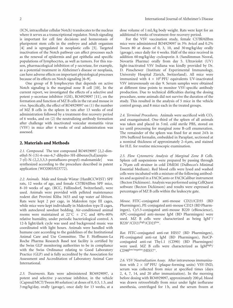
2 International Journal of Alzheimer’s Disease
(ICN, intracellular cellular Notch) translocates to the nucleuswhere it serves as a transcriptional regulator. Notch signalingis important for cell fate decisions and homeostasis ofpluripotent stem cells in the embryo and adult organism[4] and is upregulated in neoplastic cells [5]. Targetedinactivation of the Notch pathway can affect processes suchas the renewal of epidermis and gut epithelia and specificpopulations of lymphocytes, as well as tumors. For this rea-son, pharmacological inhibition of γ-secretase, for example,as a potential treatment in Alzheimer’s disease or neoplasia,can have adverse effects on important physiological processesbecause of its effects on Notch signaling [6–9].
One group of B lymphocytes that depends on activeNotch signaling is the marginal zone B cell [10]. In thecurrent report, we investigated the effects of a selective andpotent γ-secretase inhibitor (GSI), RO4929097 [11], on theformation and function of MZ B cells in the rat and mouse invivo. Specifically, the effect of RO4929097 on (1) the numberof MZ B cells in the spleen in rats after 13 weeks of oraladministration followed by a treatment-free recovery periodof 6 weeks, and on (2) the neutralizing antibody formationafter challenge with inactivated vesicular stomatitis virus(VSV) in mice after 4 weeks of oral administration wasassessed.
2. Materials and Methods
2.1. Compound. The test compound RO4929097 [2,2-dim-ethyl-N-((S)-6-oxo-6,7-dihydro-5H-dibenzo[b,d]azepin-7-yl)-N-(2,2,3,3,3-pentafluoro-propyl)-malonamide] wassynthesized according to the procedure described in patentapplication (WO2005/023772).
2.2. Animals. Male and female Wistar (HanRCCWIST) SPFrats, 12 weeks of age, and female C57Bl/6JIbm SPF mice,8–10 weeks of age, (RCC, Fullinsdorf, Switzerland), wereused. Animals were provided with pelleted maintenancerodent diet Provimi Kliba 3433 and tap water ad libitum.Rats were kept 2 per cage, in Makrolon type III cages,while mice were kept individually in Makrolon type II cages,with autoclaved sawdust bedding. Air-conditioned animalrooms were maintained at 22◦C ± 2◦C and 40%–80%relative humidity, under periodic bacteriological control. A12-h light/dark cycle was used and background music wascoordinated with light hours. Animals were handled withhumane care according to the guidelines of the InstitutionalAnimal Care and Use Committee. The F. Hoffmann-LaRoche Pharma Research Basel test facility is certified bythe Swiss GLP monitoring authorities to be in compliancewith the Swiss Ordinance relating to Good LaboratoryPractice (GLP) and is fully accredited by the Association forAssessment and Accreditation of Laboratory Animal CareInternational.
2.3. Treatments. Rats were administered RO4929097, apotent and selective γ-secretase inhibitor, in the vehicle(Capmul/MCT/Tween 80 solution) at doses of 0, 0.5, 1.5, and3 mg/kg/day, orally (gavage), once daily for 13 weeks, at a
dose volume of 1 mL/kg body weight. Rats were kept for anadditional 6 weeks of treatment-free recovery period.
For the VSV vaccination study, female C57Bl/6JIbmmice were administered RO4929097 in 3% Avicel and 0.2%Tween 80 at doses of 0, 3, 10, and 30 mg/kg/day orally(gavage), once daily for 4 weeks. Half of the mice received inaddition 60 mg/kg/day cyclosporin A (Sandimmun Neoral,Novartis Pharma) orally from day 5. Ultraviolet (UV)light-inactivated VSV Indiana was kindly provided by Dr.D. Pinschewer (Institute of Experimental Immunology,University Hospital Zurich, Switzerland). All mice wereimmunized with 4 × 10⁶ PFU equivalents UV-inactivatedVSV intravenously on day 9. Serum samples were obtainedat different time points to monitor VSV-specific antibodyproduction. Due to technical difficulties during the dosingprocedure, some animals did not survive the duration of thestudy. This resulted in the analysis of 5 mice in the vehiclecontrol group, and 8 mice each in the treated groups.
2.4. Terminal Procedures. Animals were sacrificed with CO2
and exsanguinated. One-third of the spleen of all animalswas taken and placed in 5 mL cold sterile PBS, stored onice until processing for marginal zone B-cell enumeration.The remainder of the spleen was fixed for at most 24 h in10% buffered formalin, embedded in Paraplast, sectioned ata nominal thickness of approximately 2–4 μm, and stainedfor H.E. for routine microscopic examination.
2.5. Flow Cytometric Analysis of Marginal Zone B Cells.Spleen cell suspensions were prepared by passing througha 70 μm cell strainer in cold DMEM (Dulbecco’s MinimalEssential Medium). Red blood cells were lysed and washedcells were incubated with a mixture of the following antibod-ies and acquired in a FACSCanto or FACSCalibur instrument(Becton Dickinson). Analysis was performed using CellQuestsoftware (Becton Dickinson) and results were expressed aspercentages of MZ B cells within the leukocyte gate.
Mouse. FITC-conjugated anti-mouse CD21/CD35 (BDPharmingen), PE-conjugated anti-mouse CD23 (BD Pharm-ingen), Cy5.5-conjugated anti-mouse B220 (eBioscience),APC-conjugated anti-mouse IgM (BD Pharmingen) wereused. MZ B cells were characterized as being IgM+/B220+/CD21high/CD23low.
Rat. FITC-conjugated anti-rat HIS57 (BD Pharmingen),PE-conjugated anti-rat IgM (BD Pharmingen), PerCP-conjugated anti-rat Thy1.1 (CD90) (BD Pharmingen)were used. MZ B cells were characterized as IgMhigh/CD90low/negative/HIS57+.
2.6. VSV Neutralization Assay. After intravenous immuniza-tion with 2 × 10⁶ PFU (plaque-forming units) VSV-IND,serum was collected from mice at specified times (days2, 4, 7, 14, and 20 after immunization). In the morningbefore dosing with RO4929097, approximately 200 μL bloodwas drawn retroorbitally from mice under light isofluraneanesthesia, centrifuged for 1 h, and the serum frozen at
International Journal of Alzheimer’s Disease 3
−20◦C. Thawed serum was prediluted 40-fold in DMEMcontaining 5% FCS and heat-inactivated for 30 min at 56 ◦C.Serial twofold dilutions were mixed with equal volumes ofVSV (500 PFU mL−1) and incubated for 90 min at 37 ◦Cin an atmosphere with 5% CO2. The serum-virus mixturewas transferred onto Vero cell monolayers in 96-well platesand incubated for 1 h at 37 ◦C. The monolayers were thenoverlaid with DMEM containing 1% methylcellulose andincubated for 24 h at 37 ◦C after which the monolayerswere fixed and stained with 0.5% crystal violet. The highestdilution of serum that reduced the number of plaques by50% was taken as the titer. To determine IgG titers, undilutedserum was pretreated with an equal volume of 0.1 mM β-2-mercaptoethanol in saline before heat inactivation andplating onto Vero cells. This treatment has been shown toeliminate only IgM but not IgG from serum [12]. Unreducedsamples were taken as IgM titers only, if the correspondingreduced samples had at least a fourfold lower titer, that is,when the IgG present in the unreduced sample could beneglected.
2.7. Statistics. For significance testing of dose-related effectson cellularity of MZ B cells in spleen and VSV neutralizingantibody titers in serum, Student’s t-test was used. Individualtime points (study days) were analyzed separately.
For significance testing of dose-related effects on whiteblood cell, neutrophil, and lymphocyte numbers in periph-eral blood, Jonckheere and the Mann-Whitney U tests wereused.
3. Results
3.1. 13-Week RO4929097 Treatment in Rats Followed by a6-Week Treatment-Free Recovery Period. Oral administrationof RO4929097 for 13 weeks was generally well toleratedand there were no treatment-related deaths during thecourse of the study. In the peripheral blood, moderatereductions in lymphocytes and increases in neutrophils from≥1.5 mg/kg/day in both male and female rats persisteduntil the end of the 6-week treatment-free recovery period(Tables 1(a) and 1(b)).
Flow cytometry analysis revealed a dose-dependentreduction in the percentage of MZ B cells in the spleenof both male and female rats following oral RO4929097administration for 13 weeks. Compared with the control,this reduction was statistically significant and greater than60% reduction at ≥1.5 mg/kg/day (Figure 1(a)). After therecovery period, the percentages of marginal zone B cells ofRO4929097-treated rats had normalized and were compara-ble with those of control animals (Figure 1(b)).
Histopathologic changes were recorded in the spleen ofnearly all male and female rats that received ≥1.5 mg/kg/dayRO4929097 (Figure 2). These included minimal-to-markeddecreased cellularity and infiltration of neutrophils of themarginal zone and periarteriolar lymphoid sheath areas.Individual animals showed minimally increased apoptosis inthe marginal zone. In addition, slight-to-moderate stromalchange, characterized by a focal, somewhat circumscribed
area of occasional pale staining lymphocytes, dendritic cells,eosinophilic hyaline-like deposits, early fibroplasia, necroticcell debris, and polymorphonuclear leukocyte infiltration,was noted in occasional animals. There were no RO4929097-related findings in the spleen after the 6-week treatment-freerecovery period.
3.2. VSV Vaccination Study in Mice. Mice were treated dailywith RO4929097 and vaccinated with inactivated VSV inorder to characterize the immune response to VSV in relationto a dose-dependent decrease in MZ B cellularity. Theimmunosuppressant cyclosporin A was used as a positivecontrol. Cyclosporin A is known to suppress the switch froma T-independent IgM to a T-dependent IgG response to VSVin this model [13].
Flow cytometry analysis of spleen cells at the end ofthe 4-week treatment period showed a dose-dependentreduction of relative MZ B cell counts (Figure 3). There wasno significant reduction at 3 mg/kg/day, a 51% reductionat 10 mg/kg/day, and a 95% reduction at 30 mg/kg/dayRO4929097. Vehicle or cyclosporin A treatment had no effecton MZ B cellularity.
Dosing with ≥10 mg/kg/day RO4929097 resulted in adelayed appearance of early neutralizing antibodies (presum-ably of IgM class) in response to VSV, that is, titers werebelow detection limit at 2 days after vaccination (Figure 4).Notably at 10 mg/kg with an average 51% depletion of MZB cells, the antibody response to VSV was only affected atthe earliest time point on day 2, after which no differenceto untreated animals in the immune response was noted.At 4 days after vaccination, antibody titers were detectablein all groups, but these were still reduced in animals dosedwith 30 mg/kg/day in comparison to all other groups. Inaddition, treatment with 30 mg/kg/day RO4929097 resultedin a reduction of neutralizing immunoglobulin titers ofthe IgG class at 7 days after vaccination (average 7.8-foldreduction), but titers recovered at later time points.
4. Discussion and Conclusion
Previous preclinical toxicity studies with RO4929097 in rats,mice, and dogs have shown that significant effects wereattributable to Notch impairment [11]. Similar findingswere observed following oral administration of RO4929097to male and female Wistar rats for 13 weeks. Flowcytometry analysis showed a dose-dependent reduction inrelative numbers of splenic MZ B cells that correlatedwith histopathologic atrophy of the MZ. Marginal zoneB cell depletion was completely reversible after a 6-weekrecovery period. Similarly, a dose-dependent reduction inrelative numbers of splenic MZ B cells was seen in femaleC57Bl/6JIbm mice administered RO4929097 for 4 weeks.Total early neutralizing antibodies (presumably IgM) toinactivated VSV were also reduced, and this was consideredto reflect the pharmacological effect of γ-secretase inhibitorRO4929097 on MZ B cells. At the high dose of 30 mg/kg/dayRO4929097, neutralizing IgG titers were also reduced at 7days after vaccination, but recovered at later time points.
4 International Journal of Alzheimer’s Disease
Table 1: Effects of RO4929097 on absolute numbers of white blood cells, neutrophils, and lymphocytes in male and female rats.
(a) After 13 weeks of treatment
Males Females
Dose (mg/kg/day) WBC Neutrophils Lymphocytes WBC Neutrophils Lymphocytes
(109/L) (109/L) (G/L) (109/L) (109/L) (109/L)
0
N 10 10 10 10 10 10
Mean 5.95 1.23 4.26 3.77 0.55 2.94
SD 1.23 0.42 0.92 0.60 0.20 0.47
0.5
N 10 10 10 10 10 10
Mean 5.65 1.20 4.05 3.68 0.67# 2.76
SD 1.08 0.39 0.75 0.92 0.12 0.86
1.5
N 10 10 10 9 9 9
Mean 6.31 1.78∗ 4.01 3.10 1.11∗∗ 1.70∗∗
SD 1.39 0.54 1.28 0.85 0.54 0.78
3
N 10 10 10 10 10 10
Mean 6.37 2.95∗∗ 2.70∗∗ 2.87 1.10 1.51
SD 1.58 1.29 1.17 0.47 0.24 0.47
Jonckheere test: ∗∗P ≤ 1%; ∗1% < P ≤ 5%.U test: P ≤ 1%; #1% < P ≤ 5%.N: number of animals.SD: standard deviation.
(b) After 6 weeks of recovery
Males Females
Dose (mg/kg/day) WBC Neutrophils Lymphocytes WBC Neutrophils Lymphocytes
(109/L) (109/L) (109/L) (109/L) (109/L) (109/L)
0
N 10 10 10 10 10 10
Mean 7.56 1.02 6.19 5.28 0.68 4.36
SD 1.17 0.27 1.18 0.84 0.18 0.76
0.5
N 10 10 10 10 10 10
Mean 7.28 1.07 5.85 5.06 0.68 4.14
SD 1.37 0.25 1.16 1.00 0.17 0.90
1.5
N 10 10 10 10 10 10
Mean 8.40 1.70∗∗ 6.25 4.01∗∗ 1.28∗∗ 2.45∗∗
SD 1.34 0.50 1.25 1.03 0.72 0.81
3
N 10 10 10 10 10 10
Mean 9.13∗ 3.45∗∗ 4.99 3.85∗∗ 1.48∗∗ 2.08∗∗
SD 2.32 2.10 1.73 0.78 0.54 0.59
Jonckheere test: ∗∗P ≤ 1%; ∗1% < P ≤ 5%.N: number of animals.SD: standard deviation.
International Journal of Alzheimer’s Disease 5
0
5
10
15
20
25
Vehicle control 0.5 mg/kg 1.5 mg/kg 3 mg/kg
Male animals
0
5
10
15
20
25
Vehicle control 0.5 mg/kg 1.5 mg/kg 3 mg/kg
Female animals
MZ
B c
ells
in le
uco
cyte
gat
e (%
)M
Z B
cel
ls in
leu
cocy
te g
ate
(%)
∗
∗
∗∗
(a)
0
5
10
15
20
25
Vehicle control 0.5 mg/kg 1.5 mg/kg 3 mg/kg
Male recovery animals
0
5
10
15
20
25
Vehicle control 0.5 mg/kg 1.5 mg/kg 3 mg/kg
Female recovery animals
MZ
B c
ells
in le
uco
cyte
gat
e (%
)M
Z B
cel
ls in
leu
cocy
te g
ate
(%)
(b)
Figure 1: (a) Marginal zone B cells in male and female rats dosed for 13 weeks with RO4929097. Spleen cell suspensions were analyzed byflow cytometry (FACS) using fluorescently labeled antibodies. For each rat the percentage of MZ B cells (= IgMhigh/CD90low/neg/HIS57+) inthe leukocyte gate is shown. Dots represent individual rats. - = average per group. ∗ = statistically significant difference to vehicle control(P < 0.05). n = 10 rats/sex/group. (b) Splenic MZ B cells in male and female rats dosed for 13 weeks with RO4929097 followed by a recoveryphase of 6 weeks. Dots represent individual rats. - = average per group. n = 6 rats/sex/group.
MZ
(a) Control
MZ
(b) RO4929097
Figure 2: Atrophy of the marginal zone (MZ) of the spleen of rats administered 3.0 mg/kg/day RO4929097 for 13 weeks. (hematoxylin-eosinstain).
Class switching from IgM to IgG requires T-cell help, and itis conceivable that high dose RO4929097 not only affectedMZ B-cell cellularity but also T-cell function since Notchis also expressed in T cells. Alternatively, a severely reducedMZ B-cell compartment resulted in fewer B cells switchingfrom IgM to IgG. The effect of RO4929097 on IgG antibodyformation in these mice was considered relatively mildand a threshold (i.e., 3 mg/kg/day) could be defined where
no effects on MZ B-cell counts and neutralizing antibodyresponses were seen. Originally designed for the treatmentof Alzheimer’s disease, the efficacious dose of RO4929097 onAβ reduction was found to be 3 mg/kg in the brain of Aβtransgenic mice, and 10 mg/kg in cerebrospinal fluid of naıverats. The ED50 for mouse MZ B-cell depletion was 10 mg/kg,while for the rat it was lower at 1 mg/kg, but differencesin treatment duration should be taken into account. As
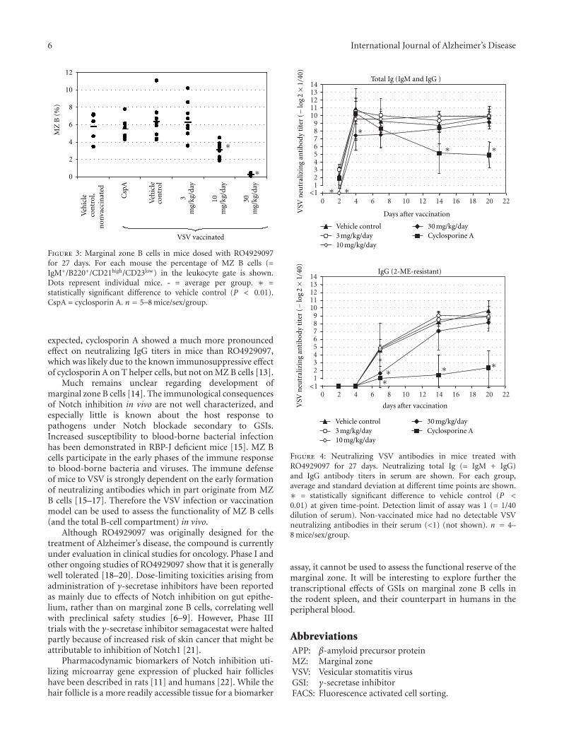
6 International Journal of Alzheimer’s DiseaseM
Z B
(%
)
0
2
4
6
8
10
12
Csp
A
Veh
icle
con
trol
∗
∗
30
mg/
kg/d
ay
10
mg/
kg/d
ay
3m
g/kg
/day
VSV vaccinated
Veh
icle
con
trol
,n
onva
ccin
ated
Figure 3: Marginal zone B cells in mice dosed with RO4929097for 27 days. For each mouse the percentage of MZ B cells (=IgM+/B220+/CD21high/CD23low) in the leukocyte gate is shown.Dots represent individual mice. - = average per group. ∗ =statistically significant difference to vehicle control (P < 0.01).CspA = cyclosporin A. n = 5–8 mice/sex/group.
expected, cyclosporin A showed a much more pronouncedeffect on neutralizing IgG titers in mice than RO4929097,which was likely due to the known immunosuppressive effectof cyclosporin A on T helper cells, but not on MZ B cells [13].
Much remains unclear regarding development ofmarginal zone B cells [14]. The immunological consequencesof Notch inhibition in vivo are not well characterized, andespecially little is known about the host response topathogens under Notch blockade secondary to GSIs.Increased susceptibility to blood-borne bacterial infectionhas been demonstrated in RBP-J deficient mice [15]. MZ Bcells participate in the early phases of the immune responseto blood-borne bacteria and viruses. The immune defenseof mice to VSV is strongly dependent on the early formationof neutralizing antibodies which in part originate from MZB cells [15–17]. Therefore the VSV infection or vaccinationmodel can be used to assess the functionality of MZ B cells(and the total B-cell compartment) in vivo.
Although RO4929097 was originally designed for thetreatment of Alzheimer’s disease, the compound is currentlyunder evaluation in clinical studies for oncology. Phase I andother ongoing studies of RO4929097 show that it is generallywell tolerated [18–20]. Dose-limiting toxicities arising fromadministration of γ-secretase inhibitors have been reportedas mainly due to effects of Notch inhibition on gut epithe-lium, rather than on marginal zone B cells, correlating wellwith preclinical safety studies [6–9]. However, Phase IIItrials with the γ-secretase inhibitor semagacestat were haltedpartly because of increased risk of skin cancer that might beattributable to inhibition of Notch1 [21].
Pharmacodynamic biomarkers of Notch inhibition uti-lizing microarray gene expression of plucked hair follicleshave been described in rats [11] and humans [22]. While thehair follicle is a more readily accessible tissue for a biomarker
0 2 4 6 8 10 12 14 16 18 20 22
123456789
1011121314
Total Ig (IgM and IgG )
Days after vaccinationVSV
neu
tral
izin
g an
tibo
dy t
iter
(−
log
2×
1/40
)
∗∗
123456789
1011121314
VSV
neu
tral
izin
g an
tibo
dy t
iter
(−
log
2×
1/40
)
Vehicle control3 mg/kg/day10 mg/kg/day
30 mg/kg/dayCyclosporine A
0 2 4 6 8 10 12 14 16 18 20 22
days after vaccination
Vehicle control3 mg/kg/day10 mg/kg/day
30 mg/kg/dayCyclosporine A
IgG (2-ME-resistant)
∗
∗ ∗
∗∗ ∗ ∗
<1
<1
Figure 4: Neutralizing VSV antibodies in mice treated withRO4929097 for 27 days. Neutralizing total Ig (= IgM + IgG)and IgG antibody titers in serum are shown. For each group,average and standard deviation at different time points are shown.∗ = statistically significant difference to vehicle control (P <0.01) at given time-point. Detection limit of assay was 1 (= 1/40dilution of serum). Non-vaccinated mice had no detectable VSVneutralizing antibodies in their serum (<1) (not shown). n = 4–8 mice/sex/group.
assay, it cannot be used to assess the functional reserve of themarginal zone. It will be interesting to explore further thetranscriptional effects of GSIs on marginal zone B cells inthe rodent spleen, and their counterpart in humans in theperipheral blood.
AbbreviationsAPP: β-amyloid precursor proteinMZ: Marginal zoneVSV: Vesicular stomatitis virusGSI: γ-secretase inhibitorFACS: Fluorescence activated cell sorting.
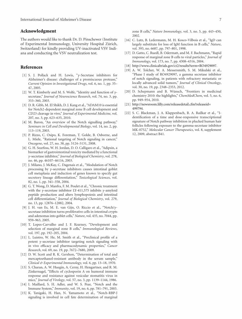
International Journal of Alzheimer’s Disease 7
Acknowledgment
The authors would like to thank Dr. D. Pinschewer (Instituteof Experimental Immunology, University Hospital Zurich,Switzerland) for kindly providing UV-inactivated VSV Indi-ana and conducting the VSV neutralization test.
References
[1] S. J. Pollack and H. Lewis, “γ-Secretase inhibitors forAlzheimer’s disease: challenges of a promiscuous protease,”Current Opinion in Investigational Drugs, vol. 6, no. 1, pp. 35–47, 2005.
[2] W. T. Kimberly and M. S. Wolfe, “Identity and function of γ-secretase,” Journal of Neuroscience Research, vol. 74, no. 3, pp.353–360, 2003.
[3] D. R. Gibb, M. El Shikh, D. J. Kang et al., “ADAM10 is essentialfor Notch2-dependent marginal zone B cell development andCD23 cleavage in vivo,” Journal of Experimental Medicine, vol.207, no. 3, pp. 623–635, 2010.
[4] M. Baron, “An overview of the Notch signalling pathway,”Seminars in Cell and Developmental Biology, vol. 14, no. 2, pp.113–119, 2003.
[5] P. Rizzo, C. Osipo, K. Foreman, T. Golde, B. Osborne, andL. Miele, “Rational targeting of Notch signaling in cancer,”Oncogene, vol. 27, no. 38, pp. 5124–5131, 2008.
[6] G. H. Searfoss, W. H. Jordan, D. O. Calligaro et al., “Adipsin, abiomarker of gastrointestinal toxicity mediated by a functionalγ-secretase inhibitor,” Journal of Biological Chemistry, vol. 278,no. 46, pp. 46107–46116, 2003.
[7] J. Milano, J. McKay, C. Dagenais et al., “Modulation of Notchprocessing by γ-secretase inhibitors causes intestinal gobletcell metaplasia and induction of genes known to specify gutsecretory lineage differentiation,” Toxicological Sciences, vol.82, no. 1, pp. 341–358, 2004.
[8] G. T. Wong, D. Manfra, F. M. Poulet et al., “Chronic treatmentwith the γ-secretase inhibitor LY-411,575 inhibits γ-amyloidpeptide production and alters lymphopoiesis and intestinalcell differentiation,” Journal of Biological Chemistry, vol. 279,no. 13, pp. 12876–12882, 2004.
[9] J. H. van Es, M. E. van Gijn, O. Riccio et al., “Notch/γ-secretase inhibition turns proliferative cells in intestinal cryptsand adenomas into goblet cells,” Nature, vol. 435, no. 7044, pp.959–963, 2005.
[10] T. Lopes-Carvalho and J. F. Kearney, “Development andselection of marginal zone B cells,” Immunological Reviews,vol. 197, pp. 192–205, 2004.
[11] L. Luistro, W. He, M. Smith et al., “Preclinical profile of apotent γ-secretase inhibitor targeting notch signaling within vivo efficacy and pharmacodynamic properties,” CancerResearch, vol. 69, no. 19, pp. 7672–7680, 2009.
[12] D. W. Scott and R. K. Gershon, “Determination of total andmercaptoethanol-resistant antibody in the serum sample,”Clinical & Experimental Immunology, vol. 6, pp. 13–18, 1970.
[13] S. Charan, A. W. Huegin, A. Cerny, H. Hengartner, and R. M.Zinkernagel, “Effects of cyclosporin A on humoral immuneresponse and resistance against vesicular stomatitis virus inmice,” Journal of Virology, vol. 57, no. 3, pp. 1139–1144, 1986.
[14] I. Maillard, S. H. Adler, and W. S. Pear, “Notch and theImmune System,” Immunity, vol. 19, no. 6, pp. 781–791, 2003.
[15] K. Tanigaki, H. Han, N. Yamamoto et al., “Notch-RBP-Jsignaling is involved in cell fate determination of marginal
zone B cells,” Nature Immunology, vol. 3, no. 5, pp. 443–450,2002.
[16] C. Lutz, B. Ledermann, M. H. Kosco-Vilbois et al., “IgD canlargely substitute for loss of IgM function in B cells,” Nature,vol. 393, no. 6687, pp. 797–801, 1998.
[17] D. Gatto, C. Ruedl, B. Odermatt, and M. F. Bachmann, “Rapidresponse of marginal zone B cells to viral particles,” Journal ofImmunology, vol. 173, no. 7, pp. 4308–4316, 2004.
[18] http://www.clinicaltrials.gov/ct2/results?term=RO4929097.[19] A. W. Tolcher, W. A. Messersmith, S. M. Mikulski et al.,
“Phase I study of RO4929097, a gamma secretase inhibitorof notch signaling, in patients with refractory metastatic orlocally advanced solid tumors,” Journal of Clinical Oncology,vol. 30, no. 19, pp. 2348–2353, 2012.
[20] D. Schepmann and B. Wunsch, “Frontiers in medicinalchemistry 2010: the highlights,” ChemMedChem, vol. 5, no. 6,pp. 949–954, 2010.
[21] http://newsroom.lilly.com/releasedetail.cfm?releaseid=499794.
[22] S. C. Blackman, J. A. Klappenbach, R. A. Railkar et al., “I-dentification of a time and dose-responsive transcriptionalsignature of Notch pathway inhibition in plucked human hairfollicles following exposure to the gamma-secretase inhibitorMK-0752,” Molecular Cancer Therapeutics, vol. 8, supplement12, 2009, abstract B41.
Hindawi Publishing CorporationInternational Journal of Alzheimer’s DiseaseVolume 2012, Article ID 295207, 10 pagesdoi:10.1155/2012/295207
Research Article
γ-Secretase Modulators: Can We Combine Potency with Safety?
Harrie J. M. Gijsen and Marc Mercken
Department of Neuroscience, Janssen Research & Development, Pharmaceutical Companies of Johnson & Johnson,Turnhoutseweg 30, B-2340 Beerse, Belgium
Correspondence should be addressed to Harrie J. M. Gijsen, [email protected]
Received 2 August 2012; Accepted 8 November 2012
Academic Editor: Jeremy Toyn
Copyright © 2012 H. J. M. Gijsen and M. Mercken. This is an open access article distributed under the Creative CommonsAttribution License, which permits unrestricted use, distribution, and reproduction in any medium, provided the original work isproperly cited.
γ-Secretase modulation has been proposed as a potential disease modifying anti-Alzheimer’s approach. γ-Secretase modulators(GSMs) cause a product shift from the longer amyloid-beta (Aβ) peptide isoforms to shorter, more soluble, and less amyloidogenicisoforms, without inhibiting APP or Notch proteolytic processing. As such, modulating γ-secretase may avoid some of the adverseeffects observed with γ-secretase inhibitors. Since the termination of the GSM tarenfurbil in 2008 due to negative phase III trialresults, a considerable progress has been made towards more potent and better brain penetrable compounds. However, an analysisof their lipophilic efficiency indices indicates that their increased potency can be largely attributed to their increased lipophilicity.The need for early and chronic dosing with GSMs will require high-safety margins. This will be a challenge to achieve with thecurrent, highly lipophilic GSMs. We will demonstrate that by focusing on the drug-like properties of GSMs, a combination of highin vitro potency and reduced lipophilicity can be achieved and does result in better tolerated compounds. The next hurdle will beto translate this knowledge into GSMs which are highly efficacious and safe in vivo.
1. Introduction
With an ageing population, the prevalence of Alzheimer’sdisease (AD) and the consequent burden to society arerapidly rising [1]. The currently approved medication forAD only offers symptomatic treatment of limited durationwithout affecting the progression of the disease. Therefore,disease modifying approaches are urgently needed. A hall-mark pathology of AD is the presence of amyloid plaquesin the brain, which are mainly aggregates of amyloid-beta(Aβ) peptides of varying length, among which Aβ42 isthe most amyloidogenic and neurotoxic. These peptidesare formed via sequential, proteolytic processing of theamyloid precursor protein (APP) by two aspartyl proteases:β-secretase (BACE) and γ-secretase (GS). Consequently,pharmacological intervention in the activity of these sec-retases has been heavily investigated for over a decade toprevent both the buildup of the amyloid plaques, as well asthe formation of toxic amyloid dimers and oligomers [2, 3].Small molecules inhibiting either BACE or GS offer a directway to reduce the production of all amyloid peptides. Wherethe development of β-secretase inhibitors has been seriously
slowed down/hampered due to the difficulty in achievingadequate brain penetration and concomitant in vivo efficacy[4], potent, centrally active γ-secretase inhibitors (GSIs)have been discovered and investigated in the clinic andtheir progress has been reviewed [5, 6]. Phase III clinicaltrials with the most advanced compound, semagacestat (1)(LY450, 139, Figure 1), were prematurely halted in 2010 [6].Instead of slowing disease progression, 1 was associated witha statistically significant decline in cognition. In addition, anincreased risk of skin cancer was reported, most likely relatedto Notch inhibition.
In comparison with GSIs, γ-secretase modulators(GSMs) cause a product shift from the longer amyloid-beta(Aβ) peptide isoforms to shorter, more soluble, and less amy-loidogenic isoforms, without inhibiting APP or Notch prote-olytic processing. As such, modulating γ-secretase may avoidthe target-related adverse effects observed upon inhibition ofGS.
The identification of a subset of NSAIDs as GSMs in 2001has led to tarenflurbil 2 (Flurizan) as the first NSAID-derivedGSM to be tested in the clinic [7, 8]. After negative results in

2 International Journal of Alzheimer’s Disease
phase III clinical trials, development was stopped in 2008.This late stage clinical failure can potentially be attributedto the very low brain penetration and weak potency oftarenflurbil [9].
Non-NSAID-derived compounds which do not contain acarboxylic acid group, but are characterized by an imidazolegroup, were described in a patent application by Neuroge-netics in 2004 [10]. Subsequent work by Eisai led to the firstcompound from this series reaching the clinic, E-2012 (3).Although the phase I clinical trial with E-2012 was suspendedafter lenticular opacity was observed in a high-dose group ofa preclinical safety study in rats, in human the compoundwas reported to reduce plasma Aβ42 levels dose-dependently,with a maximum reduction of ∼50% after a 400 mg dose[11].
Since then, many companies have elaborated on thesetwo series, and a considerable progress has been madetowards more potent and better brain penetrable compoundsin both NSAID- and imidazole-derived chemical series[12, 13]. Despite the efforts spent in this field, very fewcompounds have made it to the clinic since the terminationof tarenflurbil and E-2012. Currently, only two compoundsare reported to be in early clinical development: E-2212 andEVP-0962, both with undisclosed structures. In this report,we will provide an overview of our own work and put thisinto context of the poor drug-like properties of most of thedescribed GSMs to date, especially their high lipophilicity.The need to improve on this in order to obtain safe andefficacious GSMs, suitable for chronic treatment, will also bediscussed.
2. Methods
2.1. Calculation of Efficiency Parameters. Mathematically,ligand efficiency (LE) is calculated according to (1), whereΔG represents the binding free energy of a ligand and nis the number of heavy (nonhydrogen) atoms. ΔG can becalculated using (2), where the dissociation constant Kd canbe replaced by IC50 [14]. Calculation of LE in this paperwas done using a temperature (T) of 310 K and using themeasured or reported cellular IC50 values for the inhibitionof Aβ42. As a result, LE is given in kcal/heavy atom accordingto (3),
LE = ΔG
n, (1)
ΔG = −RT lnKd ≈ −RT ln(IC50), (2)
LE = 1.4pIC50
n. (3)
Ligand lipophilicity efficiency (LLE) and ligand effi-ciency-dependent lipophilicity (LELP) were calculated accor-ding to (4) and (5), respectively. The octanol-water partitioncoefficient (clogP) is used to describe the lipophilicity of
a compound. LogP was calculated (clogP) using biobytesoftware.
LLE = pIC50 − clogP, (4)
LELP = clogPLE
. (5)
2.2. Biology. For the cellular in vitro activity of our own com-pounds, screening was carried out using SKNBE2 humanneuroblastoma cells carrying the hAPP 695 wild-type asdescribed in [15]. For a description of the mouse in vivoexperiments, see references [15, 16]; for details on the dogin vivo experiments, see [17].
3. Discussion
For both the NSAID-derived carboxylic acid GSMs and theimidazole-derived GSMs, we have reported recently on theactivity of optimized, potent submicromolar compoundswith good in vivo activity in lowering Aβ42 levels in mousebrain and/or dog CSF. The structures of the most profiledcompounds are shown in Figure 2. Carboxylic acid JNJ-40418677 (4) has lowered Aβ42 in cells with an IC50 of200 nM and had a brain/plasma ratio of 0.5–1.0 in mice,depending on the dose [16]. Imidazole JNJ-42601572 (5)lowered Aβ42 in cells with an IC50 of 16 nM and had abrain/plasma ratio of 0.7–1.1 in mice [15, 17]. A time profileof JNJ-42601572 in mouse is shown in Figure 3. A similartime profile for JNJ-40418677 has been published [16].
Both compounds have been tested in advanced animalmodels, including repeated dosing in rat and dog. In thesestudies, early signs of liver toxicity were noted for bothcompounds, such as bilirubin and ALT/AST increases [17].Based on these findings, further development was halted.Since then, the dog model combining PK/PD data withearly observations of liver toxicity is routinely used in ourdiscovery program to triage compounds [17]. Both JNJ-40418677 and JNJ-42601572 contain a considerable numberof conjugated aromatic rings and are characterized by highlipophilicity and molecular weight. In recent years a numberof publications have appeared relating such properties witha low probability of success in clinical development [29,30]. Catchy phrases like “molecular obesity” and “escapefrom flatland” have been used to describe the issues asso-ciated with high molecular weight and number of (hetero)aromatic rings in molecules [31, 32]. Based on analysisof compound properties and their attrition or success in(pre)clinical development, guidelines have been formulatedand proposed which can be applied during optimizationefforts in medicinal chemistry programs to strive towardsmore drug-like compounds. The concept of ligand efficiency(LE) is now routinely applied in the drug discovery process[14]. It is defined as the binding energy towards a biologicaltarget per (heavy = nonhydrogen) atom in a compound.Since most assays are not directly measuring binding energy,IC50 values are often used to make a relative comparisonof the LE of compounds. By working towards a high LE,

International Journal of Alzheimer’s Disease 3
F
ON
N
ON
O
FNNH
O
NH
O O
OH
OH
1, semagacestat 2, tarenflurbil 3, E-2012
Figure 1
N
N
O
N
NH N
N
F
O
CF3
F3C
OH
4 Ab42 IC50 = 200 nM 5 Ab42 IC50 = 16 nM
Figure 2
Bra
in c
once
ntr
atio
n
0
1
2
3
4
5
1 2 4 6 8 16 20 24
JNJ-42601572 (h)
of J
NJ-
4260
1572
(μ
M)
BQLBQLBQL
(a)
0
20
40
60
80
100
120
140
160
1 2 4 6 8 16 20 24
JNJ-42601572 (h)
Vehicle
Aβ
tota
l (ve
hic
le (
%))
(b)
0
20
40
60
80
100
120
140
160
1 2 4 6 8 16 20 24
JNJ-42601572 (h)
Vehicle
Aβ
42 (
veh
icle
(%
))
(c)
0
20
40
60
80
100
120
140
160
180
200
1 2 4 6 8 16 20 24
JNJ-42601572 (h)
Vehicle
Aβ
38 (
veh
icle
(%
))
(d)
Figure 3: Effects of a single oral dose of 10 mg/kg JNJ-42601572 on Aβ levels in nontransgenic mouse brain as measured with differentialELISAs [16]. Mean (+SEM) brain Aβ levels after drug treatment are expressed as percentage of Aβ levels in brain of vehicle-treated mice.(n = 6 mice per data point). BQL: below quantifiable limits (in brain 0.07–0.12 μM).
4 International Journal of Alzheimer’s Disease
molecular weight can be kept in control while optimiz-ing pharmacological activity. Additional parameters havebeen derived, taking the lipophilicity into account: ligandlipophilicity efficiency (LLE) [33] and ligand efficiency-dependent lipophilicity (LELP) [34] are indices which havebeen proposed to drive the medicinal chemistry towardsan acceptable balance between potency and lipophilicity.Striving for optimal values for these parameters in leadoptimization should lead to an increase in potency withoutincreasing lipophilicity. For LLE, a value above 5 is desirable,and for LELP, a value below 10 [35]. In this report we willanalyze how the GSM field has evolved in regard to lipophilicefficiency and the implications for their developability.
We have calculated the efficiency indices for a number ofrepresentative optimized compounds taken from the litera-ture, including our own compounds. A set of acid-derivedGSMs are shown in Table 1, and imidazole-containing GSMsare shown in Table 2. Due to the lack of a direct bindingassay to GS, we have calculated the ligand efficiency usingthe pIC50 values for reducing Aβ42 in the cellular assay.Cellular activity can be influenced by additional factors, suchas permeability, and can therefore differ substantially fromthe actual binding of the compounds to the GS complex.Nevertheless, by comparing the various efficiency indiceswithin the two respective chemical classes, we feel thecalculated values can still be used to rank order compounds.
In Table 1, the acid-derived GSMs are shown in moreor less chronological order. Starting with tarenflurbil, theslightly more potent and brain penetrant CHF5074 isfollowed by more recently published and considerably morepotent analogues JNJ-40418677, BIIB042, and compounds 8and 9 from GSK and Merck, respectively. The optimizationefforts around tarenflurbil have led to an increase inpotency and brain penetration, but the ligand and lipophilicefficiency indices for these compounds have hardly beenimproved. LLE has remained very low for 5, 6, and 8,and although an improvement is seen for 7 and 9, thevalues are still below the desirable level (>5). For LELP,all compounds have values well above the desirable upperlimit of 10, and no compounds show improvement overtarenflurbil. The increase in potency of GSMs 5, 7, and 8 canmainly be attributed to an increase in lipophilicity. For 9, thelipophilicity has not increased compared to tarenflurbil, and9 also has a reduced aromatic ring count, compared to theother analogs. However, potency and brain penetration forthis compound remain suboptimal. Regarding these metrics,tarenflurbil is still the most drug-like molecule, and indeedthe compound was well tolerated during clinical trials despitehigh dosage [36]. In addition to tarenflurbil, of this set ofcompounds only the closest analogue, CHF5074 (6), hasprogressed into clinical trials. For all other compounds inTable 1 no further development has been reported.
A similar picture emerges for the nonacidic GSMs,mostly containing an imidazole moiety (Table 2). The earlyneurogenetic compound 10 has a relatively high ligand effi-ciency of 0.37 but is characterized by very high lipophilicity,resulting in a low LLE and high LELP value. Going from 10 toE-2012 (3), both LLE and LELP have improved. Subsequentwork around this series by various companies, as exemplified
in Table 2 by 11–15 and 5, has not resulted into a significantfurther improvement of values relevant for drug-likeness,and in most cases a deterioration of them. The medicinalchemistry program towards 15 was aimed at optimizinglipophilic efficiency [28], which has resulted in the bestvalues for clogP, LLE, and LELP within the set of compoundsshown in Table 2. However, potency of this compound isstill moderate, resulting in a 36% lowering of Aβ42 levelsin Guinea pig brain after a 100 mg/kg oral dose [28]. Fromthe compounds in Table 2, only compound 3 (E-2012) hasprogressed into the clinic. After a single dose of 80 mg/kgof 3 in dog, we have observed liver parameter changes aswell as large changes in gene-expression profile in liver tissueobtained in this study [17]. The still suboptimal values forlipophilic efficiency of 3 may have contributed to this, as wellas to the termination of 3 in phase 1 clinical trials.
From the in vivo data obtained with JNJ-40418677 [16],as well as for JNJ-42601572 shown in Figure 3, two relevantobservations can be made, which are also apparent fromall other GSMs we have tested in vivo up to now. The firstobservation is that reduction of brain Aβ42 nicely correlateswith the compound levels in brain. After the disappearanceof the compound from the brain, the Aβ levels quickly returnto baseline. This implies that compound levels need to besustained to maintain a desired change in Aβ levels. Thesecond observation is the large difference between the invitro potency and the compound concentrations requiredto reduce Aβ42 levels in vivo. A considerably higher totalcompound concentration is required than the in vitro IC50
to achieve a significant reduction of Aβ42 levels in vivo. Acommon practice is to correct the plasma and brain concen-tration for the fraction bound to plasma proteins or braintissue and to use the free compound concentrations. BothJNJ-40418677 and JNJ-42601572 are highly protein/braintissue bound, with free fractions in plasma and brain ofless than 0.1%, resulting in free compound concentrationsbelow the in vitro (cellular) IC50 values. However, it shouldbe noted that free fractions below 1% are often difficult tobe determined accurately, with minor changes in absolutevalues leading to major differences in calculated free concen-trations. After dosing of JNJ-42601572 in dog, the compoundconcentration in CSF has also been measured, which can beconsidered as a surrogate for free brain concentrations. Afteran oral dose of 20 mg/kg, compound levels were only mea-surable at the plasma Cmax of 4 h, with a CSF/plasma ratio of0.003. This corresponds to a CSF compound concentrationof 11±5 nM, in the same range as the in vitro cellular potency(IC50, 16 nM). An increase in free brain concentration asmeasured by the fraction unbound in brain (Fub) couldpotentially lead to an improved in vivo reduction in Aβ42.A good correlation exists between Fub and the lipophilicityof a compound (LogP) [37], giving another reason to aimfor a reduced lipophilicity of the compounds. The highlipophilicity required to achieve potency, as evident from allthe published GSMs and data shown in Tables 1 and 2, islikely related to the membrane embedded character of the GSproteins and the site where these modulators interact withthese proteins. This poses the question if the nature of thetarget allows for potent compounds with a low lipophilicity.
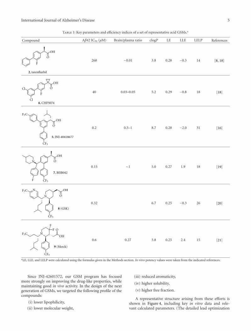
International Journal of Alzheimer’s Disease 5
Table 1: Key parameters and efficiency indices of a set of representative acid GSMs.a
Compound Aβ42 IC50 (μM) Brain/plasma ratio clogP LE LLE LELP References
F
O
2, tarenflurbil
OH
268 ∼0.01 3.8 0.28 −0.3 14 [8, 18]
F
O
Cl
Cl
6, CHF5074
OH
40 0.03–0.05 5.2 0.29 −0.8 18 [18]
O
F3C
OH
CF3
5, JNJ-40418677
0.2 0.5–1 8.7 0.28 −2.0 31 [16]
7, BIIB042
F
N O
OH
CF3
0.15 ∼1 5.0 0.27 1.9 18 [19]
N
N O
CF3
F3C
8 (GSK)
OH
0.32 6.7 0.25 −0.3 26 [20]
NOF
F
OH
9 (Merck)
F3C
CF3
0.6 0.27 3.8 0.25 2.4 15 [21]
aLE, LLE, and LELP were calculated using the formulas given in the Methods section. In vitro potency values were taken from the indicated references.
Since JNJ-42601572, our GSM program has focusedmore strongly on improving the drug-like properties, whilemaintaining good in vivo activity. In the design of the nextgeneration of GSMs, we targeted the following profile of thecompounds:
(i) lower lipophilicity,
(ii) lower molecular weight,
(iii) reduced aromaticity,
(iv) higher solubility,
(v) higher free fraction.
A representative structure arising from these efforts isshown in Figure 4, including key in vitro data and rele-vant calculated parameters. (The detailed lead optimization
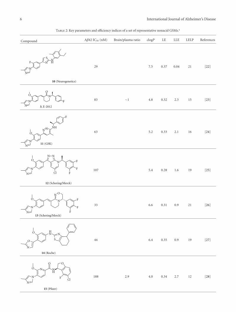
6 International Journal of Alzheimer’s Disease
Table 2: Key parameters and efficiency indices of a set of representative nonacid GSMs.a
Compound Aβ42 IC50 (nM) Brain/plasma ratio clogP LE LLE LELP References
F
N
N
NH
S
N
10 (Neurogenetics)
29 7.5 0.37 0.04 21 [22]
O
N
N
F
N
O
3, E-2012
83 ∼1 4.8 0.32 2.3 15 [23]
O
N
N
F
N N
11 (GSK)
NH
63 5.2 0.33 2.1 16 [24]
O
N
N Cl F
F
FN
N N
12 (Schering/Merck)
107 5.4 0.28 1.6 19 [25]
O
N
N F
F
FN
ON
13 (Schering/Merck)
33 6.6 0.31 0.9 21 [26]
O
N
O
N
S
NH
14 (Roche)
44 6.4 0.35 0.9 19 [27]
O
N
N
O
ClF
O
NH
N
15 (Pfizer)
188 2.9 4.0 0.34 2.7 12 [28]
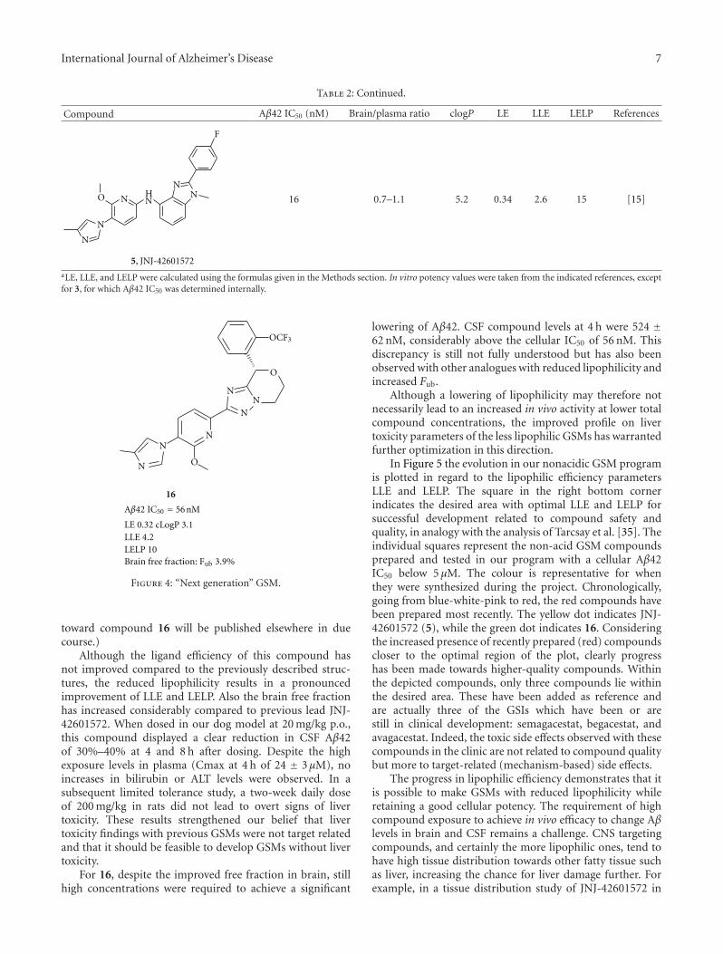
International Journal of Alzheimer’s Disease 7
Table 2: Continued.
Compound Aβ42 IC50 (nM) Brain/plasma ratio clogP LE LLE LELP References
O
N
N
F
NN
NH
N
5, JNJ-42601572
16 0.7–1.1 5.2 0.34 2.6 15 [15]
aLE, LLE, and LELP were calculated using the formulas given in the Methods section. In vitro potency values were taken from the indicated references, exceptfor 3, for which Aβ42 IC50 was determined internally.
N
N
O
N
O
NN
N
OCF3
16
Aβ42 IC50 = 56 nM
LE 0.32 cLogP 3.1LLE 4.2LELP 10Brain free fraction: Fub 3.9%
Figure 4: “Next generation” GSM.
toward compound 16 will be published elsewhere in duecourse.)
Although the ligand efficiency of this compound hasnot improved compared to the previously described struc-tures, the reduced lipophilicity results in a pronouncedimprovement of LLE and LELP. Also the brain free fractionhas increased considerably compared to previous lead JNJ-42601572. When dosed in our dog model at 20 mg/kg p.o.,this compound displayed a clear reduction in CSF Aβ42of 30%–40% at 4 and 8 h after dosing. Despite the highexposure levels in plasma (Cmax at 4 h of 24 ± 3μM), noincreases in bilirubin or ALT levels were observed. In asubsequent limited tolerance study, a two-week daily doseof 200 mg/kg in rats did not lead to overt signs of livertoxicity. These results strengthened our belief that livertoxicity findings with previous GSMs were not target relatedand that it should be feasible to develop GSMs without livertoxicity.
For 16, despite the improved free fraction in brain, stillhigh concentrations were required to achieve a significant
lowering of Aβ42. CSF compound levels at 4 h were 524 ±62 nM, considerably above the cellular IC50 of 56 nM. Thisdiscrepancy is still not fully understood but has also beenobserved with other analogues with reduced lipophilicity andincreased Fub.
Although a lowering of lipophilicity may therefore notnecessarily lead to an increased in vivo activity at lower totalcompound concentrations, the improved profile on livertoxicity parameters of the less lipophilic GSMs has warrantedfurther optimization in this direction.
In Figure 5 the evolution in our nonacidic GSM programis plotted in regard to the lipophilic efficiency parametersLLE and LELP. The square in the right bottom cornerindicates the desired area with optimal LLE and LELP forsuccessful development related to compound safety andquality, in analogy with the analysis of Tarcsay et al. [35]. Theindividual squares represent the non-acid GSM compoundsprepared and tested in our program with a cellular Aβ42IC50 below 5 μM. The colour is representative for whenthey were synthesized during the project. Chronologically,going from blue-white-pink to red, the red compounds havebeen prepared most recently. The yellow dot indicates JNJ-42601572 (5), while the green dot indicates 16. Consideringthe increased presence of recently prepared (red) compoundscloser to the optimal region of the plot, clearly progresshas been made towards higher-quality compounds. Withinthe depicted compounds, only three compounds lie withinthe desired area. These have been added as reference andare actually three of the GSIs which have been or arestill in clinical development: semagacestat, begacestat, andavagacestat. Indeed, the toxic side effects observed with thesecompounds in the clinic are not related to compound qualitybut more to target-related (mechanism-based) side effects.
The progress in lipophilic efficiency demonstrates that itis possible to make GSMs with reduced lipophilicity whileretaining a good cellular potency. The requirement of highcompound exposure to achieve in vivo efficacy to change Aβlevels in brain and CSF remains a challenge. CNS targetingcompounds, and certainly the more lipophilic ones, tend tohave high tissue distribution towards other fatty tissue suchas liver, increasing the chance for liver damage further. Forexample, in a tissue distribution study of JNJ-42601572 in
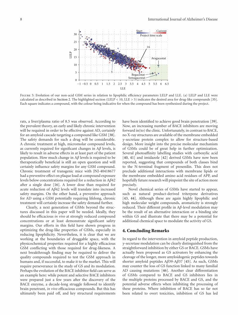
8 International Journal of Alzheimer’s Disease
30
25
20
15
10
5
00 0.5 1 1.5 2 2.5 3 3.5 4 4.5 5 5.5 6 6.5
Start Current
LELP
LLE
16
5
Avagacestat
BegacestatSemagacestat
Project time scale
−1 −0.5
Figure 5: Evolution of our non-acid GSM series in relation to lipophilic efficiency parameters LELP and LLE. (a) LELP and LLE werecalculated as described in Section 2. The highlighted section (LELP < 10, LLE > 5) indicates the desired area for drug-like compounds [35].Each square indicates a compound, with the colour being indicative for when the compound has been synthesized during the project.
rats, a liver/plasma ratio of 8.5 was observed. According tothe prevalent theory, an early and likely chronic interventionwill be required in order to be effective against AD, certainlyfor an amyloid cascade targeting a compound like GSM [38].The safety demands for such a drug will be considerable.A chronic treatment at high, micromolar compound levels,as currently required for significant changes in Aβ levels, islikely to result in adverse effects in at least part of the patientpopulation. How much change in Aβ levels is required to betherapeutically beneficial is still an open question and willcertainly influence safety margins for any GSM compound.Chronic treatment of transgenic mice with JNJ-40418677had a preventive effect on plaque load at compound exposurelevels below concentrations required for a reduction in Aβ42after a single dose [16]. A lower dose than required foracute reduction of Aβ42 levels will translate into increasedsafety margins. On the other hand, a preventive approachfor AD using a GSM potentially requiring lifelong, chronictreatment will certainly increase the safety demand further.
Clearly, a next generation of GSMs beyond the struc-tures discussed in this paper will be needed. Ideally, theyshould be efficacious in vivo at strongly reduced compoundconcentrations or at least demonstrate significant safetymargins. Our efforts in this field have shown progress inoptimizing the drug-like properties of GSMs, especially inreducing lipophilicity. Nevertheless, it is clear that we areworking at the boundaries of druggable space, with thephysicochemical properties required for a highly efficaciousGSM conflicting with those required for drug-likeness. Anext breakthrough finding may be required to deliver thequality compounds required to test the GSM approach inhumans and, if successful, to make it to the market. This willrequire perseverance in the study of GS and its modulation.Perhaps the evolution of the BACE inhibitor field can serve asan example here: while potent and selective BACE inhibitorswere prepared just a few years after the discovery of theBACE enzyme, a decade-long struggle followed to identifybrain penetrant, in vivo efficacious compounds. But this hasultimately been paid off, and key structural requirements
have been identified to achieve good brain penetration [39].Now, an increasing number of BACE inhibitors are movingforward in(to) the clinic. Unfortunately, in contrast to BACE,no X-ray structures are available of the membrane embeddedγ-secretase protein complex to allow for structure-baseddesign. More insight into the precise molecular mechanismof GSMs could be of great help in further optimization.Several photoaffinity labelling studies with carboxylic acid[40, 41] and imidazole [42] derived GSMs have now beenreported, suggesting that compounds of both classes bindto the N-terminal fragment of presenilin. This does notpreclude additional interactions with membrane lipids orthe membrane embedded amino acid residues of APP, andhopefully future research will pinpoint the site of action moreprecisely.
Novel chemical series of GSMs have started to appear,such as natural product-derived triterpene derivatives[43, 44]. Although these are again highly lipophilic andhigh molecular weight compounds, aromaticity is stronglyreduced. Their different profile in Aβ-level modulation maybe the result of an alternative interaction or a binding sitewithin GS and illustrate that there may be a potential foradditional chemical space to modulate the activity of GS.
4. Concluding Remarks
In regard to the intervention in amyloid peptide production,γ-secretase modulation can be clearly distinguished from thestraightforward inhibition by either GS or BACE. GSMs haveactually been proposed as GS activators by enhancing thecleavage of the longer, more amyloidogenic peptides towardsshorter amyloid peptides Aβ39-Aβ37 [45]. As such, GSMsmay counter the loss of GS function linked to many familialAD causing mutations [46]. Another clear differentiationof GSMs compared to BACE and GS inhibitors lies inthe multiple proteins processed by BACE and GS, and thepotential adverse effects when inhibiting the processing ofthese proteins. Where inhibition of BACE has so far notbeen related to overt toxicities, inhibition of GS has led
International Journal of Alzheimer’s Disease 9
to especially Notch-related toxicity. Based on these dis-tinctions, GS modulation deserves an optimal effort toobtain drugable, high quality GSMs. Where the principleof modulation makes on-target side effects less likely, thepotential for off-target-related side effects is considerablewith the currently required high, micromolar concentrationsof relatively lipophilic compounds. With the compoundsfrom the chemical series described in this paper, the questionin the title “can we combine potency with safety?” cannotbe answered positively yet. Nevertheless, progress towardsless lipophilic, more drug-like GSMs has been made. A nextgeneration of increased drug-likeness, potency, and/or in vivoefficacy, will be required. Further insight into the structure ofthe GS complex and the interaction with GSMs may providethe required insight on how to get to novel chemical space. Inaddition, the use of ligand efficiency parameters as discussedin this paper and other drug-likeness guidelines during theoptimization process will be crucial to ultimately deliver ahigh quality-GSM.
Acknowledgments
The authors sincerely thank the many coworkers in the neu-roscience and supporting departments who have contributedto the GSM project, including the collaborators at Cellzomeand VIB, especially Professor Bart De Strooper.
References
[1] Alzheimer’s Association, “2012 Alzheimer’s disease facts andfigures,” Alzheimer’s and Dementia, vol. 8, pp. 131–168, 2012.
[2] J. Hardy and D. J. Selkoe, “The amyloid hypothesis ofAlzheimer’s disease: progress and problems on the road to the-rapeutics,” Science, vol. 297, no. 5580, pp. 353–356, 2002.
[3] B. De Strooper, R. Vassar, and T. Golde, “The secretases:enzymes with therapeutic potential in Alzheimer disease,”Nature Reviews Neurology, vol. 6, no. 2, pp. 99–107, 2010.
[4] J. Varghese, BACE: Lead Target for Orchestrated Therapy ofAlzheimer’s Disease, John Wiley & Sons, Hoboken, NJ, USA,2010.
[5] A. F. Kreft, R. Martone, and A. Porte, “Recent advances inthe identification of γ-secretase inhibitors to clinically testthe Aβ oligomer hypothesis of Alzheimer’s disease,” Journal ofMedicinal Chemistry, vol. 52, no. 20, pp. 6169–6188, 2009.
[6] G. D’Onofrio, F. Panza, V. Frisardi et al., “Advances in theidentification of γ-secretase inhibitors for the treatment ofAlzheimer’s disease,” Expert Opinion on Drug Discovery, vol.7, no. 1, pp. 19–37, 2012.
[7] S. Weggen, J. L. Eriksen, P. Das et al., “A subset of NSAIDslower amyloidogenic Aβ42 independently of cyclooxygenaseactivity,” Nature, vol. 414, no. 6860, pp. 212–216, 2001.
[8] J. L. Eriksen, S. A. Sagi, T. E. Smith et al., “NSAIDs andenantiomers of flurbiprofen target γ-secretase and lower Aβ42in vivo,” Journal of Clinical Investigation, vol. 112, no. 3, pp.440–449, 2003.
[9] B. P. Imbimbo, “Why did tarenflurbil fail in alzheimer’s di-sease?” Journal of Alzheimer’s Disease, vol. 17, no. 4, pp. 757–760, 2009.
[10] S. Cheng, D. D. Comer, L. Mao, G. P. Balow, and D. Pleynet,“Aryl compounds and uses in modulating amyloid β,” WO2004110350, 2004.
[11] C. Nagy, E. Schuck, A. Ishibashi, Y. Nakatani, B. Rege, andV. Logovinsky, “E2012, a novel gamma-secretase modulator,decreases plasma amyloid-beta (Aβ) levels in humans,” Journalof Alzheimer’s Disease, vol. 6, no. 4, p. S574, 2010.
[12] H. Zettl, S. Weggen, P. Schneider, and G. Schneider, “Exploringthe chemical space of γ-secretase modulators,” Trends inPharmacological Sciences, vol. 31, no. 9, pp. 402–410, 2010.
[13] D. Oehlrich, D. J. C. Berthelot, and H. J. M. Gijsen, “γ-Secretase modulators as potential disease modifying anti-Alzheimer’s drugs,” Journal of Medicinal Chemistry, vol. 54, no.3, pp. 669–698, 2011.
[14] A. L. Hopkins, C. R. Groom, and A. Alex, “Ligand efficiency: auseful metric for lead selection,” Drug Discovery Today, vol. 9,no. 10, pp. 430–431, 2004.
[15] F. Bischoff, D. Berthelot, M. De Cleyn et al., “Design andsynthesis of a novel series of bicyclic heterocycles as potentgamma;-secretase modulators,” Journal of Medicinal Chem-istry, vol. 55, no. 21, pp. 9089–9106, 2012.
[16] B. Van Broeck, J. M. Chen, G. Treton et al., “Chronic treatmentwith a novel γ-secretase modulator, JNJ-40418677, inhibitsamyloid plaque formation in a mouse model of Alzheimer’sdisease,” British Journal of Pharmacology, vol. 163, no. 2, pp.375–389, 2011.
[17] H. Borghys, M. Tuefferd, B. Van Broeck et al., “A canine modelto evaluate efficacy and safety of γ-secretase inhibitors andmodulators,” Journal of Alzheimer’s Disease, vol. 28, no. 4, pp.809–822, 2012.
[18] B. P. Imbimbo, E. Del Giudice, V. Cenacchi et al., “In vitro andin vivo profiling of CHF5022 and CHF5074. Two β-amyloid1-42 lowering agents,” Pharmacological Research, vol. 55, no. 4,pp. 318–328, 2007.
[19] H. Peng, T. Talreja, Z. Xin et al., “Discovery of BIIB042, apotent, selective, and orally bioavailable γ-secretase modula-tor,” ACS Medicinal Chemistry Letters, vol. 2, no. 10, pp. 786–791, 2011.
[20] A. Hall, R. L. Elliott, G. M. P. Giblin et al., “Piperidine-derivedγ-secretase modulators,” Bioorganic and Medicinal ChemistryLetters, vol. 20, no. 3, pp. 1306–1311, 2010.
[21] M. G. Stanton, J. Hubbs, D. Sloman et al., “Fluorinatedpiperidine acetic acids as γ-secretase modulators,” Bioorganicand Medicinal Chemistry Letters, vol. 20, no. 2, pp. 755–758,2010.
[22] M. Z. Kounnas, A. M. Danks, S. Cheng et al., “Modulationof gamma-secretase reduces beta-amyloid deposition in atransgenic mouse model of Alzheimer’s disease,” Neuron, vol.67, no. 5, pp. 769–780, 2010.
[23] T. Hashimoto, A. Ishibashi, H. Hagiwara, Y. Murata, O.Takenaka, and T. Miyagawa, “E2012: a novel gamma-secretasemodulator; pharmacology part,” Alzheimer’s & Dementia, vol.6, no. 4, p. S242, 2010.
[24] Z. Wan, A. Hall, Y. Jin et al., “Pyridazine-derived γ-secretasemodulators,” Bioorganic and Medicinal Chemistry Letters, vol.21, no. 13, pp. 4016–4019, 2011.
[25] J. Qin, W. Zhou, X. Huang et al., “Discovery of a potent pyra-zolopyridine series of γ-secretase modulators,” ACS MedicinalChemistry Letters, vol. 2, no. 6, pp. 471–476, 2011.
[26] Z.-Y. Sun, T. Asberom, T. Bara et al., “Cyclic hydroxyamidinesas amide isosteres: discovery of oxadiazolines and oxadiazinesas potent and highly efficacious γ-secretase modulators invivo,” Journal of Medicinal Chemistry, vol. 55, no. 1, pp. 489–502, 2012.
[27] T. Lubbers, A. Flohr, S. Jolidon et al., “Aminothiazoles asγ-secretase modulators,” Bioorganic and Medicinal ChemistryLetters, vol. 21, no. 21, pp. 6554–6558, 2011.
10 International Journal of Alzheimer’s Disease
[28] M. Pettersson, D. S. Johnson, C. Subramanyam et al., “Designand synthesis of dihydrobenzofuran amides as orally bioavail-able, centrally active γ-secretase modulators,” Bioorganic andMedicinal Chemistry Letters, vol. 22, no. 8, pp. 2906–2911,2012.
[29] N. A. Meanwell, “Improving drug candidates by design: afocus on physicochemical properties as a means of improvingcompound disposition and safety,” Chemical Research inToxicology, vol. 24, no. 9, pp. 1420–1456, 2011.
[30] M. M. Hann and G. M. Keseu, “Finding the sweet spot: the roleof nature and nurture in medicinal chemistry,” Nature ReviewsDrug Discovery, vol. 11, no. 5, pp. 355–365, 2012.
[31] M. M. Hann, “Molecular obesity, potency and other addic-tions in drug discovery,” Medicinal Chemistry Communica-tions, vol. 2, no. 5, pp. 349–355, 2011.
[32] F. Lovering, J. Bikker, and C. Humblet, “Escape from flatland:increasing saturation as an approach to improving clinicalsuccess,” Journal of Medicinal Chemistry, vol. 52, no. 21, pp.6752–6756, 2009.
[33] P. D. Leeson and B. Springthorpe, “The influence of drug-likeconcepts on decision-making in medicinal chemistry,” NatureReviews Drug Discovery, vol. 6, no. 11, pp. 881–890, 2007.
[34] G. M. Keseru and G. M. Makara, “The influence of leaddiscovery strategies on the properties of drug candidates,”Nature Reviews Drug Discovery, vol. 8, no. 3, pp. 203–212,2009.
[35] A. Tarcsay, K. Nyıri, and G. M. Keseru, “Impact of lipophilicefficiency on compound quality,” Journal of Medicinal Chem-istry, vol. 55, no. 3, pp. 1252–1260, 2012.
[36] R. C. Green, L. S. Schneider, D. A. Amato et al., “Effect oftarenflurbil on cognitive decline and activities of daily living inpatients with mild Alzheimer disease: a randomized controlledtrial,” Journal of the American Medical Association, vol. 302, no.23, pp. 2557–2564, 2009.
[37] H. Wan, M. Rehngren, F. Giordanetto, F. Bergstrom, andA. Tunek, “High-throughput screening of drug-brain tissuebinding and in silico prediction for assessment of centralnervous system drug delivery,” Journal of Medicinal Chemistry,vol. 50, no. 19, pp. 4606–4615, 2007.
[38] E. Karran, M. Mercken, and B. D. Strooper, “The amyloidcascade hypothesis for Alzheimer’s disease: an appraisal for thedevelopment of therapeutics,” Nature Reviews Drug Discovery,vol. 10, no. 9, pp. 698–712, 2011.
[39] G. Probst and Y.-Z. Xu, “Small-molecule BACE1 inhibitors:a patent literature review (2006—2011),” Expert Opinion onTherapeutic Patents, vol. 22, no. 5, pp. 511–540, 2012.
[40] Y. Ohki, T. Higo, K. Uemura et al., “Phenylpiperidine-type γ-secretase modulators target the transmembrane domain 1 ofpresenilin 1,” EMBO Journal, vol. 30, no. 23, pp. 4815–4824,2011.
[41] T. Jumpertz, A. Rennhack, J. Ness et al., “Presenilin is themolecular target of acidic γ-secretase modulators in livingcells,” PLoS ONE, vol. 7, no. 1, Article ID e30484, 2012.
[42] A. Ebke, T. Luebbers, A. Fukumori et al., “Novel γ-secretaseenzyme modulators directly target presenilin protein,” Journalof Biological Chemistry, vol. 286, no. 43, pp. 37181–37186,2011.
[43] M. A. Findeis, F. Schroeder, T. D. McKee et al., “Discovery of anovel pharmacological and structural class of gamma secretasemodulators derived from the Extract of Actaea racemosa,” ACSChemical Neuroscience, vol. 3, no. 11, pp. 941–951, 2012.
[44] J. L. Hubbs, N. O. Fuller, W. F. Austin et al., “Optimization of anatural product-based class of gamma;-secretase modulators,”Journal of Medicinal Chemistry, vol. 55, no. 21, pp. 9270–9282,2012.
[45] L. Chevez-Gutierrez, L. Bammens, I. Benilova et al., “Themechanism of γ-Secretase dysfunction in familial Alzheimerdisease,” EMBO Journal, vol. 31, no. 10, pp. 2261–2274, 2012.
[46] B. De Strooper and W. Annaert, “Novel research horizonsfor presenilins and γ-secretases in cell biology and disease,”Annual Review of Cell and Developmental Biology, vol. 26, pp.235–260, 2010.


![Counterfactual Model for Online Systems€¦ · Definition [IPS Utility Estimator]: Given 𝑆= 1, 1,𝛿1,…, 𝑛, 𝑛,𝛿𝑛 collected under 𝜋0, Unbiased estimate of utility](https://static.fdocument.org/doc/165x107/605dfba17555fe2bcb505bac/counterfactual-model-for-online-definition-ips-utility-estimator-given-.jpg)