β-Glucan exacerbates allergic airway responses to house ...
3
LETTER TO THE EDITOR Open Access β-Glucan exacerbates allergic airway responses to house dust mite allergen Sabelo Hadebe 1 , Frank Kirstein 2 , Kaat Fierens 3 , Pierre Redelinghuys 1 , Graeme I. Murray 4 , David L. Williams 5 , Bart N. Lambrecht 3,6 , Frank Brombacher 2 and Gordon D. Brown 1,7* Abstract β-(1,3)-Glucan is present in mould cell walls and frequently detected in house dust mite (HDM) faeces. β-Glucan exposure is thought to be associated with pulmonary allergic inflammation in mouse and man, although the published data are inconsistent. Here, we show that highly purified β-glucan exacerbates HDM-induced eosinophilic, T helper 2 type airway responses by acting as an adjuvant, promoting activation, proliferation and polarisation of HDM-specific T cells (1-Derβ T cells). We therefore provide definitive evidence that β-glucan can influence allergic pulmonary inflammation. Keywords: β-glucans, Allergy, Eosinophil, T helper 2, House dust mite Results Asthma is a common chronic obstructive airway disease, which presents as episodes of wheeze, shortness of breath and chest tightness, and in extreme cases the dis- ease can be fatal [1, 2]. It is traditionally a disease of the developed world, with increasing incidence both in childhood and adulthood [3]. Asthma is widely regarded as a T helper 2 (Th2) cell-mediated disease, although other forms exist [2]. Th2 type asthma can be charac- terised by eosinophil accumulation in the alveolar space and cytokines, including interleukin (IL-) IL-4, IL-5 and IL-13, as well as by other physiological changes such as goblet cell hyperplasia [1]. The underlying factors contributing to the disease are numerous and not well understood. Environmental allergen sensitisation is known to play a major part in asthma development and exacerbation. Fungal spores are one of many environ- mental allergens encountered daily and their exposure directly correlates with increased incidence of asthma episodes and hospital admission [4]. β-(1,3)-Glucan (β- glucan) is a pathogen-associated molecular pattern (PAMP) mainly present in fungal cell walls, but also present in bacteria, plants and has been detected in house dust mite (HDM) faeces [5]. β-Glucan has been implicated in both innate and allergic respiratory inflam- matory responses, however, studies in both human and animal models are inconsistent [6]. These discrepancies are due, in large part, to the purity and solubility of the β-glucan preparations used [6, 7]. Here, we made use of highly purified particulate β- glucans of similar size to fungal spores, but without contaminating agonists [8] and have investigated their ef- fects on pulmonary inflammation in the context of HDM- induced responses. For these experiments, we sensitised C57BL/6 mice intratracheally (i.t) with HDM alone or with HDM together with β-glucan and subsequently chal- lenged these mice i.t. with HDM only or PBS as a control (Fig. 1a). Mice sensitised and challenged with HDM alone developed eosinophilic pulmonary inflammation in the bronchoalveolar lavage fluid (BALF) (Fig. 1b), as previ- ously shown [9]. However, when HDM plus β-glucan sen- sitised mice were challenged with HDM, they developed a more profound pulmonary inflammation, characterised by significantly higher numbers of eosinophils (Fig. 1b). There were also slight, but significant, increases in the numbers of neutrophils, monocytes/macrophages and T- cells (Fig. 1b; note the difference in scales). Consistent with these observations, higher levels of IL-4, IL-5, IL-13 and IL-17 were detected in the BALF of mice sensitised * Correspondence: [email protected] 1 Aberdeen Fungal Group, Infection, Immunity and Inflammation Programme, University of Aberdeen, Aberdeen, UK 7 Aberdeen Fungal Group, MRC Centre for Medical Mycology, Infection, Immunity and Inflammation Programme, School of Medicine & Dentistry, Institute of Medical Sciences, University of Aberdeen, Foresterhill, Aberdeen AB25 2ZD, UK Full list of author information is available at the end of the article © 2016 Hadebe et al. Open Access This article is distributed under the terms of the Creative Commons Attribution 4.0 International License (http://creativecommons.org/licenses/by/4.0/), which permits unrestricted use, distribution, and reproduction in any medium, provided you give appropriate credit to the original author(s) and the source, provide a link to the Creative Commons license, and indicate if changes were made. The Creative Commons Public Domain Dedication waiver (http://creativecommons.org/publicdomain/zero/1.0/) applies to the data made available in this article, unless otherwise stated. Hadebe et al. Respiratory Research (2016) 17:35 DOI 10.1186/s12931-016-0352-5
Transcript of β-Glucan exacerbates allergic airway responses to house ...
LETTER TO THE EDITOR Open Access
β-Glucan exacerbates allergic airwayresponses to house dust mite allergenSabelo Hadebe1, Frank Kirstein2, Kaat Fierens3, Pierre Redelinghuys1, Graeme I. Murray4, David L. Williams5,Bart N. Lambrecht3,6, Frank Brombacher2 and Gordon D. Brown1,7*
Abstract
β-(1,3)-Glucan is present in mould cell walls and frequently detected in house dust mite (HDM) faeces. β-Glucanexposure is thought to be associated with pulmonary allergic inflammation in mouse and man, although thepublished data are inconsistent. Here, we show that highly purified β-glucan exacerbates HDM-inducedeosinophilic, T helper 2 type airway responses by acting as an adjuvant, promoting activation, proliferation andpolarisation of HDM-specific T cells (1-Derβ T cells). We therefore provide definitive evidence that β-glucan caninfluence allergic pulmonary inflammation.
Keywords: β-glucans, Allergy, Eosinophil, T helper 2, House dust mite
ResultsAsthma is a common chronic obstructive airway disease,which presents as episodes of wheeze, shortness ofbreath and chest tightness, and in extreme cases the dis-ease can be fatal [1, 2]. It is traditionally a disease of thedeveloped world, with increasing incidence both inchildhood and adulthood [3]. Asthma is widely regardedas a T helper 2 (Th2) cell-mediated disease, althoughother forms exist [2]. Th2 type asthma can be charac-terised by eosinophil accumulation in the alveolar spaceand cytokines, including interleukin (IL-) IL-4, IL-5 andIL-13, as well as by other physiological changes such asgoblet cell hyperplasia [1]. The underlying factorscontributing to the disease are numerous and not wellunderstood. Environmental allergen sensitisation isknown to play a major part in asthma development andexacerbation. Fungal spores are one of many environ-mental allergens encountered daily and their exposuredirectly correlates with increased incidence of asthmaepisodes and hospital admission [4]. β-(1,3)-Glucan (β-glucan) is a pathogen-associated molecular pattern
(PAMP) mainly present in fungal cell walls, but alsopresent in bacteria, plants and has been detected inhouse dust mite (HDM) faeces [5]. β-Glucan has beenimplicated in both innate and allergic respiratory inflam-matory responses, however, studies in both human andanimal models are inconsistent [6]. These discrepanciesare due, in large part, to the purity and solubility of theβ-glucan preparations used [6, 7].Here, we made use of highly purified particulate β-
glucans of similar size to fungal spores, but withoutcontaminating agonists [8] and have investigated their ef-fects on pulmonary inflammation in the context of HDM-induced responses. For these experiments, we sensitisedC57BL/6 mice intratracheally (i.t) with HDM alone orwith HDM together with β-glucan and subsequently chal-lenged these mice i.t. with HDM only or PBS as a control(Fig. 1a). Mice sensitised and challenged with HDM alonedeveloped eosinophilic pulmonary inflammation in thebronchoalveolar lavage fluid (BALF) (Fig. 1b), as previ-ously shown [9]. However, when HDM plus β-glucan sen-sitised mice were challenged with HDM, they developed amore profound pulmonary inflammation, characterised bysignificantly higher numbers of eosinophils (Fig. 1b).There were also slight, but significant, increases in thenumbers of neutrophils, monocytes/macrophages and T-cells (Fig. 1b; note the difference in scales). Consistentwith these observations, higher levels of IL-4, IL-5, IL-13and IL-17 were detected in the BALF of mice sensitised
* Correspondence: [email protected] Fungal Group, Infection, Immunity and Inflammation Programme,University of Aberdeen, Aberdeen, UK7Aberdeen Fungal Group, MRC Centre for Medical Mycology, Infection,Immunity and Inflammation Programme, School of Medicine & Dentistry,Institute of Medical Sciences, University of Aberdeen, Foresterhill, AberdeenAB25 2ZD, UKFull list of author information is available at the end of the article
© 2016 Hadebe et al. Open Access This article is distributed under the terms of the Creative Commons Attribution 4.0International License (http://creativecommons.org/licenses/by/4.0/), which permits unrestricted use, distribution, andreproduction in any medium, provided you give appropriate credit to the original author(s) and the source, provide a link tothe Creative Commons license, and indicate if changes were made. The Creative Commons Public Domain Dedication waiver(http://creativecommons.org/publicdomain/zero/1.0/) applies to the data made available in this article, unless otherwise stated.
Hadebe et al. Respiratory Research (2016) 17:35 DOI 10.1186/s12931-016-0352-5
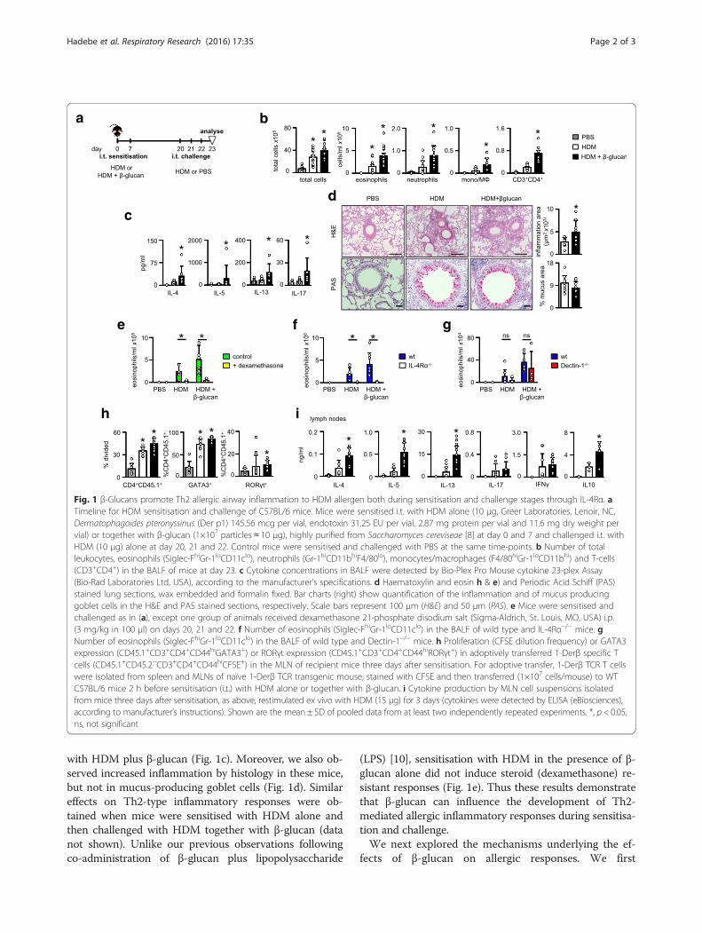
with HDM plus β-glucan (Fig. 1c). Moreover, we also ob-served increased inflammation by histology in these mice,but not in mucus-producing goblet cells (Fig. 1d). Similareffects on Th2-type inflammatory responses were ob-tained when mice were sensitised with HDM alone andthen challenged with HDM together with β-glucan (datanot shown). Unlike our previous observations followingco-administration of β-glucan plus lipopolysaccharide
(LPS) [10], sensitisation with HDM in the presence of β-glucan alone did not induce steroid (dexamethasone) re-sistant responses (Fig. 1e). Thus these results demonstratethat β-glucan can influence the development of Th2-mediated allergic inflammatory responses during sensitisa-tion and challenge.We next explored the mechanisms underlying the ef-
fects of β-glucan on allergic responses. We first
a
c
b
d
e
h
f
i
g
Fig. 1 β-Glucans promote Th2 allergic airway inflammation to HDM allergen both during sensitisation and challenge stages through IL-4Rα. aTimeline for HDM sensitisation and challenge of C57BL/6 mice. Mice were sensitised i.t. with HDM alone (10 μg, Greer Laboratories, Lenoir, NC,Dermatophagoides pteronyssinus (Der p1) 145.56 mcg per vial, endotoxin 31.25 EU per vial, 2.87 mg protein per vial and 11.6 mg dry weight pervial) or together with β-glucan (1×10⁷ particles ≈ 10 μg), highly purified from Saccharomyces cereviseae [8] at day 0 and 7 and challenged i.t. withHDM (10 μg) alone at day 20, 21 and 22. Control mice were sensitised and challenged with PBS at the same time-points. b Number of totalleukocytes, eosinophils (Siglec-FhiGr-1loCD11clo), neutrophils (Gr-1hiCD11bhiF4/80lo), monocytes/macrophages (F4/80hiGr-1loCD11bhi) and T-cells(CD3+CD4+) in the BALF of mice at day 23. c Cytokine concentrations in BALF were detected by Bio-Plex Pro Mouse cytokine 23-plex Assay(Bio-Rad Laboratories Ltd, USA), according to the manufacturer’s specifications. d Haematoxylin and eosin h & e) and Periodic Acid Schiff (PAS)stained lung sections, wax embedded and formalin fixed. Bar charts (right) show quantification of the inflammation and of mucus producinggoblet cells in the H&E and PAS stained sections, respectively. Scale bars represent 100 μm (H&E) and 50 μm (PAS). e Mice were sensitised andchallenged as in (a), except one group of animals received dexamethasone 21-phosphate disodium salt (Sigma-Aldrich, St. Louis, MO, USA) i.p.(3 mg/kg in 100 μl) on days 20, 21 and 22. f Number of eosinophils (Siglec-FhiGr-1loCD11clo) in the BALF of wild type and IL-4Rα−/− mice. gNumber of eosinophils (Siglec-FhiGr-1loCD11clo) in the BALF of wild type and Dectin-1−/− mice. h Proliferation (CFSE dilution frequency) or GATA3expression (CD45.1+CD3+CD4+CD44hiGATA3+) or RORγt expression (CD45.1+CD3+CD4+CD44hiRORγt+) in adoptively transferred 1-Derβ specific Tcells (CD45.1+CD45.2−CD3+CD4+CD44hiCFSE+) in the MLN of recipient mice three days after sensitisation. For adoptive transfer, 1-Derβ TCR T cellswere isolated from spleen and MLNs of naïve 1-Derβ TCR transgenic mouse, stained with CFSE and then transferred (1×10⁷ cells/mouse) to WTC57BL/6 mice 2 h before sensitisation (i.t.) with HDM alone or together with β-glucan. i Cytokine production by MLN cell suspensions isolatedfrom mice three days after sensitisation, as above, restimulated ex vivo with HDM (15 μg) for 3 days (cytokines were detected by ELISA (eBiosciences),according to manufacturer’s instructions). Shown are the mean ± SD of pooled data from at least two independently repeated experiments. *, p < 0.05,ns, not significant
Hadebe et al. Respiratory Research (2016) 17:35 Page 2 of 3
determined if IL-4 receptor α (IL-4Rα) was essentiallyrequired for the β-glucan-mediated effects observed inour model [11]. Indeed, loss of IL-4Rα completely abro-gated the eosinophilic airway inflammation in the pres-ence of β-glucans (Fig. 1f ). We then determined if theexacerbated responses induced by β-glucan were beingmediated by the major beta-glucan receptor, Dectin-1[12]. Unexpectedly, we found that loss of this receptorhad no significant effect on the enhanced eosinophilicresponse induced by β-glucans (Fig. 1g). This suggeststhat other systems are mediating these activities, orcompensating for the loss of Dectin-1, such as CR3 and/or complement [7, 13].To gain further insights, we next explored allergic T-cell
responses by making use of a T cell receptor (TCR) trans-genic (Tg) mouse that recognises an immuno-dominantpeptide from the HDM-derived allergen, Derp-1 (1-DerβTg) [14]. We found that adoptively transferred naïve 1-Derβ T cells proliferated in mice sensitised with HDMalone, but proliferated more in mice sensitised with HDMplus β-glucan (Fig. 1h). Moreover, in mice sensitised withHDM plus β-glucan, adoptively transferred 1-Derβ T cellsexpressed higher intracellular levels of the transcriptionalfactor GATA3 compared to 1-Derβ T cells from HDMsensitised mice or PBS controls (Fig. 1h). This enhancedTh2 polarisation of HDM-specific T cells could also bedemonstrated by the increased levels of relevant cytokines,including IL-4, IL-5 and IL-13 that were produced by ex-vivo HDM stimulated MLNs (Fig. 1i). There was a slightincrease in RORγT in 1-Derβ T cells from mice sensitisedwith HDM plus β-glucan, which did not translate into in-creased levels of IL-17 upon restimulation in vitro. Therewas no significant effect of β-glucan on IFN-γ production,but these carbohydrates did increase the production of IL-10 (Fig. 1i). Although we cannot exclude some contribu-tion from innate lymphoid cells [15], we show here thatparticulate β-glucans exacerbate airway inflammation toHDM by promoting HDM-specific T cell priming.
AbbreviationsAHR: Airway hyper-reactivity; HDM: House dust mite; IL: interleukin;PAMPs: Pathogen-associated molecular patterns; Th2: T helper 2.
Competing interestsThe authors declare that they have no competing interests.
Authors’ contributionsConceived and designed the experiments: GDB, FB. Performed the experiments:SH, FK, PR, KF. Analysed the data: GIM, SH, BL, FB, GDB. Contributed reagents/materials/analysis tools: BL, DLW. Wrote the paper: SH, GDB. All authorsdiscussed the results and commented on the manuscript. All read andapproved the final manuscript.
AcknowledgementsWe also thank Animal Unit staff for care of the animals used in this study.Funding: The authors thank the Wellcome Trust (102705) and the Universitiesof Aberdeen and Cape Town for funding. DLW is partly supported byNational Institutes of Health GM53522, GM083016 and C06RR0306551. KFand BNL are funded by the Fonds Wetenschappelijk Onderzoek, BNL is the
recipient of an European Research Commission consolidator grant andparticipates in the European Union FP7 programs EUBIOPRED and MedALL.The funders had no role in study design, data collection.
Author details1Aberdeen Fungal Group, Infection, Immunity and Inflammation Programme,University of Aberdeen, Aberdeen, UK. 2International Centre for GeneticEngineering and Biotechnology and Division of Immunology, Institute ofInfectious Disease and Molecular Medicine, Faculty of Health Science,University of Cape Town, Cape Town, South Africa. 3VIB InflammationResearch Center, Laboratory of Immunoregulation and Mucosal Immunology,University Ghent, Ghent 9000, Belgium. 4Pathology, Division of AppliedMedicine, Institute of Medical Sciences, Foresterhill, University of Aberdeen,Aberdeen AB25 2ZD, UK. 5Department of Surgery and Center forInflammation, Infectious Disease and Immunity, James H. Quillen College ofMedicine, East Tennessee State University, Johnson City, TN, USA.6Department of Pulmonary Medicine, Erasmus MC, Rotterdam, TheNetherlands. 7Aberdeen Fungal Group, MRC Centre for Medical Mycology,Infection, Immunity and Inflammation Programme, School of Medicine &Dentistry, Institute of Medical Sciences, University of Aberdeen, Foresterhill,Aberdeen AB25 2ZD, UK.
Received: 22 January 2016 Accepted: 25 March 2016
References1. Barnes PJ. Immunology of asthma and chronic obstructive pulmonary
disease. Nat Rev Immunol. 2008;8:183–92.2. Fahy JV. Type 2 inflammation in asthma–present in most, absent in many.
Nat Rev Immunol. 2015;15:57–65.3. Lambrecht BN, Hammad H. The immunology of asthma. Nat Immunol.
2015;16:45–56.4. O’Driscoll BR, Hopkinson LC, Denning DW. Mold sensitization is common
amongst patients with severe asthma requiring multiple hospitaladmissions. BMC Pulm Med. 2005;5:4.
5. Netea MG, Brown GD, Kullberg BJ, Gow NA. An integrated model of therecognition of Candida albicans by the innate immune system. Nat RevMicrobiol. 2008;6:67–78.
6. Douwes J. (1→3)-Beta-D-glucans and respiratory health: a review of thescientific evidence. Indoor Air. 2005;15:160–9.
7. Brown GD, Gordon S. Fungal beta-glucans and mammalian immunity.Immunity. 2003;19:311–5.
8. Dennehy KM, Ferwerda G, Faro-Trindade I, Pyz E, Willment JA, Taylor PR,Kerrigan A, Tsoni SV, Gordon S, Meyer-Wentrup F, et al. Syk kinase isrequired for collaborative cytokine production induced through Dectin-1and Toll-like receptors. Eur J Immunol. 2008;38:500–6.
9. Hammad H, Chieppa M, Perros F, Willart MA, Germain RN, Lambrecht BN.House dust mite allergen induces asthma via Toll-like receptor 4 triggeringof airway structural cells. Nat Med. 2009;15:410–6.
10. Hadebe S, Kirstein F, Fierens K, Chen K, Drummond RA, Vautier S, Sajaniemi S,Murray G, Williams DL, Redelinghuys P, et al. Microbial Ligand CostimulationDrives Neutrophilic Steroid-Refractory Asthma. PLoS One. 2015;10, e0134219.
11. Grunig G, Warnock M, Wakil AE, Venkayya R, Brombacher F, Rennick DM,Sheppard D, Mohrs M, Donaldson DD, Locksley RM, Corry DB. Requirement forIL-13 independently of IL-4 in experimental asthma. Science. 1998;282:2261–3.
12. Brown GD, Gordon S. Immune recognition: A new receptor for beta-glucans.Nature. 2001;413:36–7.
13. McDonald JU, Rosas M, Brown GD, Jones SA, Taylor PR. Differentialdependencies of monocytes and neutrophils on dectin-1, dectin-2 andcomplement for the recognition of fungal particles in inflammation. PLoSOne. 2012;7, e45781.
14. Plantinga M, Guilliams M, Vanheerswynghels M, Deswarte K, Branco-MadeiraF, Toussaint W, Vanhoutte L, Neyt K, Killeen N, Malissen B, et al. Conventional andMonocyte-Derived CD11b +Dendritic Cells Initiate and Maintain T Helper 2 Cell-Mediated Immunity to House Dust Mite Allergen. Immunity. 2013;38:322–35.
15. Klein Wolterink RG, Kleinjan A, Van Nimwegen M, Bergen I, De Bruijn M,Levani Y, Hendriks RW. Pulmonary innate lymphoid cells are majorproducers of IL-5 and IL-13 in murine models of allergic asthma. Eur JImmunol. 2012;42:1106–16.
Hadebe et al. Respiratory Research (2016) 17:35 Page 3 of 3


![Biochimica et Biophysica Acta - COnnecting REpositories · chronic and acute airway inflammation [2,3]. In this regard, especially terpenoids,likethe monoterpene oxide1,8-cineol](https://static.fdocument.org/doc/165x107/5f0a739a7e708231d42bb33b/biochimica-et-biophysica-acta-connecting-repositories-chronic-and-acute-airway.jpg)