Primary Cell e Newsletter Issue 18 (jun 2016) Cell Applications
Biochemical and Biophysical Research Communications 450 (2014) 1475–1480
Contents lists available at ScienceDirect
Biochemical and Biophysical Research Communications
journal homepage: www.elsevier .com/locate /ybbrc
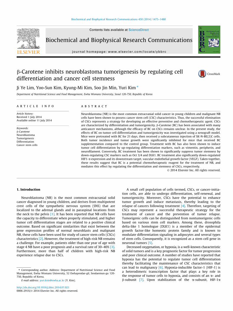
b-Carotene inhibits neuroblastoma tumorigenesis by regulating celldifferentiation and cancer cell stemness
http://dx.doi.org/10.1016/j.bbrc.2014.07.0210006-291X/� 2014 Elsevier Inc. All rights reserved.
⇑ Corresponding author. Address: Department of Nutritional Science and FoodManagement, Ewha Womans University, 52 Ewhayeodae-gil, Seodaemun-gu 120-750, Republic of Korea.
E-mail address: [email protected] (Y. Kim).
Ji Ye Lim, Yoo-Sun Kim, Kyung-Mi Kim, Soo Jin Min, Yuri Kim ⇑Department of Nutritional Science and Food Management, Ewha Womans University, Seoul 120-750, Republic of Korea
a r t i c l e i n f o
Article history:Received 1 July 2014Available online 11 July 2014
Keywords:b-CaroteneNeuroblastomaTumorigenesisDifferentiationCancer stem cells
a b s t r a c t
Neuroblastoma (NB) is the most common extracranial solid cancer in young children and malignant NBcells have been shown to possess cancer stem cell (CSC) characteristics. Thus, the successful eliminationof CSCs represents a strategy for developing an effective preventive and chemotherapeutic agent. CSCsare characterized by differentiation and tumorigenicity. b-Carotene (BC) has been associated with manyanticancer mechanisms, although the efficacy of BC on CSCs remains unclear. In the present study, theeffects of BC on tumor cell differentiation and tumorigenicity was investigated using a xenograft model.Mice were pretreated with BC for 21 days, then received a subcutaneous injection of SK-N-BE(2)C cells.Both tumor incidence and tumor growth were significantly inhibited for mice that received BCsupplementation compared to the control group. Treatment with BC has also been shown to inducetumor cell differentiation by up-regulating differentiation markers, such as vimentin, peripherin, andneurofilament. Conversely, BC treatment has been shown to significantly suppress tumor stemness bydown-regulating CSC markers such as Oct 3/4 and DLK1. BC treatment also significantly down-regulatedHIF1-a expression and its downstream target, vascular endothelial growth factor (VEGF). Taken together,these results suggest that BC is a potential chemotherapeutic reagent for the treatment of NB, andmediates this effect by regulating the differentiation and stemness of CSCs, respectively.
� 2014 Elsevier Inc. All rights reserved.
1. Introduction
Neuroblastoma (NB) is the most common extracranial solidcancer diagnosed in young children, and derives from multipotentcrest cells of the sympathetic nervous system (SNS) that arelocalized to the adrenal glands and in paraspinal locations fromthe neck to the pelvis [1]. It has been reported that NB cells havethe capacity to differentiate when properly stimulated, and highertumor cell differentiation stages are related to a positive clinicaloutcome. Based on significant similarities that exist between thegene expression profiles of normal neuroblasts and malignantNB, these cells have been used for study of cancer stem cells (CSCs)characteristics [2]. However, the treatment of high-risk NB remainsa challenge. For example, patients older than one year of age withstage 4 NB have a poor prognosis and a survival rate of 30–40% [3].Furthermore, more than half of children with high-risk NBexperience relapse due to CSCs.
A small cell population of cells termed, CSCs, or cancer-initia-tion cells, are able to undergo differentiation, self-renewal, andtumorigenicity. Moreover, CSCs have the potential to enhancetumor growth and induce metastasis, thereby leading to therelapse of cancers following treatment [4]. Therefore, targeting ofCSCs may represent a successful therapeutic strategy for thetreatment of cancer and the prevention of tumor relapse.Tumorigenic cells can be distinguished from nontumorigenic cellsbased on various stem cell markers. Among these, drosophiladelta-like 1 homologue (DLK1) is a member of the epidermalgrowth factor-like homeotic protein family and is known tomodulate differentiation signaling in adipocytes and several typesof stem cells. Consequently, it is recognized as a stem cell gene inneuronal tumors [5].
Decreased oxygenation, or hypoxia, is a well-known characteristicof solid tumors and is a key prognostic factor for tumor progressionand poor clinical outcome. A number of studies have reported thathypoxia has the potential to regulate tumor cell differentiationthereby facilitating the maintenance of CSC characteristics thatcan lead to malignancy [6]. Hypoxia-inducible factor-1 (HIF-1) isa heterodimeric transcription factor that plays a key role inthe response of tumor cells to hypoxia, and consists of an a- andb-subunit [7]. Upon stabilization of the a-subunit, HIF-1a

1476 J.Y. Lim et al. / Biochemical and Biophysical Research Communications 450 (2014) 1475–1480
translocates to the nucleus to form an HIF-1a/b heterodimer andtransactivate target gene expression. One of these targets includesvascular endothelial growth factor (VEGF) [8]. Thus, regulation ofhypoxia may represent an important therapeutic strategy fortargeting malignant cancers.
b-Carotene (BC) is a well-known vitamin A precursor that isfound in many fruits and vegetables that are dark green or orangein color. Numerous observational studies have found that a highdietary intake of fruits and vegetables rich in BC is associated witha reduced cancer risk for several types of cancers. Accumulatingevidence also suggests that BC induces the differentiation of cancercells, including melanoma and leukemia cancer cells [9,10]. In ourrecent study, BC was found to induce the differentiation, andsuppress the cancer stemness, of malignant SK-N-BE(2)C [BE(2)C]NB cells in vitro [2]. The anti-metastatic effect of BC was also dem-onstrated both in vitro and in vivo for NBs [11]. However, little isknown about the role of BC in the tumorigenesis of CSCs. Therefore,in the present study, the effect of BC on tumor cell differentiationand tumorigenicity was investigated using a xenograft model.
2. Materials and methods
2.1. Cell culture
The human NB cell line, BE(2)C, was purchased from AmericanType Culture Collection (ATCC, Rockville, MD) and cultured in a 1:1mixture of Minimum Essential Medium (MEM, Welgene, Daegu,Korea) and Ham’s F-12 (Welgene, Daegu, Korea) medium contain-ing 10% fetal bovine serum (FBS, Hyclone, Logan, UT) and 1%penicillin–streptomycin (100 U/ml and 100 lg/ml, respectively;Invitrogen, Carlsbad, CA). The cells were maintained in humidifiedair at 37 �C with 5% CO2. b-Carotene was purchased from SigmaChemical Co. (St. Louis, MO).
2.2. In vivo tumor xenograft model
Male five-week-old BALB/c nude mice were purchased fromCentral Lab, Animal Inc. (Seoul, Korea) and were maintained underspecific pathogen-free conditions (College of Pharmacy, EwhaWomans University, Seoul, Korea). Weight and food intake weremonitored twice a week. After a one-week acclimation period,mice were randomly assigned to three groups (10 animals pergroup) as follows; (a) tumor control group (Ctrl): oral supplemen-tation with corn oil as a vehicle; (b) BC 2 group (BC 2): oral supple-mentation with 2 mg/kg b.w. of BC; (c) BC 6 group (BC 6): oralsupplementation with 6 mg/kg b.w of BC, respectively. The micereceived either corn oil, or each dose of BC in corn oil, orally twicea week for seven weeks. On day 21 of the BC treatment regimen,mice were subcutaneously injected with 4.0 � 105 BE(2)C cellssuspended in growth factor-reduced matrigel (BD Bioscience Labo-ratory, Bedford, MA) into the right flank region. After the tumorswere established, tumor volume [volume = length (mm) �width2
(mm2) � 0.5] was measured twice a week using digital calipers.Mice were sacrificed four weeks later, with pimonidazole (60 mg/kg) administered 1 h prior to sacrifice. Tumors were counted andexcised. After measuring the size and weight of each tumor, theywere immediately frozen or immersed in 4% neutral bufferedformaldehyde. Animal care and experimental protocols for thisstudy were approved by the Animal Care and Use Committee ofEwha Womans University (IACUC approval No: 2013-01-028).
2.3. Reverse transcription-PCR and quantitative PCR analysis
PCR amplification of cDNA was performed using Taq polymerase(TAKARA, Tokyo, Japan). The resulting PCR products were separatedin a 2% agarose gel containing ethidium bromide. For each sample,
quantitative PCR analysis detected b-actin as an internal control.The primers used included: 50-AGCA CCC ATG GCA GAA GG-30
(forward) and 50-CTC GAT TGG ATG GCA GTA CT-30 (reverse) forhuman VEGF; and 50-ATT GGC AAT GAG CGG TTC-30 (forward) and50-GGA TGC CAC AGG ACT CCA T-30 (reverse) for b-actin.
2.4. Western blot analysis
Western blot assays were performed as described previously[12]. Primary antibodies raised against peripherin (Millipore, Bille-rica, MA), vimentin (Cell Signaling Technology, Boston, MA), Oct 3/4 (Santa Cruz Biotechnology, Santa Cruz, CA), HIF-1a (Novus, Little-ton, CO), and a-tubulin (Sigma Aldrich, St. Louis, MO) were used forthe experiment.
2.5. Immunohistochemistry
Immunohistochemistry staining for neurofilament (DAKO, Ely,UK), DLK1 (Millipore, Billerica, MA), and HIF-1a antibody (Novus,Littleton, CO) was performed as described previously [11].
2.6. Statistical analysis
All data are presented as the mean ± standard error of the mean(SEM) and were analyzed using GraphPad PRISM software (Graph-Pad Software, San Diego, CA). Pearson’s Chi-square or one-wayanalysis of variance (ANOVA) followed by Tukey’s post hoc testwas performed. A P-value less than 0.05 was considered statisti-cally significant.
3. Results
3.1. BC supplementation reduces tumor volume and tumor incidence
At the time of sacrifice, 10 out of 10 Ctrl mice developed tumors,while only 4 out of 10 mice in BC 6 groups developed tumors(Table 1). Thus, BC supplementation inhibited tumorigenesis by60%. Furthermore, the tumors formed in the BC 6 group were smal-ler than those formed in Ctrl and BC 2 mouse groups. For example,the mean tumor volume for the Ctrl group was 997.6 ± 703.0 mm3
compared with 226.2 ± 335.2 mm3 for the BC 6 group. This repre-sented a 77% decrease in tumor volume for the BC 6 group. TheBC 2 group also exhibited a smaller tumor volume compared withthe Ctrl group, however, the difference was not statistically signif-icant. These data suggest that BC supplementation may inhibittumor growth in vivo.
3.2. BC supplementation induces cell differentiation in vivo
It was previously demonstrated that BC inhibited cancer cellstemness by inducing the differentiation of NB cells in vitro [2].To investigate the anti-tumorigenic effect of BC supplementationin vivo, the cell differentiation markers, peripherin and vimentinwere assayed. In the BC 6 tumor tissues, expression of the neuronaldifferentiation marker, peripherin was significantly up-regulated(Fig. 1A). Vimentin, a marker of cytodifferentiation, was also up-regulated in the BC 6 tumor tissues compared to the Ctrl tissues(Fig. 1B). Similarly, expression of peripherin and vimentin werehigher in BC 2 tissues compared to the Ctrl tissues, although thedifferences were not statistically significant.
To confirm these results, expression of neurofilament was ana-lyzed by immunohistochemistry. Neurofilament is a protein whichexhibits limited expression only in mature neurons. In Ctrltumor tissues, levels of neurofilament were very low (Fig. 1C). Incontrast, BC 2 and BC 6 tumor tissues expressed higher levels of

Fig. 1. BC supplementation induces cell differentiation in vivo. (A and B) Expression levels of peripherin (A) and vimentin (B) were detected in tumor tissue extracts byWestern blot. Detection of a-tubulin was used as a loading control. Representative figures are shown (upper panel). Quantification of peripherin levels is shown (lower panel).(C) Immunohistochemical staining of neurofilament was performed for the tumor sections indicated. Stained slides were imaged at 400� magnification. The scale barrepresents 4 lm. Neurofilament positive cells were counted and quantified. Bars represent the mean ± SEM. Bars labeled with different letters differ significantly (P < 0.05).Ctrl, tumor control; BC 2, tumor injection + BC 2 mg/kg b.w; BC 6, tumor injection + BC 6 mg/kg b.w.
Table 1BC suppresses tumor incidence and final tumor volume1.
Treatment group Total number of mice Tumor incidence Final tumor volume (mm3)
TC 10 10/10 997.60 ± 703.02a
BC 2 8 8/8 563.90 ± 504.71a
BC 6 10 4/10 226.22 ± 335.22b
The different groups with different alphabets are significantly different (P < 0.05).1 Number of mice with tumor incidence and final tumor volume of mice injected with BE(2)C cells. Comparisons among groups were conducted by chi-square (P < 0.05).
J.Y. Lim et al. / Biochemical and Biophysical Research Communications 450 (2014) 1475–1480 1477
neurofilament. Taken together, these results suggest that BCsuppresses tumor formation by NB cells by inducing cell differen-tiation via regulation of the differentiation markers, peripherin,vimentin, and neurofilament.
3.3. BC suppresses CSC marker expression by NB cells in vivo
CSCs have the potential to induce tumor growth and progres-sion [4]. Correspondingly, it has been reported that BC inhibitsCSC characteristics, such as self-renewal, in NB cells in vitro [2].To understand the effect of BC on cancer cell stemness in NB cellsin vivo, the expression of several CSC markers were analyzed usingWestern blot and immunohistochemistry assays (Fig. 2). Expres-sion levels of Oct 3/4, a hallmark for CSC-like cells, were signifi-cantly lower in BC 6 group compared to Ctrl group (Fig. 2A). Incontrast, BC 2 group exhibited lower levels of Oct 3/4 expressioncompared to the Ctrl group, although the difference was notstatistically significant. DLK1 is a neuronal stem cell marker whichis important for the maintenance of stem cells [13]. Immunohisto-chemical detection of DLK1 showed that the number of DLK1positive-stained cells were significantly lower in both BC 2 andBC 6 group compared with those in Ctrl group (Fig. 2B). These
results suggest that BC supplementation suppresses cancer cellstemness by down-regulating several CSC markers.
3.4. BC supplementation suppresses the expression of HIF-1a and itsdownstream target VEGF in vivo
NB typically grow under hypoxic conditions and hypoxictumors exhibit more malignant characteristics [14]. Correspond-ingly, hypoxia has been shown to promote tumor growth and toinhibit differentiation [14]. To investigate whether BC supplemen-tation suppresses tumor growth by regulating levels of HIF-1a,immunohistochemistry and Western blot assays were performed.In tumors, HIF-1a positive cells were significantly higher in Ctrlgroup compared with BC 6 group (Fig. 3A). Quantification ofHIF-1a positive cells represented that BC 6 supplementationsuppressed HIF-1a expression. Consistent with the immunohisto-chemistry results, higher levels of HIF-1a expression were alsodetected in Ctrl tumor tissue extracts compared to BC 6 tissueextracts that were analyzed by Western blot (Fig. 3B). While BC2 group also exhibited lower levels of HIF-1a, the difference fromCtrl group was not statistically significant. VEGF is a hypoxia-inducible gene [13]. mRNA levels of VEGF were analyzed using
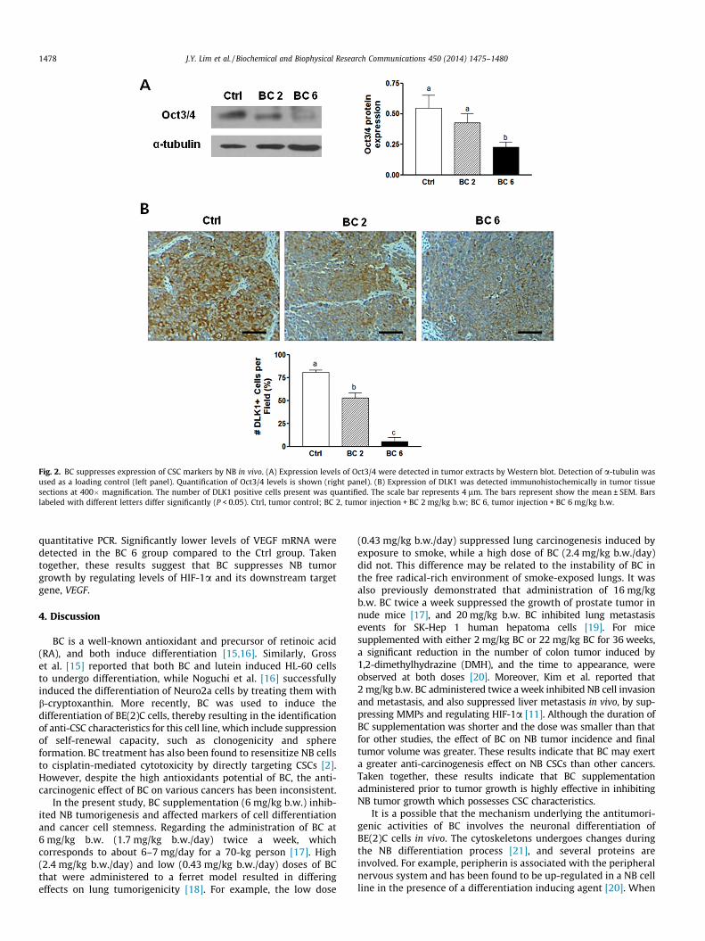
Fig. 2. BC suppresses expression of CSC markers by NB in vivo. (A) Expression levels of Oct3/4 were detected in tumor extracts by Western blot. Detection of a-tubulin wasused as a loading control (left panel). Quantification of Oct3/4 levels is shown (right panel). (B) Expression of DLK1 was detected immunohistochemically in tumor tissuesections at 400� magnification. The number of DLK1 positive cells present was quantified. The scale bar represents 4 lm. The bars represent show the mean ± SEM. Barslabeled with different letters differ significantly (P < 0.05). Ctrl, tumor control; BC 2, tumor injection + BC 2 mg/kg b.w; BC 6, tumor injection + BC 6 mg/kg b.w.
1478 J.Y. Lim et al. / Biochemical and Biophysical Research Communications 450 (2014) 1475–1480
quantitative PCR. Significantly lower levels of VEGF mRNA weredetected in the BC 6 group compared to the Ctrl group. Takentogether, these results suggest that BC suppresses NB tumorgrowth by regulating levels of HIF-1a and its downstream targetgene, VEGF.
4. Discussion
BC is a well-known antioxidant and precursor of retinoic acid(RA), and both induce differentiation [15,16]. Similarly, Grosset al. [15] reported that both BC and lutein induced HL-60 cellsto undergo differentiation, while Noguchi et al. [16] successfullyinduced the differentiation of Neuro2a cells by treating them withb-cryptoxanthin. More recently, BC was used to induce thedifferentiation of BE(2)C cells, thereby resulting in the identificationof anti-CSC characteristics for this cell line, which include suppressionof self-renewal capacity, such as clonogenicity and sphereformation. BC treatment has also been found to resensitize NB cellsto cisplatin-mediated cytotoxicity by directly targeting CSCs [2].However, despite the high antioxidants potential of BC, the anti-carcinogenic effect of BC on various cancers has been inconsistent.
In the present study, BC supplementation (6 mg/kg b.w.) inhib-ited NB tumorigenesis and affected markers of cell differentiationand cancer cell stemness. Regarding the administration of BC at6 mg/kg b.w. (1.7 mg/kg b.w./day) twice a week, whichcorresponds to about 6–7 mg/day for a 70-kg person [17]. High(2.4 mg/kg b.w./day) and low (0.43 mg/kg b.w./day) doses of BCthat were administered to a ferret model resulted in differingeffects on lung tumorigenicity [18]. For example, the low dose
(0.43 mg/kg b.w./day) suppressed lung carcinogenesis induced byexposure to smoke, while a high dose of BC (2.4 mg/kg b.w./day)did not. This difference may be related to the instability of BC inthe free radical-rich environment of smoke-exposed lungs. It wasalso previously demonstrated that administration of 16 mg/kgb.w. BC twice a week suppressed the growth of prostate tumor innude mice [17], and 20 mg/kg b.w. BC inhibited lung metastasisevents for SK-Hep 1 human hepatoma cells [19]. For micesupplemented with either 2 mg/kg BC or 22 mg/kg BC for 36 weeks,a significant reduction in the number of colon tumor induced by1,2-dimethylhydrazine (DMH), and the time to appearance, wereobserved at both doses [20]. Moreover, Kim et al. reported that2 mg/kg b.w. BC administered twice a week inhibited NB cell invasionand metastasis, and also suppressed liver metastasis in vivo, by sup-pressing MMPs and regulating HIF-1a [11]. Although the duration ofBC supplementation was shorter and the dose was smaller than thatfor other studies, the effect of BC on NB tumor incidence and finaltumor volume was greater. These results indicate that BC may exerta greater anti-carcinogenesis effect on NB CSCs than other cancers.Taken together, these results indicate that BC supplementationadministered prior to tumor growth is highly effective in inhibitingNB tumor growth which possesses CSC characteristics.
It is a possible that the mechanism underlying the antitumori-genic activities of BC involves the neuronal differentiation ofBE(2)C cells in vivo. The cytoskeletons undergoes changes duringthe NB differentiation process [21], and several proteins areinvolved. For example, peripherin is associated with the peripheralnervous system and has been found to be up-regulated in a NB cellline in the presence of a differentiation inducing agent [20]. When
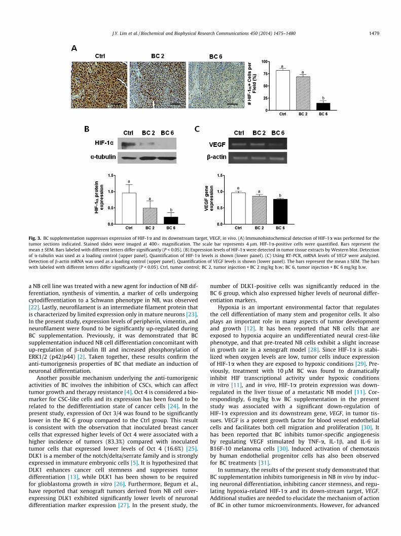
Fig. 3. BC supplementation suppresses expression of HIF-1a and its downstream target, VEGF, in vivo. (A) Immunohistochemical detection of HIF-1a was performed for thetumor sections indicated. Stained slides were imaged at 400� magnification. The scale bar represents 4 lm. HIF-1a-positive cells were quantified. Bars represent themean ± SEM. Bars labeled with different letters differ significantly (P < 0.05). (B) Expression levels of HIF-1a were detected in tumor tissue extracts by Western blot. Detectionof a-tubulin was used as a loading control (upper panel). Quantification of HIF-1a levels is shown (lower panel). (C) Using RT-PCR, mRNA levels of VEGF were analyzed.Detection of b-actin mRNA was used as a loading control (upper panel). Quantification of VEGF levels is shown (lower panel). The bars represent the mean ± SEM. The barswith labeled with different letters differ significantly (P < 0.05). Ctrl, tumor control; BC 2, tumor injection + BC 2 mg/kg b.w; BC 6, tumor injection + BC 6 mg/kg b.w.
J.Y. Lim et al. / Biochemical and Biophysical Research Communications 450 (2014) 1475–1480 1479
a NB cell line was treated with a new agent for induction of NB dif-ferentiation, synthesis of vimentin, a marker of cells undergoingcytodifferentiation to a Schwann phenotype in NB, was observed[22]. Lastly, neurofilament is an intermediate filament protein thatis characterized by limited expression only in mature neurons [23].In the present study, expression levels of peripherin, vimentin, andneurofilament were found to be significantly up-regulated duringBC supplementation. Previously, it was demonstrated that BCsupplementation induced NB cell differentiation concomitant withup-regulation of b-tubulin III and increased phosphorylation ofERK1/2 (p42/p44) [2]. Taken together, these results confirm theanti-tumorigenesis properties of BC that mediate an induction ofneuronal differentiation.
Another possible mechanism underlying the anti-tumorigenicactivities of BC involves the inhibition of CSCs, which can affecttumor growth and therapy resistance [4]. Oct 4 is considered a bio-marker for CSC-like cells and its expression has been found to berelated to the dedifferentiation state of cancer cells [24]. In thepresent study, expression of Oct 3/4 was found to be significantlylower in the BC 6 group compared to the Ctrl group. This resultis consistent with the observation that inoculated breast cancercells that expressed higher levels of Oct 4 were associated with ahigher incidence of tumors (83.3%) compared with inoculatedtumor cells that expressed lower levels of Oct 4 (16.6%) [25].DLK1 is a member of the notch/delta/serrate family and is stronglyexpressed in immature embryonic cells [5]. It is hypothesized thatDLK1 enhances cancer cell stemness and suppresses tumordifferentiation [13], while DLK1 has been shown to be requiredfor glioblastoma growth in vitro [26]. Furthermore, Begum et al.,have reported that xenograft tumors derived from NB cell over-expressing DLK1 exhibited significantly lower levels of neuronaldifferentiation marker expression [27]. In the present study, the
number of DLK1-positive cells was significantly reduced in theBC 6 group, which also expressed higher levels of neuronal differ-entiation markers.
Hypoxia is an important environmental factor that regulatesthe cell differentiation of many stem and progenitor cells. It alsoplays an important role in many aspects of tumor developmentand growth [12]. It has been reported that NB cells that areexposed to hypoxia acquire an undifferentiated neural crest-likephenotype, and that pre-treated NB cells exhibit a slight increasein growth rate in a xenograft model [28]. Since HIF-1a is stabi-lized when oxygen levels are low, tumor cells induce expressionof HIF-1a when they are exposed to hypoxic conditions [29]. Pre-viously, treatment with 10 lM BC was found to dramaticallyinhibit HIF transcriptional activity under hypoxic conditionsin vitro [11], and in vivo, HIF-1a protein expression was down-regulated in the liver tissue of a metastatic NB model [11]. Cor-respondingly, 6 mg/kg b.w BC supplementation in the presentstudy was associated with a significant down-regulation ofHIF-1a expression and its downstream gene, VEGF, in tumor tis-sues. VEGF is a potent growth factor for blood vessel endothelialcells and facilitates both cell migration and proliferation [30]. Ithas been reported that BC inhibits tumor-specific angiogenesisby regulating VEGF stimulated by TNF-a, IL-1b, and IL-6 inB16F-10 melanoma cells [30]. Induced activation of chemotaxisby human endothelial progenitor cells has also been observedfor BC treatments [31].
In summary, the results of the present study demonstrated thatBC supplementation inhibits tumorigenesis in NB in vivo by induc-ing neuronal differentiation, inhibiting cancer stemness, and regu-lating hypoxia-related HIF-1a and its down-stream target, VEGF.Additional studies are needed to elucidate the mechanism of actionof BC in other tumor microenvironments. However, for advanced

1480 J.Y. Lim et al. / Biochemical and Biophysical Research Communications 450 (2014) 1475–1480
NB, it appears that BC has the potential to mediate chemothera-peutic effects.
Acknowledgments
This research was supported by Basic Science Research Programthrough the National Research Foundation of Korea (NRF) fundedby the Ministry of Education (project no. 2011-0008169).
References
[1] B.H. Kushner, Neuroblastoma: a disease requiring a multitude of imagingstudies, J. Nucl. Med. 45 (2004) 1172–1188.
[2] H.A. Lee, S. Park, Y. Kim, Effect of beta-carotene on cancer cell stemness anddifferentiation in SK-N-BE(2)C neuroblastoma cells, Oncol. Rep. 30 (2013)1869–1877.
[3] J.M. Maris, M.D. Hogarty, R. Bagatell, S.L. Cohn, Neuroblastoma, Lancet 369(2007) 2106–2120.
[4] M.F. Clarke, J.E. Dick, P.B. Dirks, C.J. Eaves, C.H. Jamieson, D.L. Jones, J. Visvader,I.L. Weissman, G.M. Wahl, Cancer stem cells – perspectives on current statusand future directions: AACR workshop on cancer stem cells, Cancer Res. 66(2006) 9339–9344.
[5] C.M. Smas, H.S. Sul, Pref-1, a protein containing EGF-like repeats, inhibitsadipocyte differentiation, Cell 73 (1993) 725–734.
[6] Q. Lin, Z. Yun, Impact of the hypoxic tumor microenvironment on theregulation of cancer stem cell characteristics, Cancer Biol. Ther. 9 (2010)949–956.
[7] M. Puppo, F. Battaglia, C. Ottaviano, S. Delfino, D. Ribatti, L. Varesio, M.C. Bosco,Topotecan inhibits vascular endothelial growth factor production andangiogenic activity induced by hypoxia in human neuroblastoma bytargeting hypoxia-inducible factor-1alpha and -2alpha, Mol. Cancer Ther. 7(2008) 1974–1984.
[8] C. Brahimi-Horn, J. Pouyssegur, The role of the hypoxia-inducible factor intumor metabolism growth and invasion, Bull. Cancer 93 (2006) E73–E80.
[9] M.B. Hazuka, J. Edwards-Prasad, F. Newman, J.J. Kinzie, K.N. Prasad, Beta-carotene induces morphological differentiation and decreases adenylatecyclase activity in melanoma cells in culture, J. Am. Coll. Nutr. 9 (1990) 143–149.
[10] P.K. Duitsman, A.B. Barua, B. Becker, J.A. Olson, Effects of epoxycarotenoids,beta-carotene, and retinoic acid on the differentiation and viability of theleukemia cell line NB4 in vitro, Int. J. Vitam. Nutr. Res. 69 (1999) 303–308.
[11] Y.S. Kim, H.A. Lee, J.Y. Lim, Y. Kim, C.H. Jung, S.H. Yoo, Y. Kim, Beta-caroteneinhibits neuroblastoma cell invasion and metastasis in vitro and in vivo bydecreasing level of hypoxia-inducible factor-1alpha, J. Nutr. Biochem. 25(2014) 655–664.
[12] Q. Lin, Y.J. Lee, Z. Yun, Differentiation arrest by hypoxia, J. Biol. Chem. 281(2006) 30678–30683.
[13] Y. Kim, Q. Lin, D. Zelterman, Z. Yun, Hypoxia-regulated delta-like 1 homologueenhances cancer cell stemness and tumorigenicity, Cancer Res. 69 (2009)9271–9280.
[14] L. Holmquist, T. Lofstedt, S. Pahlman, Effect of hypoxia on the tumorphenotype: the neuroblastoma and breast cancer models, Adv. Exp. Med.Biol. 587 (2006) 179–193.
[15] M.D. Gross, T.D. Bishop, J.D. Belcher, D.R. Jacobs Jr., Induction of HL-60 celldifferentiation by carotenoids, Nutr. Cancer 27 (1997) 169–173.
[16] S. Noguchi, T. Sumida, H. Ogawa, M. Tada, K. Takahata, Effects of oxygenatedcarotenoid beta-cryptoxanthin on morphological differentiation and apoptosisin Neuro2a neuroblastoma cells, Biosci. Biotechnol. Biochem. 67 (2003) 2467–2469.
[17] C.M. Yang, Y.T. Yen, C.S. Huang, M.L. Hu, Growth inhibitory efficacy of lycopeneand beta-carotene against androgen-independent prostate tumor cellsxenografted in nude mice, Mol. Nutr. Food Res. 55 (2011) 606–612.
[18] C. Liu, X.D. Wang, R.T. Bronson, D.E. Smith, N.I. Krinsky, R.M. Russell, Effects ofphysiological versus pharmacological beta-carotene supplementation on cellproliferation and histopathological changes in the lungs of cigarette smoke-exposed ferrets, Carcinogenesis 21 (2000) 2245–2253.
[19] C.S. Huang, J.W. Liao, M.L. Hu, Lycopene inhibits experimental metastasis ofhuman hepatoma SK-Hep-1 cells in athymic nude mice, J. Nutr. 138 (2008)538–543.
[20] N.J. Temple, T.K. Basu, Protective effect of beta-carotene against colon tumorsin mice, J. Natl. Cancer Inst. 78 (1987) 1211–1214.
[21] J.E. Oh, K. Karlmark Raja, J.H. Shin, A. Pollak, M. Hengstschlager, G. Lubec,Cytoskeleton changes following differentiation of N1E-115 neuroblastoma cellline, Amino Acids 31 (2006) 289–298.
[22] M.M. Portier, B. Edde, F. Berthelot, B. Croizat, F. Gros, Effects on thecytoskeleton of a new inducer of the neuroblastoma morphologicaldifferentiation, Biochem. Biophys. Res. Commun. 96 (1980) 1610–1618.
[23] T. Sugimoto, T. Kato, T. Sawada, Y. Horii, J.T. Kemshead, T. Hino, H. Morioka, H.Hosoi, Schwannian cell differentiation of human neuroblastoma cell linesin vitro induced by bromodeoxyuridine, Cancer Res. 48 (1988) 2531–2537.
[24] Y. Chen, L. Shi, L. Zhang, R. Li, J. Liang, W. Yu, L. Sun, X. Yang, Y. Wang, Y. Zhang,Y. Shang, The molecular mechanism governing the oncogenic potential ofSOX2 in breast cancer, J. Biol. Chem. 283 (2008) 17969–17978.
[25] R.J. Kim, J.S. Nam, OCT4 expression enhances features of cancer stem cells in amouse model of breast cancer, Lab. Anim. Res. 27 (2011) 147–152.
[26] D. Yin, D. Xie, S. Sakajiri, C.W. Miller, H. Zhu, M.L. Popoviciu, J.W. Said, K.L.Black, H.P. Koeffler, DLK1: increased expression in gliomas and associated withoncogenic activities, Oncogene 25 (2006) 1852–1861.
[27] A. Begum, Y. Kim, Q. Lin, Z. Yun, DLK1, delta-like 1 homolog (Drosophila),regulates tumor cell differentiation in vivo, Cancer Lett. 318 (2012) 26–33.
[28] A. Jogi, I. Ora, H. Nilsson, A. Lindeheim, Y. Makino, L. Poellinger, H. Axelson, S.Pahlman, Hypoxia alters gene expression in human neuroblastoma cellstoward an immature and neural crest-like phenotype, Proc. Natl. Acad. Sci.U.S.A. 99 (2002) 7021–7026.
[29] M.S. Wiesener, H. Turley, W.E. Allen, C. Willam, K.U. Eckardt, K.L. Talks, S.M.Wood, K.C. Gatter, A.L. Harris, C.W. Pugh, P.J. Ratcliffe, P.H. Maxwell, Inductionof endothelial PAS domain protein-1 by hypoxia: characterization andcomparison with hypoxia-inducible factor-1alpha, Blood 92 (1998) 2260–2268.
[30] C. Guruvayoorappan, G. Kuttan, Beta-carotene inhibits tumor-specificangiogenesis by altering the cytokine profile and inhibits the nucleartranslocation of transcription factors in B16F-10 melanoma cells, Integr.Cancer Ther. 6 (2007) 258–270.
[31] B. Kiec-Wilk, A. Polus, J. Grzybowska, M. Mikolajczyk, J. Hartwich, J. Pryjma, J.Skrzeczynska, A. Dembinska-Kiec, Beta-carotene stimulates chemotaxis ofhuman endothelial progenitor cells, Clin. Chem. Lab. Med. 43 (2005) 488–498.